Lei Li
Carnegie Mellon University
Cross-modal Contrastive Learning for Speech Translation
May 05, 2022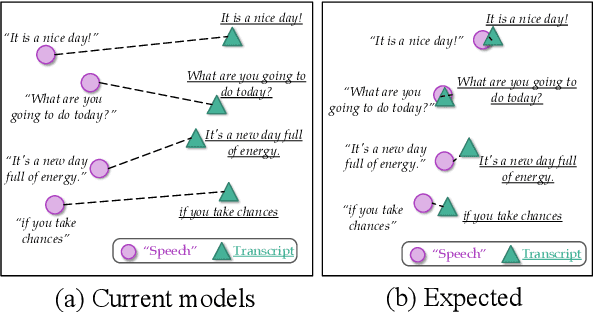
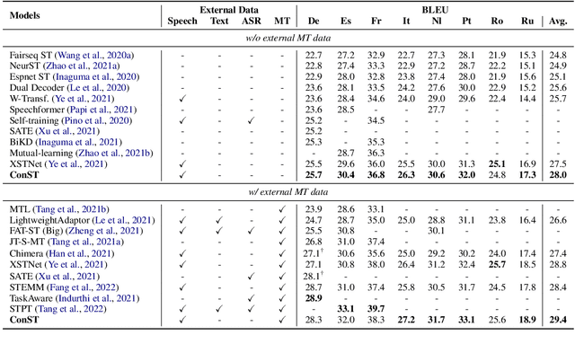
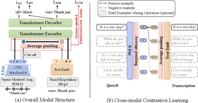
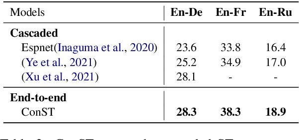
Abstract:How can we learn unified representations for spoken utterances and their written text? Learning similar representations for semantically similar speech and text is important for speech translation. To this end, we propose ConST, a cross-modal contrastive learning method for end-to-end speech-to-text translation. We evaluate ConST and a variety of previous baselines on a popular benchmark MuST-C. Experiments show that the proposed ConST consistently outperforms the previous methods on, and achieves an average BLEU of 29.4. The analysis further verifies that ConST indeed closes the representation gap of different modalities -- its learned representation improves the accuracy of cross-modal speech-text retrieval from 4% to 88%. Code and models are available at https://github.com/ReneeYe/ConST.
Hybrid Transformer with Multi-level Fusion for Multimodal Knowledge Graph Completion
May 04, 2022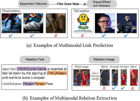
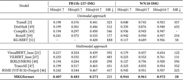
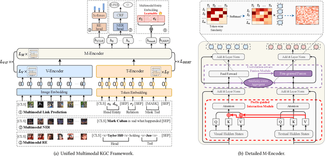
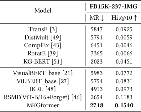
Abstract:Multimodal Knowledge Graphs (MKGs), which organize visual-text factual knowledge, have recently been successfully applied to tasks such as information retrieval, question answering, and recommendation system. Since most MKGs are far from complete, extensive knowledge graph completion studies have been proposed focusing on the multimodal entity, relation extraction and link prediction. However, different tasks and modalities require changes to the model architecture, and not all images/objects are relevant to text input, which hinders the applicability to diverse real-world scenarios. In this paper, we propose a hybrid transformer with multi-level fusion to address those issues. Specifically, we leverage a hybrid transformer architecture with unified input-output for diverse multimodal knowledge graph completion tasks. Moreover, we propose multi-level fusion, which integrates visual and text representation via coarse-grained prefix-guided interaction and fine-grained correlation-aware fusion modules. We conduct extensive experiments to validate that our MKGformer can obtain SOTA performance on four datasets of multimodal link prediction, multimodal RE, and multimodal NER. Code is available in https://github.com/zjunlp/MKGformer.
Relation Extraction as Open-book Examination: Retrieval-enhanced Prompt Tuning
May 04, 2022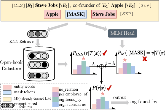

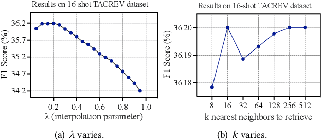
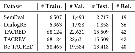
Abstract:Pre-trained language models have contributed significantly to relation extraction by demonstrating remarkable few-shot learning abilities. However, prompt tuning methods for relation extraction may still fail to generalize to those rare or hard patterns. Note that the previous parametric learning paradigm can be viewed as memorization regarding training data as a book and inference as the close-book test. Those long-tailed or hard patterns can hardly be memorized in parameters given few-shot instances. To this end, we regard RE as an open-book examination and propose a new semiparametric paradigm of retrieval-enhanced prompt tuning for relation extraction. We construct an open-book datastore for retrieval regarding prompt-based instance representations and corresponding relation labels as memorized key-value pairs. During inference, the model can infer relations by linearly interpolating the base output of PLM with the non-parametric nearest neighbor distribution over the datastore. In this way, our model not only infers relation through knowledge stored in the weights during training but also assists decision-making by unwinding and querying examples in the open-book datastore. Extensive experiments on benchmark datasets show that our method can achieve state-of-the-art in both standard supervised and few-shot settings. Code are available in https://github.com/zjunlp/PromptKG/tree/main/research/RetrievalRE.
Provably Confidential Language Modelling
May 04, 2022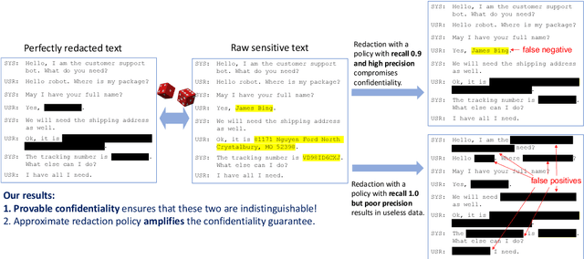

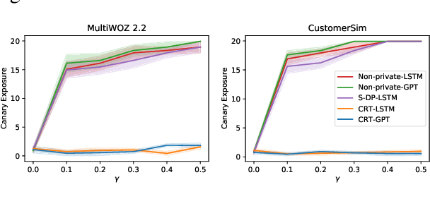
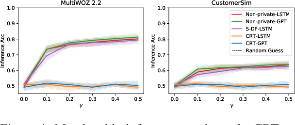
Abstract:Large language models are shown to memorize privacy information such as social security numbers in training data. Given the sheer scale of the training corpus, it is challenging to screen and filter these privacy data, either manually or automatically. In this paper, we propose Confidentially Redacted Training (CRT), a method to train language generation models while protecting the confidential segments. We borrow ideas from differential privacy (which solves a related but distinct problem) and show that our method is able to provably prevent unintended memorization by randomizing parts of the training process. Moreover, we show that redaction with an approximately correct screening policy amplifies the confidentiality guarantee. We implement the method for both LSTM and GPT language models. Our experimental results show that the models trained by CRT obtain almost the same perplexity while preserving strong confidentiality.
EasyNLP: A Comprehensive and Easy-to-use Toolkit for Natural Language Processing
Apr 30, 2022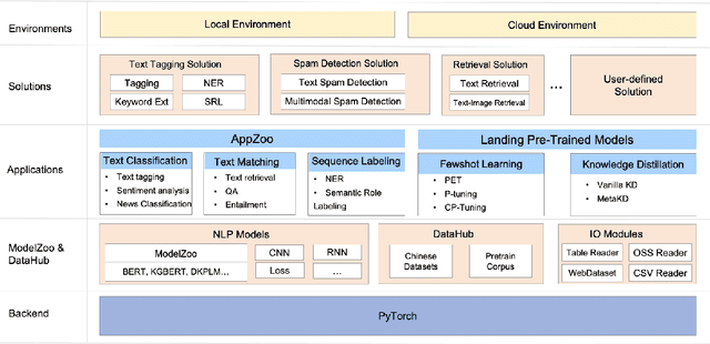
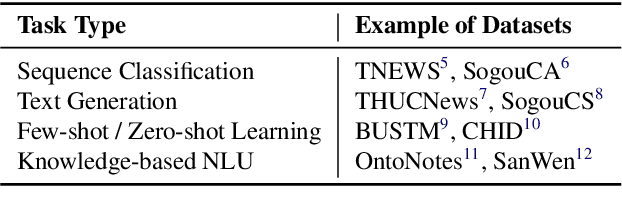


Abstract:The success of Pre-Trained Models (PTMs) has reshaped the development of Natural Language Processing (NLP). Yet, it is not easy to obtain high-performing models and deploy them online for industrial practitioners. To bridge this gap, EasyNLP is designed to make it easy to build NLP applications, which supports a comprehensive suite of NLP algorithms. It further features knowledge-enhanced pre-training, knowledge distillation and few-shot learning functionalities for large-scale PTMs, and provides a unified framework of model training, inference and deployment for real-world applications. Currently, EasyNLP has powered over ten business units within Alibaba Group and is seamlessly integrated to the Platform of AI (PAI) products on Alibaba Cloud. The source code of our EasyNLP toolkit is released at GitHub (https://github.com/alibaba/EasyNLP).
Learning Design and Construction with Varying-Sized Materials via Prioritized Memory Resets
Apr 12, 2022


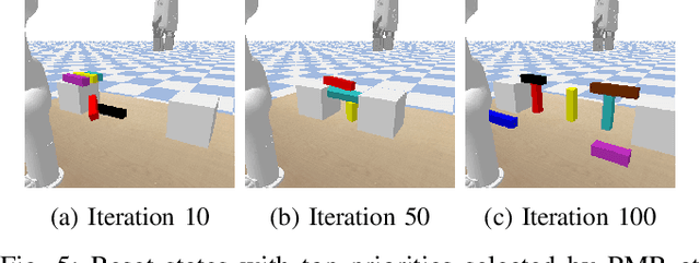
Abstract:Can a robot autonomously learn to design and construct a bridge from varying-sized blocks without a blueprint? It is a challenging task with long horizon and sparse reward -- the robot has to figure out physically stable design schemes and feasible actions to manipulate and transport blocks. Due to diverse block sizes, the state space and action trajectories are vast to explore. In this paper, we propose a hierarchical approach for this problem. It consists of a reinforcement-learning designer to propose high-level building instructions and a motion-planning-based action generator to manipulate blocks at the low level. For high-level learning, we develop a novel technique, prioritized memory resetting (PMR) to improve exploration. PMR adaptively resets the state to those most critical configurations from a replay buffer so that the robot can resume training on partial architectures instead of from scratch. Furthermore, we augment PMR with auxiliary training objectives and fine-tune the designer with the locomotion generator. Our experiments in simulation and on a real deployed robotic system demonstrate that it is able to effectively construct bridges with blocks of varying sizes at a high success rate. Demos can be found at https://sites.google.com/view/bridge-pmr.
Confidence Estimation Transformer for Long-term Renewable Energy Forecasting in Reinforcement Learning-based Power Grid Dispatching
Apr 10, 2022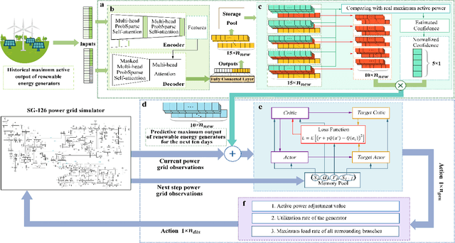
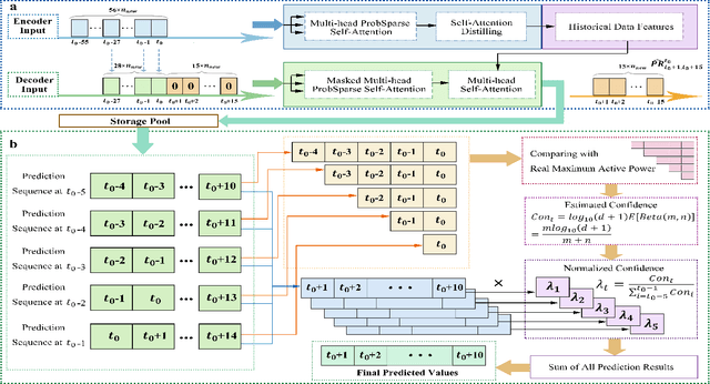
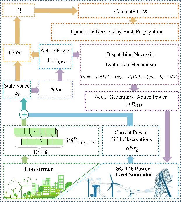

Abstract:The expansion of renewable energy could help realizing the goals of peaking carbon dioxide emissions and carbon neutralization. Some existing grid dispatching methods integrating short-term renewable energy prediction and reinforcement learning (RL) have been proved to alleviate the adverse impact of energy fluctuations risk. However, these methods omit the long-term output prediction, which leads to stability and security problems on the optimal power flow. This paper proposes a confidence estimation Transformer for long-term renewable energy forecasting in reinforcement learning-based power grid dispatching (Conformer-RLpatching). Conformer-RLpatching predicts long-term active output of each renewable energy generator with an enhanced Transformer to boost the performance of hybrid energy grid dispatching. Furthermore, a confidence estimation method is proposed to reduce the prediction error of renewable energy. Meanwhile, a dispatching necessity evaluation mechanism is put forward to decide whether the active output of a generator needs to be adjusted. Experiments carried out on the SG-126 power grid simulator show that Conformer-RLpatching achieves great improvement over the second best algorithm DDPG in security score by 25.8% and achieves a better total reward compared with the golden medal team in the power grid dispatching competition sponsored by State Grid Corporation of China under the same simulation environment. Codes are outsourced in https://github.com/buptlxh/Conformer-RLpatching.
Contextual Representation Learning beyond Masked Language Modeling
Apr 08, 2022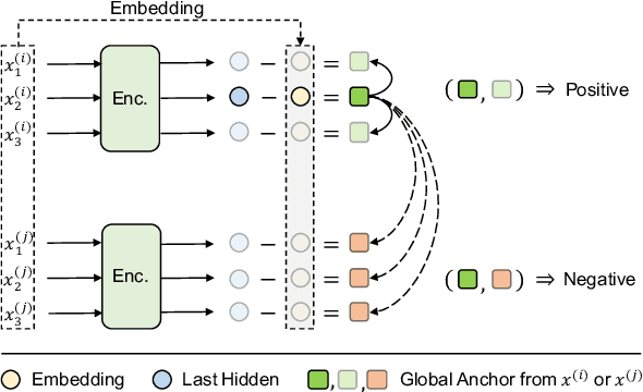

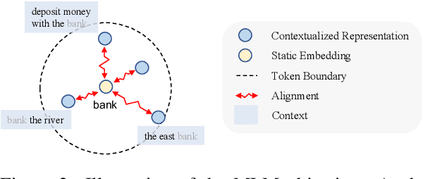
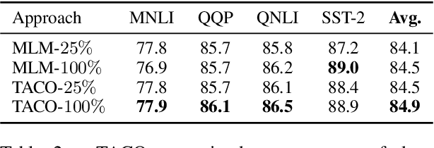
Abstract:How do masked language models (MLMs) such as BERT learn contextual representations? In this work, we analyze the learning dynamics of MLMs. We find that MLMs adopt sampled embeddings as anchors to estimate and inject contextual semantics to representations, which limits the efficiency and effectiveness of MLMs. To address these issues, we propose TACO, a simple yet effective representation learning approach to directly model global semantics. TACO extracts and aligns contextual semantics hidden in contextualized representations to encourage models to attend global semantics when generating contextualized representations. Experiments on the GLUE benchmark show that TACO achieves up to 5x speedup and up to 1.2 points average improvement over existing MLMs. The code is available at https://github.com/FUZHIYI/TACO.
$\textit{latent}$-GLAT: Glancing at Latent Variables for Parallel Text Generation
Apr 05, 2022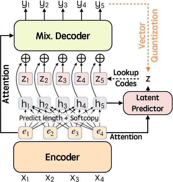

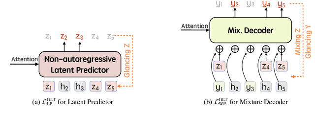
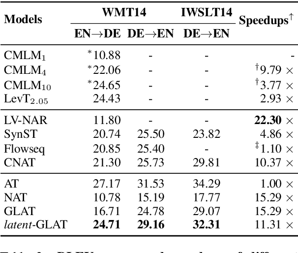
Abstract:Recently, parallel text generation has received widespread attention due to its success in generation efficiency. Although many advanced techniques are proposed to improve its generation quality, they still need the help of an autoregressive model for training to overcome the one-to-many multi-modal phenomenon in the dataset, limiting their applications. In this paper, we propose $\textit{latent}$-GLAT, which employs the discrete latent variables to capture word categorical information and invoke an advanced curriculum learning technique, alleviating the multi-modality problem. Experiment results show that our method outperforms strong baselines without the help of an autoregressive model, which further broadens the application scenarios of the parallel decoding paradigm.
STEMM: Self-learning with Speech-text Manifold Mixup for Speech Translation
Mar 20, 2022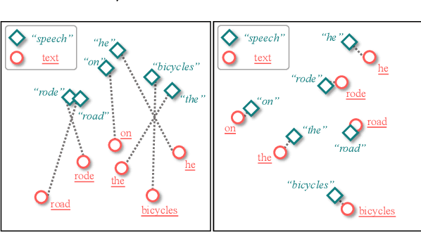
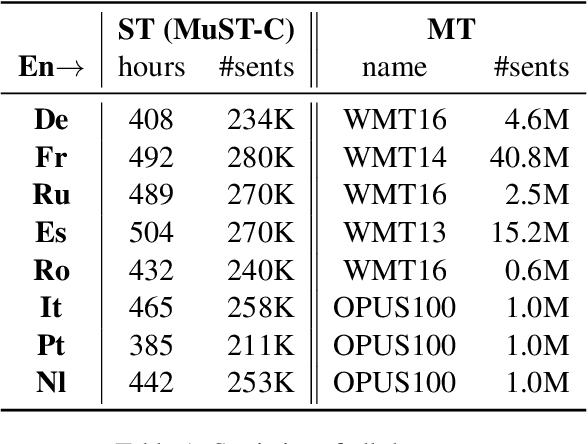
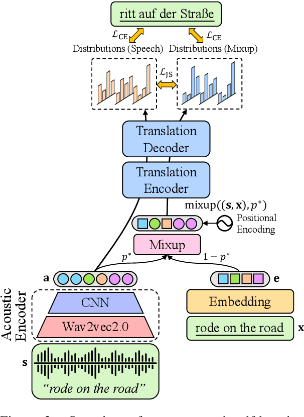
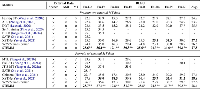
Abstract:How to learn a better speech representation for end-to-end speech-to-text translation (ST) with limited labeled data? Existing techniques often attempt to transfer powerful machine translation (MT) capabilities to ST, but neglect the representation discrepancy across modalities. In this paper, we propose the Speech-TExt Manifold Mixup (STEMM) method to calibrate such discrepancy. Specifically, we mix up the representation sequences of different modalities, and take both unimodal speech sequences and multimodal mixed sequences as input to the translation model in parallel, and regularize their output predictions with a self-learning framework. Experiments on MuST-C speech translation benchmark and further analysis show that our method effectively alleviates the cross-modal representation discrepancy, and achieves significant improvements over a strong baseline on eight translation directions.
 Add to Chrome
 Add to Firefox
 Add to Edge