Xiangde Luo
University of Electronic Science and Technology of China, Chengdu, China, ShangAI Laboratory, Shanghai, China
3D TransUNet: Advancing Medical Image Segmentation through Vision Transformers
Oct 11, 2023Abstract:Medical image segmentation plays a crucial role in advancing healthcare systems for disease diagnosis and treatment planning. The u-shaped architecture, popularly known as U-Net, has proven highly successful for various medical image segmentation tasks. However, U-Net's convolution-based operations inherently limit its ability to model long-range dependencies effectively. To address these limitations, researchers have turned to Transformers, renowned for their global self-attention mechanisms, as alternative architectures. One popular network is our previous TransUNet, which leverages Transformers' self-attention to complement U-Net's localized information with the global context. In this paper, we extend the 2D TransUNet architecture to a 3D network by building upon the state-of-the-art nnU-Net architecture, and fully exploring Transformers' potential in both the encoder and decoder design. We introduce two key components: 1) A Transformer encoder that tokenizes image patches from a convolution neural network (CNN) feature map, enabling the extraction of global contexts, and 2) A Transformer decoder that adaptively refines candidate regions by utilizing cross-attention between candidate proposals and U-Net features. Our investigations reveal that different medical tasks benefit from distinct architectural designs. The Transformer encoder excels in multi-organ segmentation, where the relationship among organs is crucial. On the other hand, the Transformer decoder proves more beneficial for dealing with small and challenging segmented targets such as tumor segmentation. Extensive experiments showcase the significant potential of integrating a Transformer-based encoder and decoder into the u-shaped medical image segmentation architecture. TransUNet outperforms competitors in various medical applications.
Dual-Reference Source-Free Active Domain Adaptation for Nasopharyngeal Carcinoma Tumor Segmentation across Multiple Hospitals
Sep 23, 2023Abstract:Nasopharyngeal carcinoma (NPC) is a prevalent and clinically significant malignancy that predominantly impacts the head and neck area. Precise delineation of the Gross Tumor Volume (GTV) plays a pivotal role in ensuring effective radiotherapy for NPC. Despite recent methods that have achieved promising results on GTV segmentation, they are still limited by lacking carefully-annotated data and hard-to-access data from multiple hospitals in clinical practice. Although some unsupervised domain adaptation (UDA) has been proposed to alleviate this problem, unconditionally mapping the distribution distorts the underlying structural information, leading to inferior performance. To address this challenge, we devise a novel Sourece-Free Active Domain Adaptation (SFADA) framework to facilitate domain adaptation for the GTV segmentation task. Specifically, we design a dual reference strategy to select domain-invariant and domain-specific representative samples from a specific target domain for annotation and model fine-tuning without relying on source-domain data. Our approach not only ensures data privacy but also reduces the workload for oncologists as it just requires annotating a few representative samples from the target domain and does not need to access the source data. We collect a large-scale clinical dataset comprising 1057 NPC patients from five hospitals to validate our approach. Experimental results show that our method outperforms the UDA methods and achieves comparable results to the fully supervised upper bound, even with few annotations, highlighting the significant medical utility of our approach. In addition, there is no public dataset about multi-center NPC segmentation, we will release code and dataset for future research.
Scribble-based 3D Multiple Abdominal Organ Segmentation via Triple-branch Multi-dilated Network with Pixel- and Class-wise Consistency
Sep 18, 2023Abstract:Multi-organ segmentation in abdominal Computed Tomography (CT) images is of great importance for diagnosis of abdominal lesions and subsequent treatment planning. Though deep learning based methods have attained high performance, they rely heavily on large-scale pixel-level annotations that are time-consuming and labor-intensive to obtain. Due to its low dependency on annotation, weakly supervised segmentation has attracted great attention. However, there is still a large performance gap between current weakly-supervised methods and fully supervised learning, leaving room for exploration. In this work, we propose a novel 3D framework with two consistency constraints for scribble-supervised multiple abdominal organ segmentation from CT. Specifically, we employ a Triple-branch multi-Dilated network (TDNet) with one encoder and three decoders using different dilation rates to capture features from different receptive fields that are complementary to each other to generate high-quality soft pseudo labels. For more stable unsupervised learning, we use voxel-wise uncertainty to rectify the soft pseudo labels and then supervise the outputs of each decoder. To further regularize the network, class relationship information is exploited by encouraging the generated class affinity matrices to be consistent across different decoders under multi-view projection. Experiments on the public WORD dataset show that our method outperforms five existing scribble-supervised methods.
Asymmetric Co-Training with Explainable Cell Graph Ensembling for Histopathological Image Classification
Aug 24, 2023


Abstract:Convolutional neural networks excel in histopathological image classification, yet their pixel-level focus hampers explainability. Conversely, emerging graph convolutional networks spotlight cell-level features and medical implications. However, limited by their shallowness and suboptimal use of high-dimensional pixel data, GCNs underperform in multi-class histopathological image classification. To make full use of pixel-level and cell-level features dynamically, we propose an asymmetric co-training framework combining a deep graph convolutional network and a convolutional neural network for multi-class histopathological image classification. To improve the explainability of the entire framework by embedding morphological and topological distribution of cells, we build a 14-layer deep graph convolutional network to handle cell graph data. For the further utilization and dynamic interactions between pixel-level and cell-level information, we also design a co-training strategy to integrate the two asymmetric branches. Notably, we collect a private clinically acquired dataset termed LUAD7C, including seven subtypes of lung adenocarcinoma, which is rare and more challenging. We evaluated our approach on the private LUAD7C and public colorectal cancer datasets, showcasing its superior performance, explainability, and generalizability in multi-class histopathological image classification.
ScribbleVC: Scribble-supervised Medical Image Segmentation with Vision-Class Embedding
Jul 30, 2023Abstract:Medical image segmentation plays a critical role in clinical decision-making, treatment planning, and disease monitoring. However, accurate segmentation of medical images is challenging due to several factors, such as the lack of high-quality annotation, imaging noise, and anatomical differences across patients. In addition, there is still a considerable gap in performance between the existing label-efficient methods and fully-supervised methods. To address the above challenges, we propose ScribbleVC, a novel framework for scribble-supervised medical image segmentation that leverages vision and class embeddings via the multimodal information enhancement mechanism. In addition, ScribbleVC uniformly utilizes the CNN features and Transformer features to achieve better visual feature extraction. The proposed method combines a scribble-based approach with a segmentation network and a class-embedding module to produce accurate segmentation masks. We evaluate ScribbleVC on three benchmark datasets and compare it with state-of-the-art methods. The experimental results demonstrate that our method outperforms existing approaches in terms of accuracy, robustness, and efficiency. The datasets and code are released on GitHub.
MIS-FM: 3D Medical Image Segmentation using Foundation Models Pretrained on a Large-Scale Unannotated Dataset
Jun 29, 2023Abstract:Pretraining with large-scale 3D volumes has a potential for improving the segmentation performance on a target medical image dataset where the training images and annotations are limited. Due to the high cost of acquiring pixel-level segmentation annotations on the large-scale pretraining dataset, pretraining with unannotated images is highly desirable. In this work, we propose a novel self-supervised learning strategy named Volume Fusion (VF) for pretraining 3D segmentation models. It fuses several random patches from a foreground sub-volume to a background sub-volume based on a predefined set of discrete fusion coefficients, and forces the model to predict the fusion coefficient of each voxel, which is formulated as a self-supervised segmentation task without manual annotations. Additionally, we propose a novel network architecture based on parallel convolution and transformer blocks that is suitable to be transferred to different downstream segmentation tasks with various scales of organs and lesions. The proposed model was pretrained with 110k unannotated 3D CT volumes, and experiments with different downstream segmentation targets including head and neck organs, thoracic/abdominal organs showed that our pretrained model largely outperformed training from scratch and several state-of-the-art self-supervised training methods and segmentation models. The code and pretrained model are available at https://github.com/openmedlab/MIS-FM.
PyMIC: A deep learning toolkit for annotation-efficient medical image segmentation
Aug 19, 2022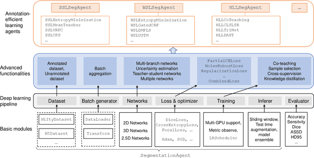
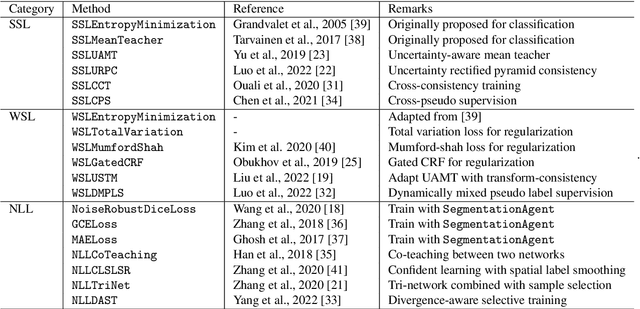
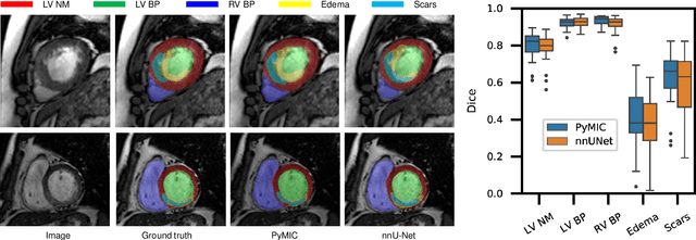

Abstract:Background and Objective: Existing deep learning platforms for medical image segmentation mainly focus on fully supervised segmentation that assumes full and accurate pixel-level annotations are available. We aim to develop a new deep learning toolkit to support annotation-efficient learning for medical image segmentation, which can accelerate and simply the development of deep learning models with limited annotation budget, e.g., learning from partial, sparse or noisy annotations. Methods: Our proposed toolkit named PyMIC is a modular deep learning platform for medical image segmentation tasks. In addition to basic components that support development of high-performance models for fully supervised segmentation, it contains several advanced components that are tailored for learning from imperfect annotations, such as loading annotated and unannounced images, loss functions for unannotated, partially or inaccurately annotated images, and training procedures for co-learning between multiple networks, etc. PyMIC is built on the PyTorch framework and supports development of semi-supervised, weakly supervised and noise-robust learning methods for medical image segmentation. Results: We present four illustrative medical image segmentation tasks based on PyMIC: (1) Achieving competitive performance on fully supervised learning; (2) Semi-supervised cardiac structure segmentation with only 10% training images annotated; (3) Weakly supervised segmentation using scribble annotations; and (4) Learning from noisy labels for chest radiograph segmentation. Conclusions: The PyMIC toolkit is easy to use and facilitates efficient development of medical image segmentation models with imperfect annotations. It is modular and flexible, which enables researchers to develop high-performance models with low annotation cost. The source code is available at: https://github.com/HiLab-git/PyMIC.
PA-Seg: Learning from Point Annotations for 3D Medical Image Segmentation using Contextual Regularization and Cross Knowledge Distillation
Aug 11, 2022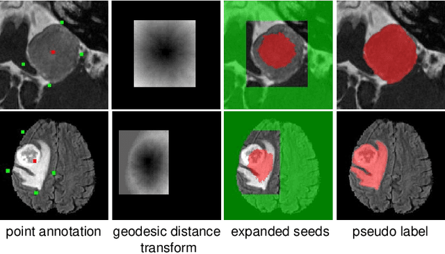

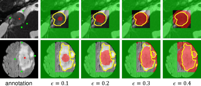
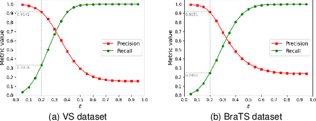
Abstract:The success of Convolutional Neural Networks (CNNs) in 3D medical image segmentation relies on massive fully annotated 3D volumes for training that are time-consuming and labor-intensive to acquire. In this paper, we propose to annotate a segmentation target with only seven points in 3D medical images, and design a two-stage weakly supervised learning framework PA-Seg. In the first stage, we employ geodesic distance transform to expand the seed points to provide more supervision signal. To further deal with unannotated image regions during training, we propose two contextual regularization strategies, i.e., multi-view Conditional Random Field (mCRF) loss and Variance Minimization (VM) loss, where the first one encourages pixels with similar features to have consistent labels, and the second one minimizes the intensity variance for the segmented foreground and background, respectively. In the second stage, we use predictions obtained by the model pre-trained in the first stage as pseudo labels. To overcome noises in the pseudo labels, we introduce a Self and Cross Monitoring (SCM) strategy, which combines self-training with Cross Knowledge Distillation (CKD) between a primary model and an auxiliary model that learn from soft labels generated by each other. Experiments on public datasets for Vestibular Schwannoma (VS) segmentation and Brain Tumor Segmentation (BraTS) demonstrated that our model trained in the first stage outperforms existing state-of-the-art weakly supervised approaches by a large margin, and after using SCM for additional training, the model can achieve competitive performance compared with the fully supervised counterpart on the BraTS dataset.
Source-free Domain Adaptation for Multi-site and Lifespan Brain Skull Stripping
Mar 11, 2022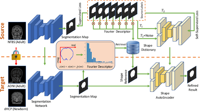


Abstract:Skull stripping is a crucial prerequisite step in the analysis of brain magnetic resonance (MR) images. Although many excellent works or tools have been proposed, they suffer from low generalization capability. For instance, the model trained on a dataset with specific imaging parameters (source domain) cannot be well applied to other datasets with different imaging parameters (target domain). Especially, for the lifespan datasets, the model trained on an adult dataset is not applicable to an infant dataset due to the large domain difference. To address this issue, numerous domain adaptation (DA) methods have been proposed to align the extracted features between the source and target domains, requiring concurrent access to the input images of both domains. Unfortunately, it is problematic to share the images due to privacy. In this paper, we design a source-free domain adaptation framework (SDAF) for multi-site and lifespan skull stripping that can accomplish domain adaptation without access to source domain images. Our method only needs to share the source labels as shape dictionaries and the weights trained on the source data, without disclosing private information from source domain subjects. To deal with the domain shift between multi-site lifespan datasets, we take advantage of the brain shape prior which is invariant to imaging parameters and ages. Experiments demonstrate that our framework can significantly outperform the state-of-the-art methods on multi-site lifespan datasets.
Scribble-Supervised Medical Image Segmentation via Dual-Branch Network and Dynamically Mixed Pseudo Labels Supervision
Mar 04, 2022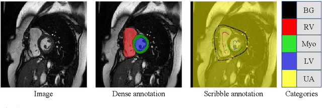
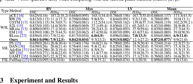
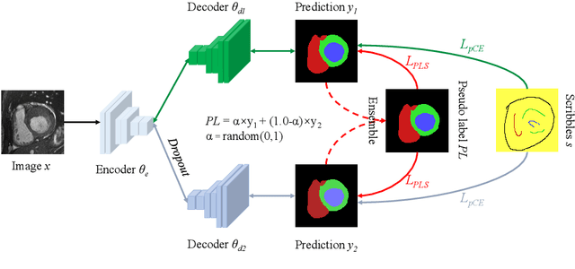

Abstract:Medical image segmentation plays an irreplaceable role in computer-assisted diagnosis, treatment planning, and following-up. Collecting and annotating a large-scale dataset is crucial to training a powerful segmentation model, but producing high-quality segmentation masks is an expensive and time-consuming procedure. Recently, weakly-supervised learning that uses sparse annotations (points, scribbles, bounding boxes) for network training has achieved encouraging performance and shown the potential for annotation cost reduction. However, due to the limited supervision signal of sparse annotations, it is still challenging to employ them for networks training directly. In this work, we propose a simple yet efficient scribble-supervised image segmentation method and apply it to cardiac MRI segmentation. Specifically, we employ a dual-branch network with one encoder and two slightly different decoders for image segmentation and dynamically mix the two decoders' predictions to generate pseudo labels for auxiliary supervision. By combining the scribble supervision and auxiliary pseudo labels supervision, the dual-branch network can efficiently learn from scribble annotations end-to-end. Experiments on the public ACDC dataset show that our method performs better than current scribble-supervised segmentation methods and also outperforms several semi-supervised segmentation methods.
 Add to Chrome
 Add to Firefox
 Add to Edge