 Add to Chrome
 Add to Firefox
 Add to Edge
Medical world models in healthcare: foundations, applications, and challenges for trustworthy clinical translation
Jul 28, 2026
Medical world models offer a framework for extending medical artificial intelligence beyond static prediction by representing evolving patient states and modelling how they change over time and in response to clinical interventions. This Review defines the conceptual boundaries, technical foundations, application domains, and evidence requirements of the field through a structured narrative synthesis with reproducible evidence mapping.We screened 1,455 unique records and assembled a corpus of 98 sources, including 14 studies that met a strict empirical definition of a medical world model. The field is organised around four capabilities: patient state representation, temporal dynamics modelling, intervention-conditioned simulation, and clinician-supervised planning. Evidence spans medical imaging, longitudinal electronic health records, treatment response modelling, physiological and multimodal state modelling, ultrasound and surgical interaction, and population and health-system simulation; clinical digital twins are treated as a cross-cutting integration framework.Current studies provide early evidence of technical feasibility for trajectory forecasting and comparison of candidate interventions, but most remain retrospective, task-specific, or preclinical. The evidence base is further limited by incomplete longitudinal intervention data, inconsistent action semantics, limited causal identifiability, long-horizon error accumulation, inadequate uncertainty estimation, and limited external validation. Clinical translation will therefore depend on precise intervention representations, robust causal and mechanistic grounding, calibrated trajectory-level uncertainty, safety-constrained planning, and prospective multicentre validation against clinically meaningful endpoints.

Agentic AI in medicine: architectures, applications, evaluation, and challenges for clinical translation
Jul 28, 2026Large language models and multimodal foundation models are enabling medical artificial intelligence (AI) systems to move beyond isolated prediction and undertake multistep clinical tasks that require planning, tool use, memory, iterative correction, and coordination among specialized agents. However, the scope of agentic AI in medicine remains unsettled, and current evaluation practices are not yet aligned with the requirements of clinical use. We conducted a scoping review with systematic evidence mapping across five electronic sources, screened 1,649 exportable records, and provisionally included 557 unique studies that met predefined criteria for goal-directed task execution, tool use, interaction with external resources, feedback-based refinement, or multi-agent collaboration. The included studies describe single agents that use external tools, workflows supported by retrieval and external knowledge, multimodal agents, and multi-agent systems applied to medical question answering, image interpretation, electronic health record analysis, drug safety, and clinical trial prediction. The evidence base remains dominated by public benchmarks, simulated settings, retrospective datasets, and small-scale expert evaluation. Process reliability, evidence traceability, uncertainty, safety, workflow impact, and external validity are evaluated less consistently. Clinical translation will depend on clearer definitions, reproducible evaluation, auditable oversight, interoperable system design, and prospective validation in real-world clinical workflows.
Distortion-Corrected Diffusion MRI Using Rotated-View EPI and Joint Field-Map/Image Estimation with Gaussian Primitives
Jun 30, 2026Echo Planar Imaging (EPI) is the standard acquisition technique for diffusion and functional neuroimaging, enabling rapid imaging but suffering from geometric distortions caused by B0 field inhomogeneities. Existing correction methods first reconstruct distorted images using parallel imaging, then estimate the B0 field and correct the distortion in the image domain. In this sequential process, reconstruction artifacts at high acceleration factors and low SNR at high diffusion b-values degrade B0 estimation and limit the overall correction quality. We propose a physics-informed framework that jointly estimates the B0 field and distortion-free image directly from k-space data, without depending on an intermediate parallel-imaging reconstruction for the correction. The image and the B0 field are each represented as a superposition of Gaussian primitives embedded within an MRI physics forward model. The explicit, continuous parameterization captures both smooth regions and tissue boundaries and supports rotated-view EPI acquisitions without interpolation. The diffusion-weighted image is modeled as real and non-negative, with the image phase absorbed into a per-shot phase factor. Rotated views distribute distortions across multiple phase-encoding orientations, improving point spread function isotropy and providing stronger constraints for B0 estimation. On in vivo brain diffusion EPI, the proposed method attains the closest brain-boundary agreement with a distortion-free structural reference, with the largest improvement over sequential methods at high b-value and high acceleration. Extensive visual comparisons further show improved detail fidelity and noise suppression.
A physics-informed foundation model for quantitative diffusion MRI
May 29, 2026Understanding the human brain requires access to its microscopic tissue architecture. Diffusion magnetic resonance imaging (MRI) provides the only noninvasive window into whole-brain microstructure in vivo, yet reliable quantitative mapping remains confined to specialized research settings requiring dense sampling and optimized acquisition protocols. To address this gap, we present a physics-informed generative microstructure network (PIGMENT) that learns a universal generative prior of human brain microstructure and adapts it zero-shot to each participant's measured data to recover subject-specific maps. Trained on 11375 scans spanning multiple sites, vendors, and field strengths, PIGMENT enabled reliable quantitative mapping for tensor, kurtosis, and NODDI models across external datasets from five independent centers. It remains effective where conventional fitting becomes unreliable, recovering meaningful maps from extremely sparse acquisitions while supporting downstream tractography and structural connectivity mapping. PIGMENT estimates demonstrated strong biological validity, preserving submillimeter cortical microarchitectural patterns and early-childhood white matter developmental trajectories from 10-fold accelerated scans. Furthermore, PIGMENT enables reliable quantitative tensor mapping on cost-efficient low-field systems and the extraction of tumor-related biomarkers using ultra-fast clinical protocols. Together, these results establish PIGMENT as a physics-informed foundation model that extends quantitative diffusion MRI into regimes traditionally too sparse, heterogeneous, or clinically constrained for reliable analysis.
Enhancing Diffusion-Weighted Images (DWI) for Diffusion MRI: Is it Enough without Non-Diffusion-Weighted B=0 Reference?
May 19, 2025Diffusion MRI (dMRI) is essential for studying brain microstructure, but high-resolution imaging remains challenging due to the inherent trade-offs between acquisition time and signal-to-noise ratio (SNR). Conventional methods often optimize only the diffusion-weighted images (DWIs) without considering their relationship with the non-diffusion-weighted (b=0) reference images. However, calculating diffusion metrics, such as the apparent diffusion coefficient (ADC) and diffusion tensor with its derived metrics like fractional anisotropy (FA) and mean diffusivity (MD), relies on the ratio between each DWI and the b=0 image, which is crucial for clinical observation and diagnostics. In this study, we demonstrate that solely enhancing DWIs using a conventional pixel-wise mean squared error (MSE) loss is insufficient, as the error in ratio between generated DWIs and b=0 diverges. We propose a novel ratio loss, defined as the MSE loss between the predicted and ground-truth log of DWI/b=0 ratios. Our results show that incorporating the ratio loss significantly improves the convergence of this ratio error, achieving lower ratio MSE and slightly enhancing the peak signal-to-noise ratio (PSNR) of generated DWIs. This leads to improved dMRI super-resolution and better preservation of b=0 ratio-based features for the derivation of diffusion metrics.
Artificial Intelligence for Neuro MRI Acquisition: A Review
Jun 10, 2024Magnetic resonance imaging (MRI) has significantly benefited from the resurgence of artificial intelligence (AI). By leveraging AI's capabilities in large-scale optimization and pattern recognition, innovative methods are transforming the MRI acquisition workflow, including planning, sequence design, and correction of acquisition artifacts. These emerging algorithms demonstrate substantial potential in enhancing the efficiency and throughput of acquisition steps. This review discusses several pivotal AI-based methods in neuro MRI acquisition, focusing on their technological advances, impact on clinical practice, and potential risks.
Advancing low-field MRI with a universal denoising imaging transformer: Towards fast and high-quality imaging
Apr 30, 2024Recent developments in low-field (LF) magnetic resonance imaging (MRI) systems present remarkable opportunities for affordable and widespread MRI access. A robust denoising method to overcome the intrinsic low signal-noise-ratio (SNR) barrier is critical to the success of LF MRI. However, current data-driven MRI denoising methods predominantly handle magnitude images and rely on customized models with constrained data diversity and quantity, which exhibit limited generalizability in clinical applications across diverse MRI systems, pulse sequences, and organs. In this study, we present ImT-MRD: a complex-valued imaging transformer trained on a vast number of clinical MRI scans aiming at universal MR denoising at LF systems. Compared with averaging multiple-repeated scans for higher image SNR, the model obtains better image quality from fewer repetitions, demonstrating its capability for accelerating scans under various clinical settings. Moreover, with its complex-valued image input, the model can denoise intermediate results before advanced post-processing and prepare high-quality data for further MRI research. By delivering universal and accurate denoising across clinical and research tasks, our model holds great promise to expedite the evolution of LF MRI for accessible and equal biomedical applications.
An Efficient Algorithm for Spatial-Spectral Partial Volume Compartment Mapping with Applications to Multicomponent Diffusion and Relaxation MRI
Jan 23, 2024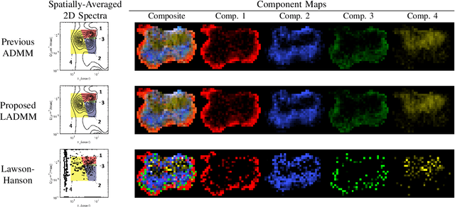
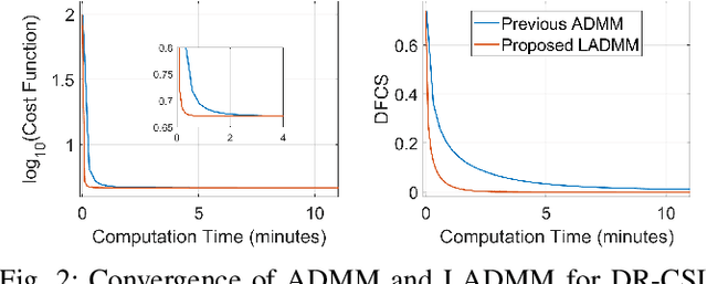


It has been previously shown that high-quality partial volume tissue compartment maps can be obtained by combining multiparametric contrast-encoded MRI data acquisition methods with spatially-regularized spectroscopic image estimation techniques. However, the advantages of this combined approach generally come at the expense of substantial computational complexity. In this work, we propose a new algorithm to solve this kind of estimation problem more efficiently. Our algorithm is based on the linearized alternating directions method of multipliers (LADMM), and relies on the introduction of novel quadratic penalty terms to substantially simplify the subproblems that must be solved at each iteration. We evaluate this algorithm on a variety of different estimation problems (diffusion-relaxation, relaxation-relaxation, relaxometry, and magnetic resonance fingerprinting), where we consistently observe substantial (roughly 5$\times$-80$\times$) speed improvements. We expect that this new faster algorithm will lower practical barriers to using spatial regularization and multiparametric contrast-encoded MRI data acquisition methods for partial volume compartment mapping.
High-resolution myelin-water fraction and quantitative relaxation mapping using 3D ViSTa-MR fingerprinting
Dec 21, 2023Purpose: This study aims to develop a high-resolution whole-brain multi-parametric quantitative MRI approach for simultaneous mapping of myelin-water fraction (MWF), T1, T2, and proton-density (PD), all within a clinically feasible scan time. Methods: We developed 3D ViSTa-MRF, which combined Visualization of Short Transverse relaxation time component (ViSTa) technique with MR Fingerprinting (MRF), to achieve high-fidelity whole-brain MWF and T1/T2/PD mapping on a clinical 3T scanner. To achieve fast acquisition and memory-efficient reconstruction, the ViSTa-MRF sequence leverages an optimized 3D tiny-golden-angle-shuffling spiral-projection acquisition and joint spatial-temporal subspace reconstruction with optimized preconditioning algorithm. With the proposed ViSTa-MRF approach, high-fidelity direct MWF mapping was achieved without a need for multi-compartment fitting that could introduce bias and/or noise from additional assumptions or priors. Results: The in-vivo results demonstrate the effectiveness of the proposed acquisition and reconstruction framework to provide fast multi-parametric mapping with high SNR and good quality. The in-vivo results of 1mm- and 0.66mm-iso datasets indicate that the MWF values measured by the proposed method are consistent with standard ViSTa results that are 30x slower with lower SNR. Furthermore, we applied the proposed method to enable 5-minute whole-brain 1mm-iso assessment of MWF and T1/T2/PD mappings for infant brain development and for post-mortem brain samples. Conclusions: In this work, we have developed a 3D ViSTa-MRF technique that enables the acquisition of whole-brain MWF, quantitative T1, T2, and PD maps at 1mm and 0.66mm isotropic resolution in 5 and 15 minutes, respectively. This advancement allows for quantitative investigations of myelination changes in the brain.
Rapid Non-cartesian Reconstruction Using an Implicit Representation of GROG Kernels
Oct 16, 2023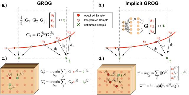
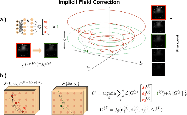

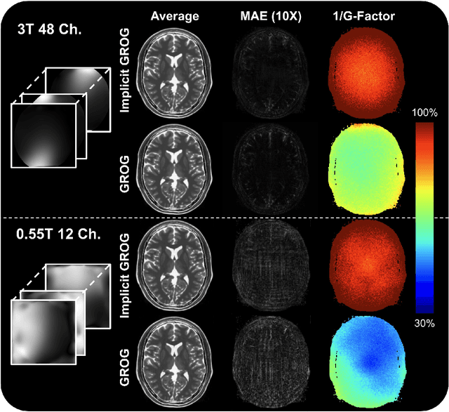
MRI data is acquired in Fourier space. Data acquisition is typically performed on a Cartesian grid in this space to enable the use of a fast Fourier transform algorithm to achieve fast and efficient reconstruction. However, it has been shown that for multiple applications, non-Cartesian data acquisition can improve the performance of MR imaging by providing fast and more efficient data acquisition, and improving motion robustness. Nonetheless, the image reconstruction process of non-Cartesian data is more involved and can be time-consuming, even through the use of efficient algorithms such as non-uniform FFT (NUFFT). This work provides an efficient approach (iGROG) to transform the non-Cartesian data into Cartesian data, to achieve simpler and faster reconstruction which should help enable non-Cartesian data sampling to be performed more widely in MRI.