 Add to Chrome
 Add to Firefox
 Add to Edge
Fleet: Few Shots Lead Effective AI-generated Image Detection
Jun 30, 2026
AI-generated image (AIGI) detection is undergoing a critical transition from laboratory benchmarks to open-world adversarial defense. The prevalent paradigm focuses on finding static feature spaces, assuming that some invariant artifacts learned from historical data can achieve universal zero-shot generalization. While achieving saturation on several AIGI benchmarks, this static hypothesis suffers a severe performance drop against rapidly evolving generators (e.g., SD3, Nano Banana Pro). To address these limitations, we propose that the field should expand beyond "static generalization" to a new paradigm of "dynamic adaptation". We introduce Fleet, a framework that pioneers a dynamic paradigm of continuous few-shot evolution, enabling rapid alignment with emerging generative threats. Fleet improves few-shot adaptation by replacing unconstrained feature updates with constrained routing correction, where avoidance routing redirects novel AI samples away from Non-AI-dominated routes within decoupled subspaces. To validate this, we present Treasure, a benchmark spanning 64 models and 360k images, featuring diverse architectures and 20 closed-source commercial engines. Experiments reveal that while static SOTA methods fail catastrophically on modern generators, Fleet restores performance from 20.4% to 73.1% with only 10-shot adaptation on "Doubao Seedream 4.0". Code and data are available at https://github.com/ICTMCG/Fleet .

Intracranial Aneurysm Classification and Segmentation via Tri-Axial ROI and Multi-Task Learning
Jun 25, 2026Intracranial aneurysms are often asymptomatic until rupture, which carries high mortality. Rupture risk assessment and treatment planning depend on both aneurysm morphology and anatomical location, yet existing automated methods remain limited to binary detection without fine-grained anatomical classification or multi-class segmentation. We present a multi-task framework that simultaneously performs multi-label classification, multi-class aneurysm segmentation, and multi-class vessel segmentation across 13 anatomical locations and four imaging modalities (CTA, MRA, T2, T1-post). Our two-stage approach combines a fast 2D tri-axial Region of Interest (ROI) extraction method with a 3D multi-task nnU-Net backbone. A dual-decoder design mitigates the extreme volume imbalance between aneurysm and vessel classes, while cross-attention pooling and modality-specific auxiliary heads improve feature learning across heterogeneous inputs. Our two-fold ensemble achieved 2nd place in the RSNA 2025 Intracranial Aneurysm Detection challenge. Code, model weights, and a 3D Slicer plugin are publicly available.
Multimodal Fusion of Skeleton Dynamics and Clinical Gait Features for Video-Based Cerebral Palsy Severity Assessment
Mar 23, 2026Video-based gait analysis has become a promising approach for assessing motor impairment in children with cerebral palsy (CP). However, existing methods usually rely on either pose sequences or handcrafted gait features alone, making it difficult to simultaneously capture spatiotemporal motion patterns and clinically meaningful biomechanical information. To address this gap, we propose a multimodal fusion framework that integrates skeleton dynamics with contribution-guided clinically meaningful gait features. First, Grad-CAM analysis on a pre-trained ST-GCN backbone identified the most discriminative body keypoints, providing an interpretable basis for subsequent gait feature extraction. We then build a dual-stream architecture, with one stream modeling skeleton dynamics using ST-GCN and the other encoding gait geatures derived from the identified keypoints. By fusing the two streams through feature cross-attention improved four-level CP motor severity classification to 70.86%, outperforming the baseline by 5.6 percentage points. Overall, this work suggests that integrating skeleton dynamics with clinically meaningful gait descriptors can improve both prediction performance and biomechanical interpretability for video-based CP severity assessment.
PowerGenie: Analytically-Guided Evolutionary Discovery of Superior Reconfigurable Power Converters
Jan 29, 2026Discovering superior circuit topologies requires navigating an exponentially large design space-a challenge traditionally reserved for human experts. Existing AI methods either select from predefined templates or generate novel topologies at a limited scale without rigorous verification, leaving large-scale performance-driven discovery underexplored. We present PowerGenie, a framework for automated discovery of higher-performance reconfigurable power converters at scale. PowerGenie introduces: (1) an automated analytical framework that determines converter functionality and theoretical performance limits without component sizing or SPICE simulation, and (2) an evolutionary finetuning method that co-evolves a generative model with its training distribution through fitness selection and uniqueness verification. Unlike existing methods that suffer from mode collapse and overfitting, our approach achieves higher syntax validity, function validity, novelty rate, and figure-of-merit (FoM). PowerGenie discovers a novel 8-mode reconfigurable converter with 23% higher FoM than the best training topology. SPICE simulations confirm average absolute efficiency gains of 10% across 8 modes and up to 17% at a single mode. Code is available at https://github.com/xz-group/PowerGenie.
Compressive Sensing Photoacoustic Imaging Receiver with Matrix-Vector-Multiplication SAR ADC
Nov 09, 2025Wearable photoacoustic imaging devices hold great promise for continuous health monitoring and point-of-care diagnostics. However, the large data volume generated by high-density transducer arrays presents a major challenge for realizing compact and power-efficient wearable systems. This paper presents a photoacoustic imaging receiver (RX) that embeds compressive sensing directly into the hardware to address this bottleneck. The RX integrates 16 AFEs and four matrix-vector-multiplication (MVM) SAR ADCs that perform energy- and area-efficient analog-domain compression. The architecture achieves a 4-8x reduction in output data rate while preserving low-loss full-array information. The MVM SAR ADC executes passive and accurate MVM using user-defined programmable ternary weights. Two signal reconstruction methods are implemented: (1) an optimization approach using the fast iterative shrinkage-thresholding algorithm, and (2) a learning-based approach employing implicit neural representation. Fabricated in 65 nm CMOS, the chip achieves an ADC's SNDR of 57.5 dB at 20.41 MS/s, with an AFE input-referred noise of 3.5 nV/sqrt(Hz). MVM linearity measurements show R^2 > 0.999 across a wide range of weights and input amplitudes. The system is validated through phantom imaging experiments, demonstrating high-fidelity image reconstruction under up to 8x compression. The RX consumes 5.83 mW/channel and supports a general ternary-weighted measurement matrix, offering a compelling solution for next-generation miniaturized, wearable PA imaging systems.
Parametric shape models for vessels learned from segmentations via differentiable voxelization
Jul 03, 2025Vessels are complex structures in the body that have been studied extensively in multiple representations. While voxelization is the most common of them, meshes and parametric models are critical in various applications due to their desirable properties. However, these representations are typically extracted through segmentations and used disjointly from each other. We propose a framework that joins the three representations under differentiable transformations. By leveraging differentiable voxelization, we automatically extract a parametric shape model of the vessels through shape-to-segmentation fitting, where we learn shape parameters from segmentations without the explicit need for ground-truth shape parameters. The vessel is parametrized as centerlines and radii using cubic B-splines, ensuring smoothness and continuity by construction. Meshes are differentiably extracted from the learned shape parameters, resulting in high-fidelity meshes that can be manipulated post-fit. Our method can accurately capture the geometry of complex vessels, as demonstrated by the volumetric fits in experiments on aortas, aneurysms, and brain vessels.
FlexDuo: A Pluggable System for Enabling Full-Duplex Capabilities in Speech Dialogue Systems
Feb 19, 2025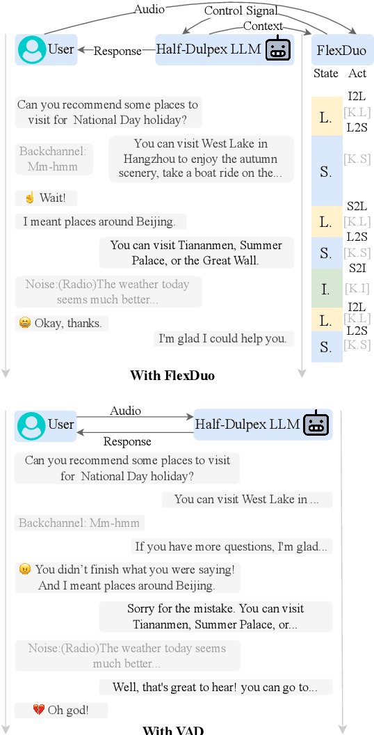
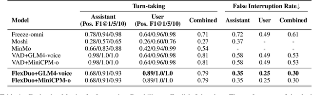
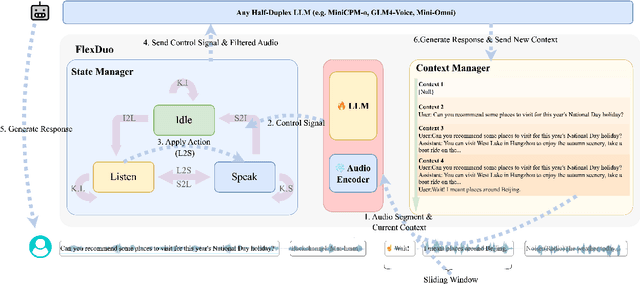

Full-Duplex Speech Dialogue Systems (Full-Duplex SDS) have significantly enhanced the naturalness of human-machine interaction by enabling real-time bidirectional communication. However, existing approaches face challenges such as difficulties in independent module optimization and contextual noise interference due to highly coupled architectural designs and oversimplified binary state modeling. This paper proposes FlexDuo, a flexible full-duplex control module that decouples duplex control from spoken dialogue systems through a plug-and-play architectural design. Furthermore, inspired by human information-filtering mechanisms in conversations, we introduce an explicit Idle state. On one hand, the Idle state filters redundant noise and irrelevant audio to enhance dialogue quality. On the other hand, it establishes a semantic integrity-based buffering mechanism, reducing the risk of mutual interruptions while ensuring accurate response transitions. Experimental results on the Fisher corpus demonstrate that FlexDuo reduces the false interruption rate by 24.9% and improves response accuracy by 7.6% compared to integrated full-duplex dialogue system baselines. It also outperforms voice activity detection (VAD) controlled baseline systems in both Chinese and English dialogue quality. The proposed modular architecture and state-based dialogue model provide a novel technical pathway for building flexible and efficient duplex dialogue systems.
SELMA3D challenge: Self-supervised learning for 3D light-sheet microscopy image segmentation
Jan 07, 2025Recent innovations in light sheet microscopy, paired with developments in tissue clearing techniques, enable the 3D imaging of large mammalian tissues with cellular resolution. Combined with the progress in large-scale data analysis, driven by deep learning, these innovations empower researchers to rapidly investigate the morphological and functional properties of diverse biological samples. Segmentation, a crucial preliminary step in the analysis process, can be automated using domain-specific deep learning models with expert-level performance. However, these models exhibit high sensitivity to domain shifts, leading to a significant drop in accuracy when applied to data outside their training distribution. To address this limitation, and inspired by the recent success of self-supervised learning in training generalizable models, we organized the SELMA3D Challenge during the MICCAI 2024 conference. SELMA3D provides a vast collection of light-sheet images from cleared mice and human brains, comprising 35 large 3D images-each with over 1000^3 voxels-and 315 annotated small patches for finetuning, preliminary testing and final testing. The dataset encompasses diverse biological structures, including vessel-like and spot-like structures. Five teams participated in all phases of the challenge, and their proposed methods are reviewed in this paper. Quantitative and qualitative results from most participating teams demonstrate that self-supervised learning on large datasets improves segmentation model performance and generalization. We will continue to support and extend SELMA3D as an inaugural MICCAI challenge focused on self-supervised learning for 3D microscopy image segmentation.
ISLES'24: Improving final infarct prediction in ischemic stroke using multimodal imaging and clinical data
Aug 20, 2024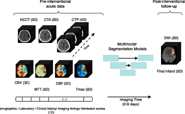
Accurate estimation of core (irreversibly damaged tissue) and penumbra (salvageable tissue) volumes is essential for ischemic stroke treatment decisions. Perfusion CT, the clinical standard, estimates these volumes but is affected by variations in deconvolution algorithms, implementations, and thresholds. Core tissue expands over time, with growth rates influenced by thrombus location, collateral circulation, and inherent patient-specific factors. Understanding this tissue growth is crucial for determining the need to transfer patients to comprehensive stroke centers, predicting the benefits of additional reperfusion attempts during mechanical thrombectomy, and forecasting final clinical outcomes. This work presents the ISLES'24 challenge, which addresses final post-treatment stroke infarct prediction from pre-interventional acute stroke imaging and clinical data. ISLES'24 establishes a unique 360-degree setting where all feasibly accessible clinical data are available for participants, including full CT acute stroke imaging, sub-acute follow-up MRI, and clinical tabular data. The contributions of this work are two-fold: first, we introduce a standardized benchmarking of final stroke infarct segmentation algorithms through the ISLES'24 challenge; second, we provide insights into infarct segmentation using multimodal imaging and clinical data strategies by identifying outperforming methods on a finely curated dataset. The outputs of this challenge are anticipated to enhance clinical decision-making and improve patient outcome predictions. All ISLES'24 materials, including data, performance evaluation scripts, and leading algorithmic strategies, are available to the research community following \url{https://isles-24.grand-challenge.org/}.
ISLES 2024: The first longitudinal multimodal multi-center real-world dataset in (sub-)acute stroke
Aug 20, 2024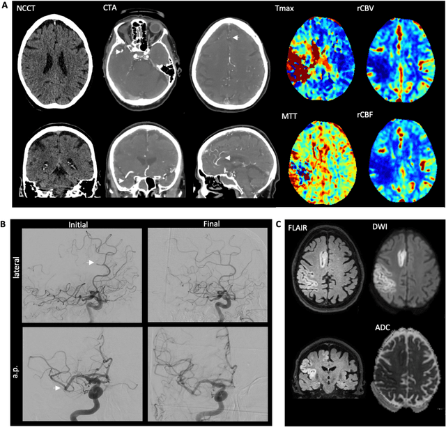
Stroke remains a leading cause of global morbidity and mortality, placing a heavy socioeconomic burden. Over the past decade, advances in endovascular reperfusion therapy and the use of CT and MRI imaging for treatment guidance have significantly improved patient outcomes and are now standard in clinical practice. To develop machine learning algorithms that can extract meaningful and reproducible models of brain function for both clinical and research purposes from stroke images - particularly for lesion identification, brain health quantification, and prognosis - large, diverse, and well-annotated public datasets are essential. While only a few datasets with (sub-)acute stroke data were previously available, several large, high-quality datasets have recently been made publicly accessible. However, these existing datasets include only MRI data. In contrast, our dataset is the first to offer comprehensive longitudinal stroke data, including acute CT imaging with angiography and perfusion, follow-up MRI at 2-9 days, as well as acute and longitudinal clinical data up to a three-month outcome. The dataset includes a training dataset of n = 150 and a test dataset of n = 100 scans. Training data is publicly available, while test data will be used exclusively for model validation. We are making this dataset available as part of the 2024 edition of the Ischemic Stroke Lesion Segmentation (ISLES) challenge (https://www.isles-challenge.org/), which continuously aims to establish benchmark methods for acute and sub-acute ischemic stroke lesion segmentation, aiding in creating open stroke imaging datasets and evaluating cutting-edge image processing algorithms.