 Add to Chrome
 Add to Firefox
 Add to Edge
CoFiDA-M: Concept-Aware Feature Modulation for Cross-Domain Adaptation with Image-Only Inference
May 29, 2026Models for AI-based skin cancer screening suffer a severe performance drop when shifting from expert dermoscopic (source) images to consumer-grade clinical (target) images, hindering real-world deployment. Existing domain adaptation methods often ignore crucial semantic invariants, such as clinical concepts. While new foundation models like MONET can provide this semantic information as dense, probabilistic scores, this metadata is unavailable at test time, creating a deployment paradox for practical image-only screening tools. We address this gap by proposing CoFiDA-M, a privileged information framework that learns from concepts at training time but deploys as an image-only model. Our method trains a teacher network that uses MONET concept probabilities to guide a FiLM modulator, transforming visual features into a semantically ``edited" feature space. A lightweight, image-only student is then trained to reproduce this edited representation, not just the teacher's final predictions. This distillation ``bakes" the clinical reasoning into the student's weights. On a challenging multi-dataset benchmark, our image-only student significantly outperforms state-of-the-art approaches, especially in melanoma recall. Our work provides a practical and generalizable framework for leveraging noisy, probabilistic metadata as privileged information, demonstrating strong cross-dataset robustness and potential for real-world deployment beyond dermatology. Implementation code is available at: https://github.com/mmu-dermatology-research/CoFiDA.git
SemCovNet: Towards Fair and Semantic Coverage-Aware Learning for Underrepresented Visual Concepts
Feb 18, 2026Modern vision models increasingly rely on rich semantic representations that extend beyond class labels to include descriptive concepts and contextual attributes. However, existing datasets exhibit Semantic Coverage Imbalance (SCI), a previously overlooked bias arising from the long-tailed semantic representations. Unlike class imbalance, SCI occurs at the semantic level, affecting how models learn and reason about rare yet meaningful semantics. To mitigate SCI, we propose Semantic Coverage-Aware Network (SemCovNet), a novel model that explicitly learns to correct semantic coverage disparities. SemCovNet integrates a Semantic Descriptor Map (SDM) for learning semantic representations, a Descriptor Attention Modulation (DAM) module that dynamically weights visual and concept features, and a Descriptor-Visual Alignment (DVA) loss that aligns visual features with descriptor semantics. We quantify semantic fairness using a Coverage Disparity Index (CDI), which measures the alignment between coverage and error. Extensive experiments across multiple datasets demonstrate that SemCovNet enhances model reliability and substantially reduces CDI, achieving fairer and more equitable performance. This work establishes SCI as a measurable and correctable bias, providing a foundation for advancing semantic fairness and interpretable vision learning.
SACB-Net: Spatial-awareness Convolutions for Medical Image Registration
Mar 25, 2025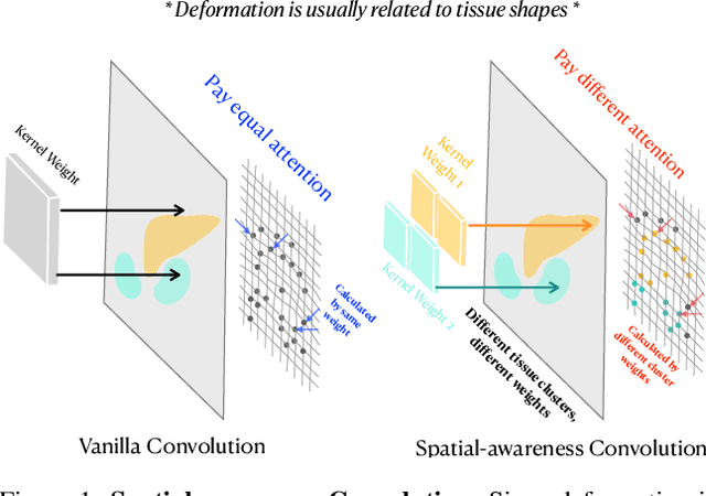
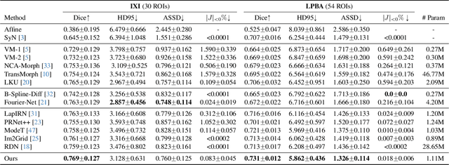
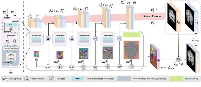
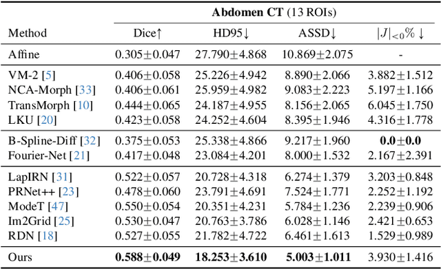
Deep learning-based image registration methods have shown state-of-the-art performance and rapid inference speeds. Despite these advances, many existing approaches fall short in capturing spatially varying information in non-local regions of feature maps due to the reliance on spatially-shared convolution kernels. This limitation leads to suboptimal estimation of deformation fields. In this paper, we propose a 3D Spatial-Awareness Convolution Block (SACB) to enhance the spatial information within feature representations. Our SACB estimates the spatial clusters within feature maps by leveraging feature similarity and subsequently parameterizes the adaptive convolution kernels across diverse regions. This adaptive mechanism generates the convolution kernels (weights and biases) tailored to spatial variations, thereby enabling the network to effectively capture spatially varying information. Building on SACB, we introduce a pyramid flow estimator (named SACB-Net) that integrates SACBs to facilitate multi-scale flow composition, particularly addressing large deformations. Experimental results on the brain IXI and LPBA datasets as well as Abdomen CT datasets demonstrate the effectiveness of SACB and the superiority of SACB-Net over the state-of-the-art learning-based registration methods. The code is available at https://github.com/x-xc/SACB_Net .
WiNet: Wavelet-based Incremental Learning for Efficient Medical Image Registration
Jul 18, 2024Deep image registration has demonstrated exceptional accuracy and fast inference. Recent advances have adopted either multiple cascades or pyramid architectures to estimate dense deformation fields in a coarse-to-fine manner. However, due to the cascaded nature and repeated composition/warping operations on feature maps, these methods negatively increase memory usage during training and testing. Moreover, such approaches lack explicit constraints on the learning process of small deformations at different scales, thus lacking explainability. In this study, we introduce a model-driven WiNet that incrementally estimates scale-wise wavelet coefficients for the displacement/velocity field across various scales, utilizing the wavelet coefficients derived from the original input image pair. By exploiting the properties of the wavelet transform, these estimated coefficients facilitate the seamless reconstruction of a full-resolution displacement/velocity field via our devised inverse discrete wavelet transform (IDWT) layer. This approach avoids the complexities of cascading networks or composition operations, making our WiNet an explainable and efficient competitor with other coarse-to-fine methods. Extensive experimental results from two 3D datasets show that our WiNet is accurate and GPU efficient. The code is available at https://github.com/x-xc/WiNet .
Development of Automated Neural Network Prediction for Echocardiographic Left ventricular Ejection Fraction
Mar 18, 2024


The echocardiographic measurement of left ventricular ejection fraction (LVEF) is fundamental to the diagnosis and classification of patients with heart failure (HF). In order to quantify LVEF automatically and accurately, this paper proposes a new pipeline method based on deep neural networks and ensemble learning. Within the pipeline, an Atrous Convolutional Neural Network (ACNN) was first trained to segment the left ventricle (LV), before employing the area-length formulation based on the ellipsoid single-plane model to calculate LVEF values. This formulation required inputs of LV area, derived from segmentation using an improved Jeffrey's method, as well as LV length, derived from a novel ensemble learning model. To further improve the pipeline's accuracy, an automated peak detection algorithm was used to identify end-diastolic and end-systolic frames, avoiding issues with human error. Subsequently, single-beat LVEF values were averaged across all cardiac cycles to obtain the final LVEF. This method was developed and internally validated in an open-source dataset containing 10,030 echocardiograms. The Pearson's correlation coefficient was 0.83 for LVEF prediction compared to expert human analysis (p<0.001), with a subsequent area under the receiver operator curve (AUROC) of 0.98 (95% confidence interval 0.97 to 0.99) for categorisation of HF with reduced ejection (HFrEF; LVEF<40%). In an external dataset with 200 echocardiograms, this method achieved an AUC of 0.90 (95% confidence interval 0.88 to 0.91) for HFrEF assessment. This study demonstrates that an automated neural network-based calculation of LVEF is comparable to expert clinicians performing time-consuming, frame-by-frame manual evaluation of cardiac systolic function.
Decoder-Only Image Registration
Feb 05, 2024


In unsupervised medical image registration, the predominant approaches involve the utilization of a encoder-decoder network architecture, allowing for precise prediction of dense, full-resolution displacement fields from given paired images. Despite its widespread use in the literature, we argue for the necessity of making both the encoder and decoder learnable in such an architecture. For this, we propose a novel network architecture, termed LessNet in this paper, which contains only a learnable decoder, while entirely omitting the utilization of a learnable encoder. LessNet substitutes the learnable encoder with simple, handcrafted features, eliminating the need to learn (optimize) network parameters in the encoder altogether. Consequently, this leads to a compact, efficient, and decoder-only architecture for 3D medical image registration. Evaluated on two publicly available brain MRI datasets, we demonstrate that our decoder-only LessNet can effectively and efficiently learn both dense displacement and diffeomorphic deformation fields in 3D. Furthermore, our decoder-only LessNet can achieve comparable registration performance to state-of-the-art methods such as VoxelMorph and TransMorph, while requiring significantly fewer computational resources. Our code and pre-trained models are available at https://github.com/xi-jia/LessNet.
Optimizing ADMM and Over-Relaxed ADMM Parameters for Linear Quadratic Problems
Jan 01, 2024


The Alternating Direction Method of Multipliers (ADMM) has gained significant attention across a broad spectrum of machine learning applications. Incorporating the over-relaxation technique shows potential for enhancing the convergence rate of ADMM. However, determining optimal algorithmic parameters, including both the associated penalty and relaxation parameters, often relies on empirical approaches tailored to specific problem domains and contextual scenarios. Incorrect parameter selection can significantly hinder ADMM's convergence rate. To address this challenge, in this paper we first propose a general approach to optimize the value of penalty parameter, followed by a novel closed-form formula to compute the optimal relaxation parameter in the context of linear quadratic problems (LQPs). We then experimentally validate our parameter selection methods through random instantiations and diverse imaging applications, encompassing diffeomorphic image registration, image deblurring, and MRI reconstruction.
Fourier-Net+: Leveraging Band-Limited Representation for Efficient 3D Medical Image Registration
Jul 06, 2023


U-Net style networks are commonly utilized in unsupervised image registration to predict dense displacement fields, which for high-resolution volumetric image data is a resource-intensive and time-consuming task. To tackle this challenge, we first propose Fourier-Net, which replaces the costly U-Net style expansive path with a parameter-free model-driven decoder. Instead of directly predicting a full-resolution displacement field, our Fourier-Net learns a low-dimensional representation of the displacement field in the band-limited Fourier domain which our model-driven decoder converts to a full-resolution displacement field in the spatial domain. Expanding upon Fourier-Net, we then introduce Fourier-Net+, which additionally takes the band-limited spatial representation of the images as input and further reduces the number of convolutional layers in the U-Net style network's contracting path. Finally, to enhance the registration performance, we propose a cascaded version of Fourier-Net+. We evaluate our proposed methods on three datasets, on which our proposed Fourier-Net and its variants achieve comparable results with current state-of-the art methods, while exhibiting faster inference speeds, lower memory footprint, and fewer multiply-add operations. With such small computational cost, our Fourier-Net+ enables the efficient training of large-scale 3D registration on low-VRAM GPUs. Our code is publicly available at \url{https://github.com/xi-jia/Fourier-Net}.
Fourier-Net: Fast Image Registration with Band-limited Deformation
Nov 29, 2022


Unsupervised image registration commonly adopts U-Net style networks to predict dense displacement fields in the full-resolution spatial domain. For high-resolution volumetric image data, this process is however resource intensive and time-consuming. To tackle this problem, we propose the Fourier-Net, replacing the expansive path in a U-Net style network with a parameter-free model-driven decoder. Specifically, instead of our Fourier-Net learning to output a full-resolution displacement field in the spatial domain, we learn its low-dimensional representation in a band-limited Fourier domain. This representation is then decoded by our devised model-driven decoder (consisting of a zero padding layer and an inverse discrete Fourier transform layer) to the dense, full-resolution displacement field in the spatial domain. These changes allow our unsupervised Fourier-Net to contain fewer parameters and computational operations, resulting in faster inference speeds. Fourier-Net is then evaluated on two public 3D brain datasets against various state-of-the-art approaches. For example, when compared to a recent transformer-based method, i.e., TransMorph, our Fourier-Net, only using 0.22$\%$ of its parameters and 6.66$\%$ of the mult-adds, achieves a 0.6\% higher Dice score and an 11.48$\times$ faster inference speed. Code is available at \url{https://github.com/xi-jia/Fourier-Net}.
U-Net vs Transformer: Is U-Net Outdated in Medical Image Registration?
Aug 13, 2022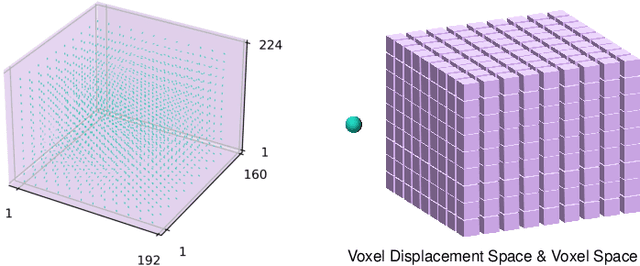
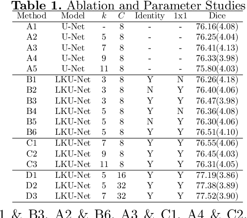
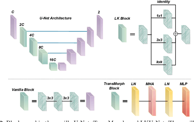
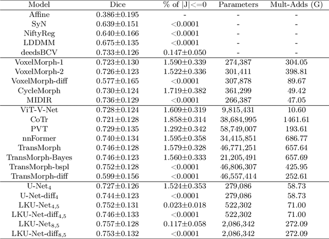
Due to their extreme long-range modeling capability, vision transformer-based networks have become increasingly popular in deformable image registration. We believe, however, that the receptive field of a 5-layer convolutional U-Net is sufficient to capture accurate deformations without needing long-range dependencies. The purpose of this study is therefore to investigate whether U-Net-based methods are outdated compared to modern transformer-based approaches when applied to medical image registration. For this, we propose a large kernel U-Net (LKU-Net) by embedding a parallel convolutional block to a vanilla U-Net in order to enhance the effective receptive field. On the public 3D IXI brain dataset for atlas-based registration, we show that the performance of the vanilla U-Net is already comparable with that of state-of-the-art transformer-based networks (such as TransMorph), and that the proposed LKU-Net outperforms TransMorph by using only 1.12% of its parameters and 10.8% of its mult-adds operations. We further evaluate LKU-Net on a MICCAI Learn2Reg 2021 challenge dataset for inter-subject registration, our LKU-Net also outperforms TransMorph on this dataset and ranks first on the public leaderboard as of the submission of this work. With only modest modifications to the vanilla U-Net, we show that U-Net can outperform transformer-based architectures on inter-subject and atlas-based 3D medical image registration. Code is available at https://github.com/xi-jia/LKU-Net.