 Add to Chrome
 Add to Firefox
 Add to Edge
Dual-Manifold Geometry Guided Representation Learning: Adaptive Coupling between Kernel and Data Spaces
Aug 13, 2026
Deep representation learning has primarily focused on how features evolve across network layers, while largely overlooking the structured geometry embedded in network parameters. We introduce a dual-manifold perspective in which each convolutional layer contains two coupled geometric spaces: a Kernel Manifold induced by convolutional filters and a Data Manifold characterized by intermediate feature representations. Because these manifolds share the same channel space, parameter geometry can provide complementary structural information to guide feature evolution. Based on this insight, we propose Kernel-Guided Feature Transform (KGFT), a lightweight module that derives a geometric guidance matrix from the kernel Gram matrix and uses it to transform the covariance structure of feature representations. Unlike conventional attention mechanisms that reweight feature responses, KGFT explicitly reshapes feature relationships by transferring geometric information from the kernel manifold to the data manifold. To accommodate network hierarchy, we further introduce Exploit and Explore modes with a depth-aware scheduling strategy and a learnable guidance strength that adaptively controls the contribution of geometric transformation. This design promotes geometric alignment in shallow layers while encouraging feature diversity in deeper layers, without imposing excessive constraints on representation learning. Theoretical analysis establishes the validity of the proposed transformation and characterizes its effect on feature covariance. Extensive experiments across CNN- and Transformer-based architectures, including ResNet, ViT, and LLaMA-7B, demonstrate consistent improvements on image classification and arithmetic reasoning tasks, validating the generality and effectiveness of kernel-guided dual-manifold representation learning. Code will be publicly available.

Building artificial intelligence virtual tissue (AIVT) for tissue state representation, feature prediction, and dynamic simulation
Jun 29, 2026Modeling tissue states and their transitions is essential for understanding tissue homeostasis in health and pathological remodeling in disease. However, conventional computational modeling approaches are inadequate to capture the complexity of tissues as spatially organized, multiscale biological systems. Artificial intelligence (AI) has shown a remarkable ability for representing intricate systems, creating new opportunities to characterize tissue states and their transitions. Here, we propose the concept of AI virtual tissue (AIVT), an AI framework grounded in spatial multimodal data for modeling tissues in health and disease. AIVT is designed to learn unified, spatially resolved, and dynamically manipulatable representations of tissue state, enabling tissue state representation and analysis, molecular and morphological feature prediction, and simulation of spatiotemporal tissue dynamics. We outline the fundamental assumptions, core capabilities, architectural components, as well as data and algorithm foundations of AIVT as a framework for AI-driven tissue modeling.
EDU-Net: Retinal Pathological Fluid Segmentation in OCT Images with Multiscale Feature Fusion and Boundary Optimization
Apr 22, 2026Objective: Diabetic macular edema (DME) is the leading cause of severe visual impairment in patients with diabetes. Quantification of retinal fluid, particularly intraretinal fluid (IRF) and subretinal fluid (SRF), plays a critical role in the management of DME. Although optical coherence tomography (OCT) can be used for detection, the variable morphology of fluid accumulation and the blurred boundaries caused by noise interference still limit the accuracy of OCT's automatic segmentation. Methods: Retrospective model development and validation study. This study proposes a novel edge-guided dual-branch encoder-decoder network (EDU-Net) to achieve accurate and efficient automatic segmentation of OCT liquid lesions. The local feature extraction branch is based on the EfficientNet model, which precisely captures tiny lesions by leveraging its lightweight separable convolution and high-resolution feature preservation strategy. The global feature extraction branch is based on the large-kernel efficient convolution (LKEC) module and the downsampling layer design to enhance long-range dependencies and global semantics. EDU-Net applies a multi-category edge-guided attention module to fuse high-frequency boundary detail information to each resolution feature to optimize the boundary segmentation performance. Results: Extensive results on the in-house and public datasets demonstrate that EDU-Net achieves state-of-the-art DSC segmentation performance in terms of efficiency and robustness, especially in the segmentation of IRF lesions. Conclusions: EDU-Net integrates local details with global context and optimizes boundaries, achieving an improvement in the accuracy of automatic segmentation of retinal fluid.
Cross-modal ultra-scale learning with tri-modalities of renal biopsy images for glomerular multi-disease auxiliary diagnosis
Dec 17, 2025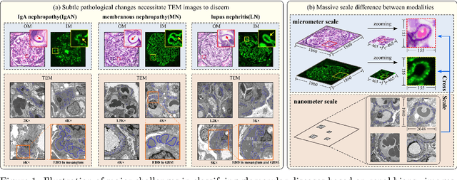
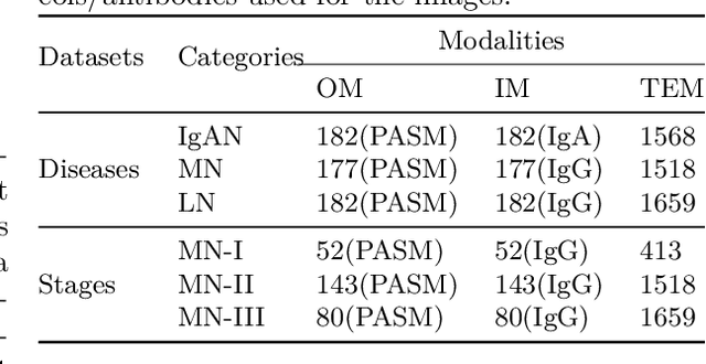
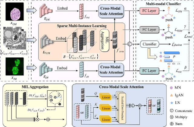
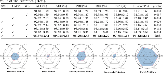
Constructing a multi-modal automatic classification model based on three types of renal biopsy images can assist pathologists in glomerular multi-disease identification. However, the substantial scale difference between transmission electron microscopy (TEM) image features at the nanoscale and optical microscopy (OM) or immunofluorescence microscopy (IM) images at the microscale poses a challenge for existing multi-modal and multi-scale models in achieving effective feature fusion and improving classification accuracy. To address this issue, we propose a cross-modal ultra-scale learning network (CMUS-Net) for the auxiliary diagnosis of multiple glomerular diseases. CMUS-Net utilizes multiple ultrastructural information to bridge the scale difference between nanometer and micrometer images. Specifically, we introduce a sparse multi-instance learning module to aggregate features from TEM images. Furthermore, we design a cross-modal scale attention module to facilitate feature interaction, enhancing pathological semantic information. Finally, multiple loss functions are combined, allowing the model to weigh the importance among different modalities and achieve precise classification of glomerular diseases. Our method follows the conventional process of renal biopsy pathology diagnosis and, for the first time, performs automatic classification of multiple glomerular diseases including IgA nephropathy (IgAN), membranous nephropathy (MN), and lupus nephritis (LN) based on images from three modalities and two scales. On an in-house dataset, CMUS-Net achieves an ACC of 95.37+/-2.41%, an AUC of 99.05+/-0.53%, and an F1-score of 95.32+/-2.41%. Extensive experiments demonstrate that CMUS-Net outperforms other well-known multi-modal or multi-scale methods and show its generalization capability in staging MN. Code is available at https://github.com/SMU-GL-Group/MultiModal_lkx/tree/main.
DCMIL: A Progressive Representation Learning Model of Whole Slide Images for Cancer Prognosis Analysis
Oct 16, 2025The burgeoning discipline of computational pathology shows promise in harnessing whole slide images (WSIs) to quantify morphological heterogeneity and develop objective prognostic modes for human cancers. However, progress is impeded by the computational bottleneck of gigapixel-size inputs and the scarcity of dense manual annotations. Current methods often overlook fine-grained information across multi-magnification WSIs and variations in tumor microenvironments. Here, we propose an easy-to-hard progressive representation learning model, termed dual-curriculum contrastive multi-instance learning (DCMIL), to efficiently process WSIs for cancer prognosis. The model does not rely on dense annotations and enables the direct transformation of gigapixel-size WSIs into outcome predictions. Extensive experiments on twelve cancer types (5,954 patients, 12.54 million tiles) demonstrate that DCMIL outperforms standard WSI-based prognostic models. Additionally, DCMIL identifies fine-grained prognosis-salient regions, provides robust instance uncertainty estimation, and captures morphological differences between normal and tumor tissues, with the potential to generate new biological insights. All codes have been made publicly accessible at https://github.com/tuuuc/DCMIL.
SpineMamba: Enhancing 3D Spinal Segmentation in Clinical Imaging through Residual Visual Mamba Layers and Shape Priors
Aug 28, 2024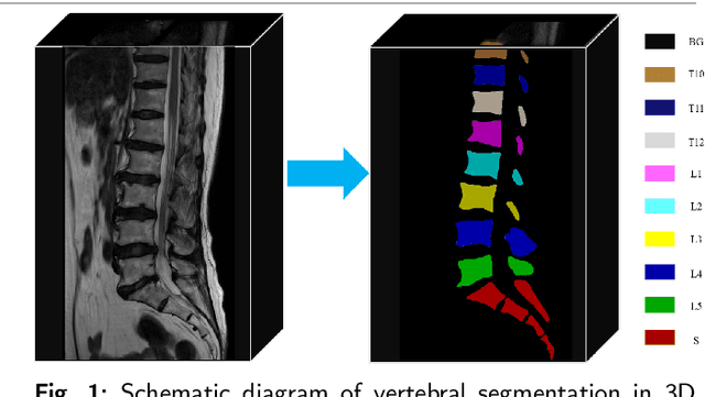
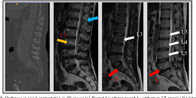

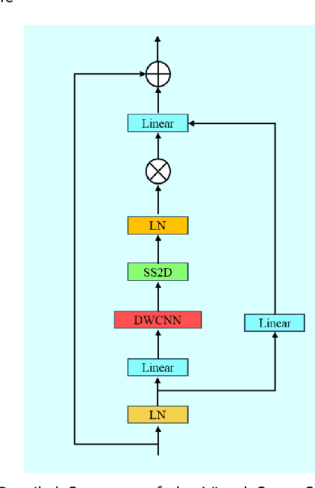
Accurate segmentation of 3D clinical medical images is critical in the diagnosis and treatment of spinal diseases. However, the inherent complexity of spinal anatomy and uncertainty inherent in current imaging technologies, poses significant challenges for semantic segmentation of spinal images. Although convolutional neural networks (CNNs) and Transformer-based models have made some progress in spinal segmentation, their limitations in handling long-range dependencies hinder further improvements in segmentation accuracy.To address these challenges, we introduce a residual visual Mamba layer to effectively capture and model the deep semantic features and long-range spatial dependencies of 3D spinal data. To further enhance the structural semantic understanding of the vertebrae, we also propose a novel spinal shape prior module that captures specific anatomical information of the spine from medical images, significantly enhancing the model's ability to extract structural semantic information of the vertebrae. Comparative and ablation experiments on two datasets demonstrate that SpineMamba outperforms existing state-of-the-art models. On the CT dataset, the average Dice similarity coefficient for segmentation reaches as high as 94.40, while on the MR dataset, it reaches 86.95. Notably, compared to the renowned nnU-Net, SpineMamba achieves superior segmentation performance, exceeding it by up to 2 percentage points. This underscores its accuracy, robustness, and excellent generalization capabilities.
Performance of Medical Image Fusion in High-level Analysis Tasks: A Mutual Enhancement Framework for Unaligned PAT and MRI Image Fusion
Jul 04, 2024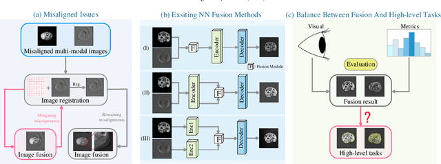
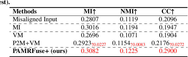
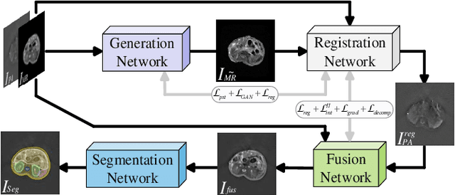
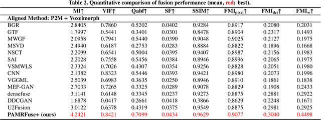
Photoacoustic tomography (PAT) offers optical contrast, whereas magnetic resonance imaging (MRI) excels in imaging soft tissue and organ anatomy. The fusion of PAT with MRI holds promising application prospects due to their complementary advantages. Existing image fusion have made considerable progress in pre-registered images, yet spatial deformations are difficult to avoid in medical imaging scenarios. More importantly, current algorithms focus on visual quality and statistical metrics, thus overlooking the requirements of high-level tasks. To address these challenges, we proposes a unsupervised fusion model, termed PAMRFuse+, which integrates image generation and registration. Specifically, a cross-modal style transfer network is introduced to simplify cross-modal registration to single-modal registration. Subsequently, a multi-level registration network is employed to predict displacement vector fields. Furthermore, a dual-branch feature decomposition fusion network is proposed to address the challenges of cross-modal feature modeling and decomposition by integrating modality-specific and modality-shared features. PAMRFuse+ achieves satisfactory results in registering and fusing unaligned PAT-MRI datasets. Moreover, for the first time, we evaluate the performance of medical image fusion with contour segmentation and multi-organ instance segmentation. Extensive experimental demonstrations reveal the advantages of PAMRFuse+ in improving the performance of medical image analysis tasks.
A multi-stage semi-supervised learning for ankle fracture classification on CT images
Mar 29, 2024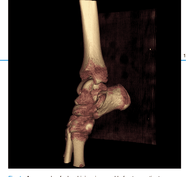
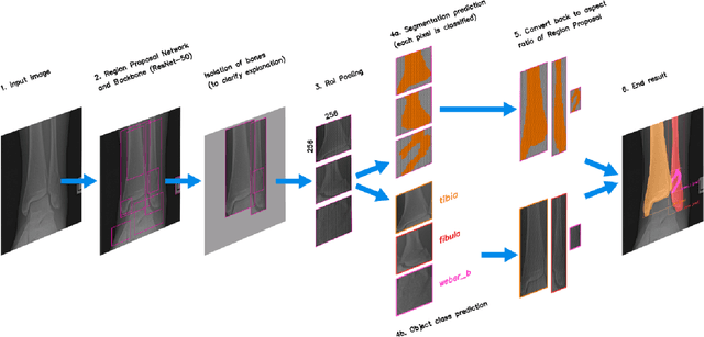
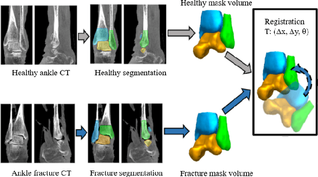
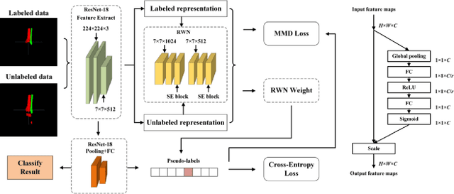
Because of the complicated mechanism of ankle injury, it is very difficult to diagnose ankle fracture in clinic. In order to simplify the process of fracture diagnosis, an automatic diagnosis model of ankle fracture was proposed. Firstly, a tibia-fibula segmentation network is proposed for the joint tibiofibular region of the ankle joint, and the corresponding segmentation dataset is established on the basis of fracture data. Secondly, the image registration method is used to register the bone segmentation mask with the normal bone mask. Finally, a semi-supervised classifier is constructed to make full use of a large number of unlabeled data to classify ankle fractures. Experiments show that the proposed method can segment fractures with fracture lines accurately and has better performance than the general method. At the same time, this method is superior to classification network in several indexes.
Decomposing and Coupling Saliency Map for Lesion Segmentation in Ultrasound Images
Aug 02, 2023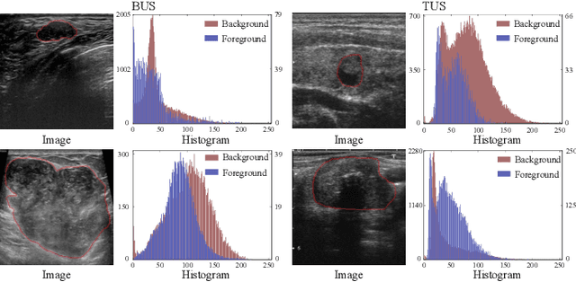
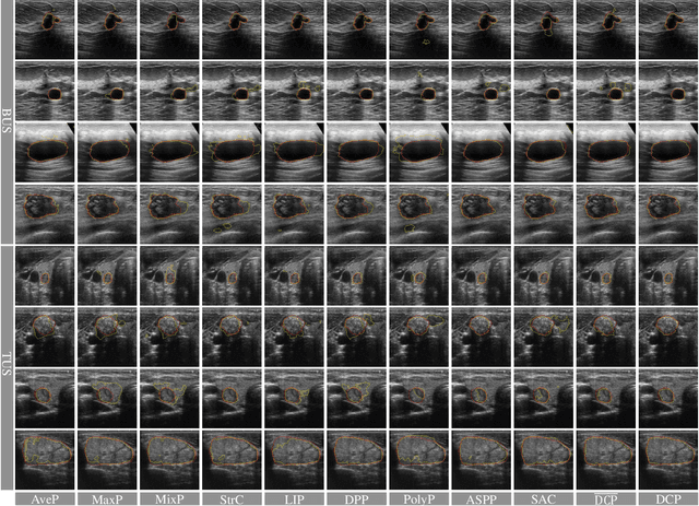
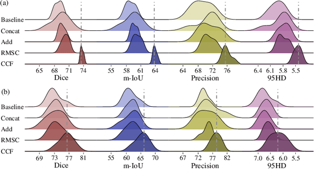
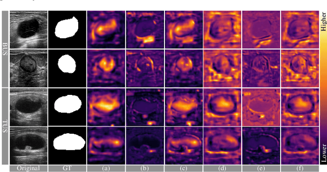
Complex scenario of ultrasound image, in which adjacent tissues (i.e., background) share similar intensity with and even contain richer texture patterns than lesion region (i.e., foreground), brings a unique challenge for accurate lesion segmentation. This work presents a decomposition-coupling network, called DC-Net, to deal with this challenge in a (foreground-background) saliency map disentanglement-fusion manner. The DC-Net consists of decomposition and coupling subnets, and the former preliminarily disentangles original image into foreground and background saliency maps, followed by the latter for accurate segmentation under the assistance of saliency prior fusion. The coupling subnet involves three aspects of fusion strategies, including: 1) regional feature aggregation (via differentiable context pooling operator in the encoder) to adaptively preserve local contextual details with the larger receptive field during dimension reduction; 2) relation-aware representation fusion (via cross-correlation fusion module in the decoder) to efficiently fuse low-level visual characteristics and high-level semantic features during resolution restoration; 3) dependency-aware prior incorporation (via coupler) to reinforce foreground-salient representation with the complementary information derived from background representation. Furthermore, a harmonic loss function is introduced to encourage the network to focus more attention on low-confidence and hard samples. The proposed method is evaluated on two ultrasound lesion segmentation tasks, which demonstrates the remarkable performance improvement over existing state-of-the-art methods.
Multi-View Imputation and Cross-Attention Network Based on Incomplete Longitudinal and Multi-Modal Data for Alzheimer's Disease Prediction
Jun 16, 2022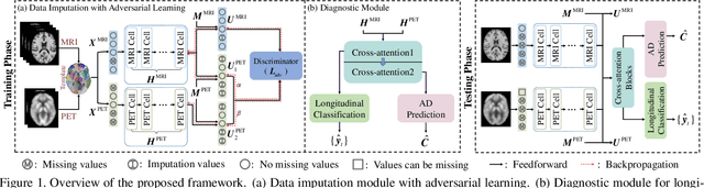
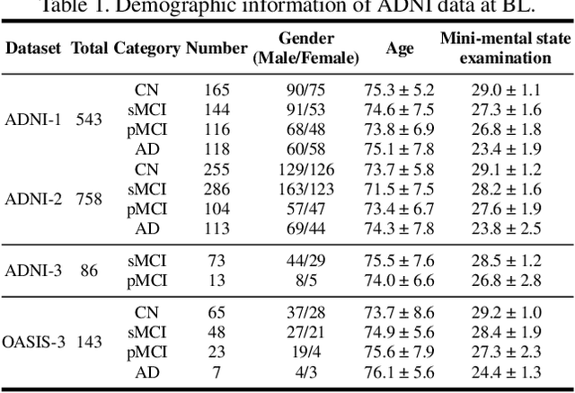
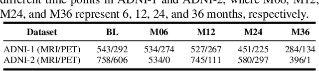
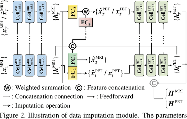
Longitudinal variations and complementary information inherent in longitudinal and multi-modal data play an important role in Alzheimer's disease (AD) prediction, particularly in identifying subjects with mild cognitive impairment who are about to have AD. However, longitudinal and multi-modal data may have missing data, which hinders the effective application of these data. Additionally, previous longitudinal studies require existing longitudinal data to achieve prediction, but AD prediction is expected to be conducted at patients' baseline visit (BL) in clinical practice. Thus, we proposed a multi-view imputation and cross-attention network (MCNet) to integrate data imputation and AD prediction in a unified framework and achieve accurate AD prediction. First, a multi-view imputation method combined with adversarial learning, which can handle a wide range of missing data situations and reduce imputation errors, was presented. Second, two cross-attention blocks were introduced to exploit the potential associations in longitudinal and multi-modal data. Finally, a multi-task learning model was built for data imputation, longitudinal classification, and AD prediction tasks. When the model was properly trained, the disease progression information learned from longitudinal data can be leveraged by BL data to improve AD prediction. The proposed method was tested on two independent testing sets and single-model data at BL to verify its effectiveness and flexibility on AD prediction. Results showed that MCNet outperformed several state-of-the-art methods. Moreover, the interpretability of MCNet was presented. Thus, our MCNet is a tool with a great application potential in longitudinal and multi-modal data analysis for AD prediction. Codes are available at https://github.com/Meiyan88/MCNET.