 Add to Chrome
 Add to Firefox
 Add to Edge
Towards Clinically Interpretable Ophthalmic VQA via Spatially-Grounded Lesion Evidence
May 21, 2026Visual Question Answering (VQA) holds great promise for clinical support, particularly in ophthalmology, where retinal fundus photography is essential for diagnosis. However, ophthalmic VQA benchmarks primarily emphasize answer accuracy, neglecting the explicit visual evidence necessary for clinical interpretability. In this work, we introduce FundusGround, a new benchmark for clinically interpretable ophthalmic VQA with spatially-grounded lesion evidence. Specifically, we propose a three-stage pipeline that collects 10,719 fundus images with 15,595 image-level meticulously annotated lesions. To ensure anatomical consistency and clinical validity, all lesions are spatially localized using the Early Treatment Diabetic Retinopathy Study (ETDRS) grid, enabling standardized mapping to nine clinically meaningful retinal regions. Built upon this structured lesion evidence, 72,706 questions are then generated spanning four formats: open-ended, closed-ended, single-choice, and multiple-choice. We further benchmark multiple general- and medical- large vision-language models using dual metrics for answer accuracy and lesion-level reasoning. The experiments demonstrate that incorporating lesion-level visual evidence consistently improves model performance and transparency, highlighting the necessity of explicit spatial grounding for reliable and explainable ophthalmic VQA.
Awakening the Hydra: Stabilizing Multi-Concept Backdoor Injection in Text-to-Image Diffusion Models
May 19, 2026Text-to-image diffusion models are increasingly developed through open-source reuse and repeated downstream fine-tuning, where reused checkpoints are difficult to verify and thus more susceptible to hidden backdoor behaviors. In such ecosystems, a single pretrained model may be sequentially adapted and redistributed by multiple independent parties, allowing multiple concept-specific trigger-target associations to accumulate in the same model. When these associations coexist, semantic conflicts can be amplified in the shared representation space, leading to cross-concept entanglement and degraded generation quality. Notably, instead of strengthening the attack, such accumulation can destabilize previously injected behaviors and reduce attack reliability. In this work, we systematically investigate backdoor attacks under this interference-prone setting and propose Hydra, a unified framework for robust and controlled multi-concept backdoor injection under cumulative and decentralized reuse. Our core insight is that stable backdoor injection under large-scale multi-concept settings requires explicitly constraining trigger semantics while coordinating cross-task interactions during optimization. Specifically, Hydra performs evolutionary trigger search in the text encoder space to identify triggers that are semantically aligned with their target concepts while remaining stable across other injected concepts. It further combines multi-task fine-tuning with trigger-clean regularization to improve training stability under dense multi-concept injection. Extensive experiments across multiple diffusion backbones under rigorous multi-concept settings show that Hydra maintains effective backdoor activation while preserving clean generation fidelity and image quality. For instance, across 8 attackers and 500 concept pairs, Hydra maintains ~95% ASR and strong clean generation.
Meta-FC: Meta-Learning with Feature Consistency for Robust and Generalizable Watermarking
Feb 25, 2026Deep learning-based watermarking has made remarkable progress in recent years. To achieve robustness against various distortions, current methods commonly adopt a training strategy where a \underline{\textbf{s}}ingle \underline{\textbf{r}}andom \underline{\textbf{d}}istortion (SRD) is chosen as the noise layer in each training batch. However, the SRD strategy treats distortions independently within each batch, neglecting the inherent relationships among different types of distortions and causing optimization conflicts across batches. As a result, the robustness and generalizability of the watermarking model are limited. To address this issue, we propose a novel training strategy that enhances robustness and generalization via \underline{\textbf{meta}}-learning with \underline{\textbf{f}}eature \underline{\textbf{c}}onsistency (Meta-FC). Specifically, we randomly sample multiple distortions from the noise pool to construct a meta-training task, while holding out one distortion as a simulated ``unknown'' distortion for the meta-testing phase. Through meta-learning, the model is encouraged to identify and utilize neurons that exhibit stable activations across different types of distortions, mitigating the optimization conflicts caused by the random sampling of diverse distortions in each batch. To further promote the transformation of stable activations into distortion-invariant representations, we introduce a feature consistency loss that constrains the decoded features of the same image subjected to different distortions to remain consistent. Extensive experiments demonstrate that, compared to the SRD training strategy, Meta-FC improves the robustness and generalization of various watermarking models by an average of 1.59\%, 4.71\%, and 2.38\% under high-intensity, combined, and unknown distortions.
Graph Federated Learning for Personalized Privacy Recommendation
Aug 08, 2025Federated recommendation systems (FedRecs) have gained significant attention for providing privacy-preserving recommendation services. However, existing FedRecs assume that all users have the same requirements for privacy protection, i.e., they do not upload any data to the server. The approaches overlook the potential to enhance the recommendation service by utilizing publicly available user data. In real-world applications, users can choose to be private or public. Private users' interaction data is not shared, while public users' interaction data can be shared. Inspired by the issue, this paper proposes a novel Graph Federated Learning for Personalized Privacy Recommendation (GFed-PP) that adapts to different privacy requirements while improving recommendation performance. GFed-PP incorporates the interaction data of public users to build a user-item interaction graph, which is then used to form a user relationship graph. A lightweight graph convolutional network (GCN) is employed to learn each user's user-specific personalized item embedding. To protect user privacy, each client learns the user embedding and the scoring function locally. Additionally, GFed-PP achieves optimization of the federated recommendation framework through the initialization of item embedding on clients and the aggregation of the user relationship graph on the server. Experimental results demonstrate that GFed-PP significantly outperforms existing methods for five datasets, offering superior recommendation accuracy without compromising privacy. This framework provides a practical solution for accommodating varying privacy preferences in federated recommendation systems.
HiD-VAE: Interpretable Generative Recommendation via Hierarchical and Disentangled Semantic IDs
Aug 06, 2025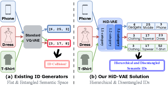
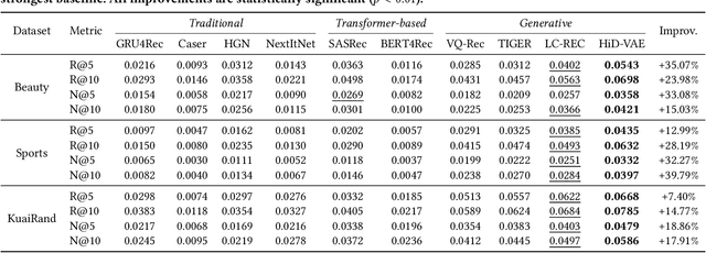
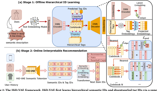
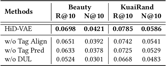
Recommender systems are indispensable for helping users navigate the immense item catalogs of modern online platforms. Recently, generative recommendation has emerged as a promising paradigm, unifying the conventional retrieve-and-rank pipeline into an end-to-end model capable of dynamic generation. However, existing generative methods are fundamentally constrained by their unsupervised tokenization, which generates semantic IDs suffering from two critical flaws: (1) they are semantically flat and uninterpretable, lacking a coherent hierarchy, and (2) they are prone to representation entanglement (i.e., ``ID collisions''), which harms recommendation accuracy and diversity. To overcome these limitations, we propose HiD-VAE, a novel framework that learns hierarchically disentangled item representations through two core innovations. First, HiD-VAE pioneers a hierarchically-supervised quantization process that aligns discrete codes with multi-level item tags, yielding more uniform and disentangled IDs. Crucially, the trained codebooks can predict hierarchical tags, providing a traceable and interpretable semantic path for each recommendation. Second, to combat representation entanglement, HiD-VAE incorporates a novel uniqueness loss that directly penalizes latent space overlap. This mechanism not only resolves the critical ID collision problem but also promotes recommendation diversity by ensuring a more comprehensive utilization of the item representation space. These high-quality, disentangled IDs provide a powerful foundation for downstream generative models. Extensive experiments on three public benchmarks validate HiD-VAE's superior performance against state-of-the-art methods. The code is available at https://anonymous.4open.science/r/HiD-VAE-84B2.
Fine-tuning is Not Fine: Mitigating Backdoor Attacks in GNNs with Limited Clean Data
Jan 10, 2025


Graph Neural Networks (GNNs) have achieved remarkable performance through their message-passing mechanism. However, recent studies have highlighted the vulnerability of GNNs to backdoor attacks, which can lead the model to misclassify graphs with attached triggers as the target class. The effectiveness of recent promising defense techniques, such as fine-tuning or distillation, is heavily contingent on having comprehensive knowledge of the sufficient training dataset. Empirical studies have shown that fine-tuning methods require a clean dataset of 20% to reduce attack accuracy to below 25%, while distillation methods require a clean dataset of 15%. However, obtaining such a large amount of clean data is commonly impractical. In this paper, we propose a practical backdoor mitigation framework, denoted as GRAPHNAD, which can capture high-quality intermediate-layer representations in GNNs to enhance the distillation process with limited clean data. To achieve this, we address the following key questions: How to identify the appropriate attention representations in graphs for distillation? How to enhance distillation with limited data? By adopting the graph attention transfer method, GRAPHNAD can effectively align the intermediate-layer attention representations of the backdoored model with that of the teacher model, forcing the backdoor neurons to transform into benign ones. Besides, we extract the relation maps from intermediate-layer transformation and enforce the relation maps of the backdoored model to be consistent with that of the teacher model, thereby ensuring model accuracy while further reducing the influence of backdoors. Extensive experimental results show that by fine-tuning a teacher model with only 3% of the clean data, GRAPHNAD can reduce the attack success rate to below 5%.
DMGNN: Detecting and Mitigating Backdoor Attacks in Graph Neural Networks
Oct 18, 2024Recent studies have revealed that GNNs are highly susceptible to multiple adversarial attacks. Among these, graph backdoor attacks pose one of the most prominent threats, where attackers cause models to misclassify by learning the backdoored features with injected triggers and modified target labels during the training phase. Based on the features of the triggers, these attacks can be categorized into out-of-distribution (OOD) and in-distribution (ID) graph backdoor attacks, triggers with notable differences from the clean sample feature distributions constitute OOD backdoor attacks, whereas the triggers in ID backdoor attacks are nearly identical to the clean sample feature distributions. Existing methods can successfully defend against OOD backdoor attacks by comparing the feature distribution of triggers and clean samples but fail to mitigate stealthy ID backdoor attacks. Due to the lack of proper supervision signals, the main task accuracy is negatively affected in defending against ID backdoor attacks. To bridge this gap, we propose DMGNN against OOD and ID graph backdoor attacks that can powerfully eliminate stealthiness to guarantee defense effectiveness and improve the model performance. Specifically, DMGNN can easily identify the hidden ID and OOD triggers via predicting label transitions based on counterfactual explanation. To further filter the diversity of generated explainable graphs and erase the influence of the trigger features, we present a reverse sampling pruning method to screen and discard the triggers directly on the data level. Extensive experimental evaluations on open graph datasets demonstrate that DMGNN far outperforms the state-of-the-art (SOTA) defense methods, reducing the attack success rate to 5% with almost negligible degradation in model performance (within 3.5%).
"No Matter What You Do!": Mitigating Backdoor Attacks in Graph Neural Networks
Oct 02, 2024


Recent studies have exposed that GNNs are vulnerable to several adversarial attacks, among which backdoor attack is one of the toughest. Similar to Deep Neural Networks (DNNs), backdoor attacks in GNNs lie in the fact that the attacker modifies a portion of graph data by embedding triggers and enforces the model to learn the trigger feature during the model training process. Despite the massive prior backdoor defense works on DNNs, defending against backdoor attacks in GNNs is largely unexplored, severely hindering the widespread application of GNNs in real-world tasks. To bridge this gap, we present GCleaner, the first backdoor mitigation method on GNNs. GCleaner can mitigate the presence of the backdoor logic within backdoored GNNs by reversing the backdoor learning procedure, aiming to restore the model performance to a level similar to that is directly trained on the original clean dataset. To achieve this objective, we ask: How to recover universal and hard backdoor triggers in GNNs? How to unlearn the backdoor trigger feature while maintaining the model performance? We conduct the graph trigger recovery via the explanation method to identify optimal trigger locations, facilitating the search of universal and hard backdoor triggers in the feature space of the backdoored model through maximal similarity. Subsequently, we introduce the backdoor unlearning mechanism, which combines knowledge distillation and gradient-based explainable knowledge for fine-grained backdoor erasure. Extensive experimental evaluations on four benchmark datasets demonstrate that GCleaner can reduce the backdoor attack success rate to 10% with only 1% of clean data, and has almost negligible degradation in model performance, which far outperforms the state-of-the-art (SOTA) defense methods.
Benchmarking the CoW with the TopCoW Challenge: Topology-Aware Anatomical Segmentation of the Circle of Willis for CTA and MRA
Dec 29, 2023


The Circle of Willis (CoW) is an important network of arteries connecting major circulations of the brain. Its vascular architecture is believed to affect the risk, severity, and clinical outcome of serious neuro-vascular diseases. However, characterizing the highly variable CoW anatomy is still a manual and time-consuming expert task. The CoW is usually imaged by two angiographic imaging modalities, magnetic resonance angiography (MRA) and computed tomography angiography (CTA), but there exist limited public datasets with annotations on CoW anatomy, especially for CTA. Therefore we organized the TopCoW Challenge in 2023 with the release of an annotated CoW dataset and invited submissions worldwide for the CoW segmentation task, which attracted over 140 registered participants from four continents. TopCoW dataset was the first public dataset with voxel-level annotations for CoW's 13 vessel components, made possible by virtual-reality (VR) technology. It was also the first dataset with paired MRA and CTA from the same patients. TopCoW challenge aimed to tackle the CoW characterization problem as a multiclass anatomical segmentation task with an emphasis on topological metrics. The top performing teams managed to segment many CoW components to Dice scores around 90%, but with lower scores for communicating arteries and rare variants. There were also topological mistakes for predictions with high Dice scores. Additional topological analysis revealed further areas for improvement in detecting certain CoW components and matching CoW variant's topology accurately. TopCoW represented a first attempt at benchmarking the CoW anatomical segmentation task for MRA and CTA, both morphologically and topologically.
nnDetection for Intracranial Aneurysms Detection and Localization
May 22, 2023


Intracranial aneurysms are a commonly occurring and life-threatening condition, affecting approximately 3.2% of the general population. Consequently, detecting these aneurysms plays a crucial role in their management. Lesion detection involves the simultaneous localization and categorization of abnormalities within medical images. In this study, we employed the nnDetection framework, a self-configuring framework specifically designed for 3D medical object detection, to detect and localize the 3D coordinates of aneurysms effectively. To capture and extract diverse features associated with aneurysms, we utilized TOF-MRA and structural MRI, both obtained from the ADAM dataset. The performance of our proposed deep learning model was assessed through the utilization of free-response receiver operative characteristics for evaluation purposes. The model's weights and 3D prediction of the bounding box of TOF-MRA are publicly available at https://github.com/orouskhani/AneurysmDetection.