 Add to Chrome
 Add to Firefox
 Add to Edge
Liver Segmentation
Papers and Code
FourierLoss: Shape-Aware Loss Function with Fourier Descriptors
Sep 21, 2023
Encoder-decoder networks become a popular choice for various medical image segmentation tasks. When they are trained with a standard loss function, these networks are not explicitly enforced to preserve the shape integrity of an object in an image. However, this ability of the network is important to obtain more accurate results, especially when there is a low-contrast difference between the object and its surroundings. In response to this issue, this work introduces a new shape-aware loss function, which we name FourierLoss. This loss function relies on quantifying the shape dissimilarity between the ground truth and the predicted segmentation maps through the Fourier descriptors calculated on their objects, and penalizing this dissimilarity in network training. Different than the previous studies, FourierLoss offers an adaptive loss function with trainable hyperparameters that control the importance of the level of the shape details that the network is enforced to learn in the training process. This control is achieved by the proposed adaptive loss update mechanism, which end-to-end learns the hyperparameters simultaneously with the network weights by backpropagation. As a result of using this mechanism, the network can dynamically change its attention from learning the general outline of an object to learning the details of its contour points, or vice versa, in different training epochs. Working on 2879 computed tomography images of 93 subjects, our experiments revealed that the proposed adaptive shape-aware loss function led to statistically significantly better results for liver segmentation, compared to its counterparts.

Autonomous Robotic Ultrasound System for Liver Follow-up Diagnosis: Pilot Phantom Study
May 09, 2024
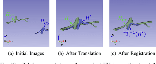
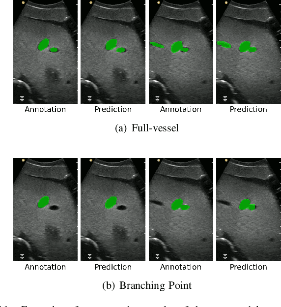

The paper introduces a novel autonomous robot ultrasound (US) system targeting liver follow-up scans for outpatients in local communities. Given a computed tomography (CT) image with specific target regions of interest, the proposed system carries out the autonomous follow-up scan in three steps: (i) initial robot contact to surface, (ii) coordinate mapping between CT image and robot, and (iii) target US scan. Utilizing 3D US-CT registration and deep learning-based segmentation networks, we can achieve precise imaging of 3D hepatic veins, facilitating accurate coordinate mapping between CT and the robot. This enables the automatic localization of follow-up targets within the CT image, allowing the robot to navigate precisely to the target's surface. Evaluation of the ultrasound phantom confirms the quality of the US-CT registration and shows the robot reliably locates the targets in repeated trials. The proposed framework holds the potential to significantly reduce time and costs for healthcare providers, clinicians, and follow-up patients, thereby addressing the increasing healthcare burden associated with chronic disease in local communities.
Synthetic Data as Validation
Oct 24, 2023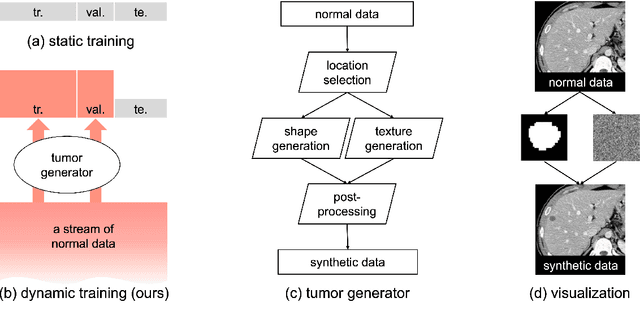
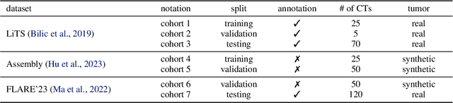
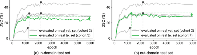
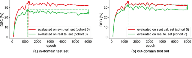
This study leverages synthetic data as a validation set to reduce overfitting and ease the selection of the best model in AI development. While synthetic data have been used for augmenting the training set, we find that synthetic data can also significantly diversify the validation set, offering marked advantages in domains like healthcare, where data are typically limited, sensitive, and from out-domain sources (i.e., hospitals). In this study, we illustrate the effectiveness of synthetic data for early cancer detection in computed tomography (CT) volumes, where synthetic tumors are generated and superimposed onto healthy organs, thereby creating an extensive dataset for rigorous validation. Using synthetic data as validation can improve AI robustness in both in-domain and out-domain test sets. Furthermore, we establish a new continual learning framework that continuously trains AI models on a stream of out-domain data with synthetic tumors. The AI model trained and validated in dynamically expanding synthetic data can consistently outperform models trained and validated exclusively on real-world data. Specifically, the DSC score for liver tumor segmentation improves from 26.7% (95% CI: 22.6%-30.9%) to 34.5% (30.8%-38.2%) when evaluated on an in-domain dataset and from 31.1% (26.0%-36.2%) to 35.4% (32.1%-38.7%) on an out-domain dataset. Importantly, the performance gain is particularly significant in identifying very tiny liver tumors (radius < 5mm) in CT volumes, with Sensitivity improving from 33.1% to 55.4% on an in-domain dataset and 33.9% to 52.3% on an out-domain dataset, justifying the efficacy in early detection of cancer. The application of synthetic data, from both training and validation perspectives, underlines a promising avenue to enhance AI robustness when dealing with data from varying domains.
Attention-based CT Scan Interpolation for Lesion Segmentation of Colorectal Liver Metastases
Aug 30, 2023Small liver lesions common to colorectal liver metastases (CRLMs) are challenging for convolutional neural network (CNN) segmentation models, especially when we have a wide range of slice thicknesses in the computed tomography (CT) scans. Slice thickness of CT images may vary by clinical indication. For example, thinner slices are used for presurgical planning when fine anatomic details of small vessels are required. While keeping the effective radiation dose in patients as low as possible, various slice thicknesses are employed in CRLMs due to their limitations. However, differences in slice thickness across CTs lead to significant performance degradation in CT segmentation models based on CNNs. This paper proposes a novel unsupervised attention-based interpolation model to generate intermediate slices from consecutive triplet slices in CT scans. We integrate segmentation loss during the interpolation model's training to leverage segmentation labels in existing slices to generate middle ones. Unlike common interpolation techniques in CT volumes, our model highlights the regions of interest (liver and lesions) inside the abdominal CT scans in the interpolated slice. Moreover, our model's outputs are consistent with the original input slices while increasing the segmentation performance in two cutting-edge 3D segmentation pipelines. We tested the proposed model on the CRLM dataset to upsample subjects with thick slices and create isotropic volume for our segmentation model. The produced isotropic dataset increases the Dice score in the segmentation of lesions and outperforms other interpolation approaches in terms of interpolation metrics.
Towards Generalizable Tumor Synthesis
Feb 29, 2024Tumor synthesis enables the creation of artificial tumors in medical images, facilitating the training of AI models for tumor detection and segmentation. However, success in tumor synthesis hinges on creating visually realistic tumors that are generalizable across multiple organs and, furthermore, the resulting AI models being capable of detecting real tumors in images sourced from different domains (e.g., hospitals). This paper made a progressive stride toward generalizable tumor synthesis by leveraging a critical observation: early-stage tumors (< 2cm) tend to have similar imaging characteristics in computed tomography (CT), whether they originate in the liver, pancreas, or kidneys. We have ascertained that generative AI models, e.g., Diffusion Models, can create realistic tumors generalized to a range of organs even when trained on a limited number of tumor examples from only one organ. Moreover, we have shown that AI models trained on these synthetic tumors can be generalized to detect and segment real tumors from CT volumes, encompassing a broad spectrum of patient demographics, imaging protocols, and healthcare facilities.
Dimensionality Reduction for Improving Out-of-Distribution Detection in Medical Image Segmentation
Aug 07, 2023Clinically deployed segmentation models are known to fail on data outside of their training distribution. As these models perform well on most cases, it is imperative to detect out-of-distribution (OOD) images at inference to protect against automation bias. This work applies the Mahalanobis distance post hoc to the bottleneck features of a Swin UNETR model that segments the liver on T1-weighted magnetic resonance imaging. By reducing the dimensions of the bottleneck features with principal component analysis, OOD images were detected with high performance and minimal computational load.
When SAM Meets Medical Images: An Investigation of Segment Anything Model (SAM) on Multi-phase Liver Tumor Segmentation
May 04, 2023


Learning to segmentation without large-scale samples is an inherent capability of human. Recently, Segment Anything Model (SAM) performs the significant zero-shot image segmentation, attracting considerable attention from the computer vision community. Here, we investigate the capability of SAM for medical image analysis, especially for multi-phase liver tumor segmentation (MPLiTS), in terms of prompts, data resolution, phases. Experimental results demonstrate that there might be a large gap between SAM and expected performance. Fortunately, the qualitative results show that SAM is a powerful annotation tool for the community of interactive medical image segmentation.
Trustworthy Multi-phase Liver Tumor Segmentation via Evidence-based Uncertainty
May 09, 2023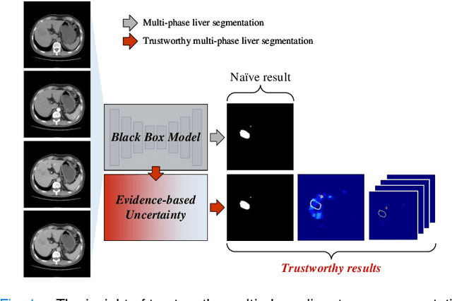
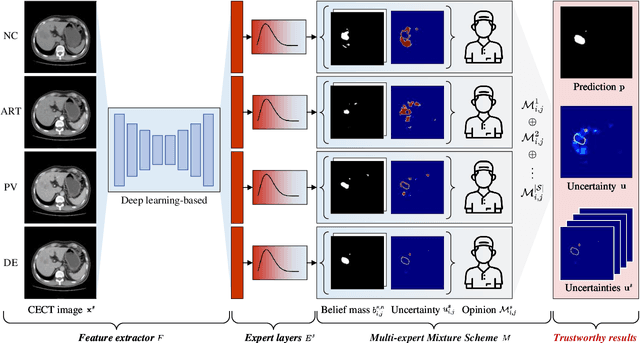
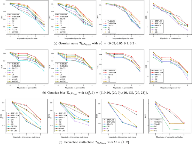
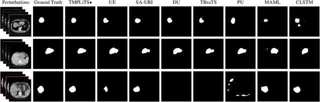
Multi-phase liver contrast-enhanced computed tomography (CECT) images convey the complementary multi-phase information for liver tumor segmentation (LiTS), which are crucial to assist the diagnosis of liver cancer clinically. However, the performances of existing multi-phase liver tumor segmentation (MPLiTS)-based methods suffer from redundancy and weak interpretability, % of the fused result, resulting in the implicit unreliability of clinical applications. In this paper, we propose a novel trustworthy multi-phase liver tumor segmentation (TMPLiTS), which is a unified framework jointly conducting segmentation and uncertainty estimation. The trustworthy results could assist the clinicians to make a reliable diagnosis. Specifically, Dempster-Shafer Evidence Theory (DST) is introduced to parameterize the segmentation and uncertainty as evidence following Dirichlet distribution. The reliability of segmentation results among multi-phase CECT images is quantified explicitly. Meanwhile, a multi-expert mixture scheme (MEMS) is proposed to fuse the multi-phase evidences, which can guarantee the effect of fusion procedure based on theoretical analysis. Experimental results demonstrate the superiority of TMPLiTS compared with the state-of-the-art methods. Meanwhile, the robustness of TMPLiTS is verified, where the reliable performance can be guaranteed against the perturbations.
$\mathrm{SAM^{Med}}$: A medical image annotation framework based on large vision model
Jul 11, 2023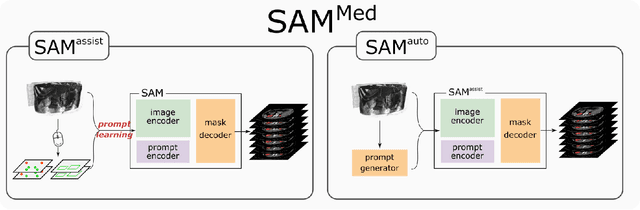
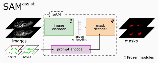

Recently, large vision model, Segment Anything Model (SAM), has revolutionized the computer vision field, especially for image segmentation. SAM presented a new promptable segmentation paradigm that exhibit its remarkable zero-shot generalization ability. An extensive researches have explore the potential and limits of SAM in various downstream tasks. In this study, we presents $\mathrm{SAM^{Med}}$, an enhanced framework for medical image annotation that leverages the capabilities of SAM. $\mathrm{SAM^{Med}}$ framework consisted of two submodules, namely $\mathrm{SAM^{assist}}$ and $\mathrm{SAM^{auto}}$. The $\mathrm{SAM^{assist}}$ demonstrates the generalization ability of SAM to the downstream medical segmentation task using the prompt-learning approach. Results show a significant improvement in segmentation accuracy with only approximately 5 input points. The $\mathrm{SAM^{auto}}$ model aims to accelerate the annotation process by automatically generating input prompts. The proposed SAP-Net model achieves superior segmentation performance with only five annotated slices, achieving an average Dice coefficient of 0.80 and 0.82 for kidney and liver segmentation, respectively. Overall, $\mathrm{SAM^{Med}}$ demonstrates promising results in medical image annotation. These findings highlight the potential of leveraging large-scale vision models in medical image annotation tasks.
TCuPGAN: A novel framework developed for optimizing human-machine interactions in citizen science
Nov 23, 2023In the era of big data in scientific research, there is a necessity to leverage techniques which reduce human effort in labeling and categorizing large datasets by involving sophisticated machine tools. To combat this problem, we present a novel, general purpose model for 3D segmentation that leverages patch-wise adversariality and Long Short-Term Memory to encode sequential information. Using this model alongside citizen science projects which use 3D datasets (image cubes) on the Zooniverse platforms, we propose an iterative human-machine optimization framework where only a fraction of the 2D slices from these cubes are seen by the volunteers. We leverage the patch-wise discriminator in our model to provide an estimate of which slices within these image cubes have poorly generalized feature representations, and correspondingly poor machine performance. These images with corresponding machine proposals would be presented to volunteers on Zooniverse for correction, leading to a drastic reduction in the volunteer effort on citizen science projects. We trained our model on ~2300 liver tissue 3D electron micrographs. Lipid droplets were segmented within these images through human annotation via the `Etch A Cell - Fat Checker' citizen science project, hosted on the Zooniverse platform. In this work, we demonstrate this framework and the selection methodology which resulted in a measured reduction in volunteer effort by more than 60%. We envision this type of joint human-machine partnership will be of great use on future Zooniverse projects.