Junde Wu
Uncertainty-Aware Adapter: Adapting Segment Anything Model (SAM) for Ambiguous Medical Image Segmentation
Mar 19, 2024


Abstract:The Segment Anything Model (SAM) gained significant success in natural image segmentation, and many methods have tried to fine-tune it to medical image segmentation. An efficient way to do so is by using Adapters, specialized modules that learn just a few parameters to tailor SAM specifically for medical images. However, unlike natural images, many tissues and lesions in medical images have blurry boundaries and may be ambiguous. Previous efforts to adapt SAM ignore this challenge and can only predict distinct segmentation. It may mislead clinicians or cause misdiagnosis, especially when encountering rare variants or situations with low model confidence. In this work, we propose a novel module called the Uncertainty-aware Adapter, which efficiently fine-tuning SAM for uncertainty-aware medical image segmentation. Utilizing a conditional variational autoencoder, we encoded stochastic samples to effectively represent the inherent uncertainty in medical imaging. We designed a new module on a standard adapter that utilizes a condition-based strategy to interact with samples to help SAM integrate uncertainty. We evaluated our method on two multi-annotated datasets with different modalities: LIDC-IDRI (lung abnormalities segmentation) and REFUGE2 (optic-cup segmentation). The experimental results show that the proposed model outperforms all the previous methods and achieves the new state-of-the-art (SOTA) on both benchmarks. We also demonstrated that our method can generate diverse segmentation hypotheses that are more realistic as well as heterogeneous.
Not just Birds and Cars: Generic, Scalable and Explainable Models for Professional Visual Recognition
Mar 08, 2024


Abstract:Some visual recognition tasks are more challenging then the general ones as they require professional categories of images. The previous efforts, like fine-grained vision classification, primarily introduced models tailored to specific tasks, like identifying bird species or car brands with limited scalability and generalizability. This paper aims to design a scalable and explainable model to solve Professional Visual Recognition tasks from a generic standpoint. We introduce a biologically-inspired structure named Pro-NeXt and reveal that Pro-NeXt exhibits substantial generalizability across diverse professional fields such as fashion, medicine, and art-areas previously considered disparate. Our basic-sized Pro-NeXt-B surpasses all preceding task-specific models across 12 distinct datasets within 5 diverse domains. Furthermore, we find its good scaling property that scaling up Pro-NeXt in depth and width with increasing GFlops can consistently enhances its accuracy. Beyond scalability and adaptability, the intermediate features of Pro-NeXt achieve reliable object detection and segmentation performance without extra training, highlighting its solid explainability. We will release the code to foster further research in this area.
PromptUNet: Toward Interactive Medical Image Segmentation
May 17, 2023


Abstract:Prompt-based segmentation, also known as interactive segmentation, has recently become a popular approach in image segmentation. A well-designed prompt-based model called Segment Anything Model (SAM) has demonstrated its ability to segment a wide range of natural images, which has sparked a lot of discussion in the community. However, recent studies have shown that SAM performs poorly on medical images. This has motivated us to design a new prompt-based segmentation model specifically for medical image segmentation. In this paper, we combine the prompted-based segmentation paradigm with UNet, which is a widly-recognized successful architecture for medical image segmentation. We have named the resulting model PromptUNet. In order to adapt the real-world clinical use, we expand the existing prompt types in SAM to include novel Supportive Prompts and En-face Prompts. We have evaluated the capabilities of PromptUNet on 19 medical image segmentation tasks using a variety of image modalities, including CT, MRI, ultrasound, fundus, and dermoscopic images. Our results show that PromptUNet outperforms a wide range of state-of-the-art (SOTA) medical image segmentation methods, including nnUNet, TransUNet, UNetr, MedSegDiff, and MSA. Code will be released at: https://github.com/WuJunde/PromptUNet.
PALM: Open Fundus Photograph Dataset with Pathologic Myopia Recognition and Anatomical Structure Annotation
May 13, 2023Abstract:Pathologic myopia (PM) is a common blinding retinal degeneration suffered by highly myopic population. Early screening of this condition can reduce the damage caused by the associated fundus lesions and therefore prevent vision loss. Automated diagnostic tools based on artificial intelligence methods can benefit this process by aiding clinicians to identify disease signs or to screen mass populations using color fundus photographs as inputs. This paper provides insights about PALM, our open fundus imaging dataset for pathological myopia recognition and anatomical structure annotation. Our databases comprises 1200 images with associated labels for the pathologic myopia category and manual annotations of the optic disc, the position of the fovea and delineations of lesions such as patchy retinal atrophy (including peripapillary atrophy) and retinal detachment. In addition, this paper elaborates on other details such as the labeling process used to construct the database, the quality and characteristics of the samples and provides other relevant usage notes.
Medical SAM Adapter: Adapting Segment Anything Model for Medical Image Segmentation
May 11, 2023


Abstract:The Segment Anything Model (SAM) has recently gained popularity in the field of image segmentation. Thanks to its impressive capabilities in all-round segmentation tasks and its prompt-based interface, SAM has sparked intensive discussion within the community. It is even said by many prestigious experts that image segmentation task has been "finished" by SAM. However, medical image segmentation, although an important branch of the image segmentation family, seems not to be included in the scope of Segmenting "Anything". Many individual experiments and recent studies have shown that SAM performs subpar in medical image segmentation. A natural question is how to find the missing piece of the puzzle to extend the strong segmentation capability of SAM to medical image segmentation. In this paper, instead of fine-tuning the SAM model, we propose Med SAM Adapter, which integrates the medical specific domain knowledge to the segmentation model, by a simple yet effective adaptation technique. Although this work is still one of a few to transfer the popular NLP technique Adapter to computer vision cases, this simple implementation shows surprisingly good performance on medical image segmentation. A medical image adapted SAM, which we have dubbed Medical SAM Adapter (MSA), shows superior performance on 19 medical image segmentation tasks with various image modalities including CT, MRI, ultrasound image, fundus image, and dermoscopic images. MSA outperforms a wide range of state-of-the-art (SOTA) medical image segmentation methods, such as nnUNet, TransUNet, UNetr, MedSegDiff, and also outperforms the fully fine-turned MedSAM with a considerable performance gap. Code will be released at: https://github.com/WuJunde/Medical-SAM-Adapter.
A Fast and Lightweight Network for Low-Light Image Enhancement
Apr 06, 2023


Abstract:Low-light images often suffer from severe noise, low brightness, low contrast, and color deviation. While several low-light image enhancement methods have been proposed, there remains a lack of efficient methods that can simultaneously solve all of these problems. In this paper, we introduce FLW-Net, a Fast and LightWeight Network for low-light image enhancement that significantly improves processing speed and overall effect. To achieve efficient low-light image enhancement, we recognize the challenges of the lack of an absolute reference and the need for a large receptive field to obtain global contrast. Therefore, we propose an efficient global feature information extraction component and design loss functions based on relative information to overcome these challenges. Finally, we conduct comparative experiments to demonstrate the effectiveness of the proposed method, and the results confirm that FLW-Net can significantly reduce the complexity of supervised low-light image enhancement networks while improving processing effect. Code is available at https://github.com/hitzhangyu/FLW-Net
Multi-rater Prism: Learning self-calibrated medical image segmentation from multiple raters
Dec 01, 2022


Abstract:In medical image segmentation, it is often necessary to collect opinions from multiple experts to make the final decision. This clinical routine helps to mitigate individual bias. But when data is multiply annotated, standard deep learning models are often not applicable. In this paper, we propose a novel neural network framework, called Multi-Rater Prism (MrPrism) to learn the medical image segmentation from multiple labels. Inspired by the iterative half-quadratic optimization, the proposed MrPrism will combine the multi-rater confidences assignment task and calibrated segmentation task in a recurrent manner. In this recurrent process, MrPrism can learn inter-observer variability taking into account the image semantic properties, and finally converges to a self-calibrated segmentation result reflecting the inter-observer agreement. Specifically, we propose Converging Prism (ConP) and Diverging Prism (DivP) to process the two tasks iteratively. ConP learns calibrated segmentation based on the multi-rater confidence maps estimated by DivP. DivP generates multi-rater confidence maps based on the segmentation masks estimated by ConP. The experimental results show that by recurrently running ConP and DivP, the two tasks can achieve mutual improvement. The final converged segmentation result of MrPrism outperforms state-of-the-art (SOTA) strategies on a wide range of medical image segmentation tasks.
ExpNet: A unified network for Expert-Level Classification
Nov 29, 2022


Abstract:Different from the general visual classification, some classification tasks are more challenging as they need the professional categories of the images. In the paper, we call them expert-level classification. Previous fine-grained vision classification (FGVC) has made many efforts on some of its specific sub-tasks. However, they are difficult to expand to the general cases which rely on the comprehensive analysis of part-global correlation and the hierarchical features interaction. In this paper, we propose Expert Network (ExpNet) to address the unique challenges of expert-level classification through a unified network. In ExpNet, we hierarchically decouple the part and context features and individually process them using a novel attentive mechanism, called Gaze-Shift. In each stage, Gaze-Shift produces a focal-part feature for the subsequent abstraction and memorizes a context-related embedding. Then we fuse the final focal embedding with all memorized context-related embedding to make the prediction. Such an architecture realizes the dual-track processing of partial and global information and hierarchical feature interactions. We conduct the experiments over three representative expert-level classification tasks: FGVC, disease classification, and artwork attributes classification. In these experiments, superior performance of our ExpNet is observed comparing to the state-of-the-arts in a wide range of fields, indicating the effectiveness and generalization of our ExpNet. The code will be made publicly available.
MedSegDiff: Medical Image Segmentation with Diffusion Probabilistic Model
Nov 16, 2022


Abstract:Diffusion probabilistic model (DPM) recently becomes one of the hottest topic in computer vision. Its image generation application such as Imagen, Latent Diffusion Models and Stable Diffusion have shown impressive generation capabilities, which aroused extensive discussion in the community. Many recent studies also found it useful in many other vision tasks, like image deblurring, super-resolution and anomaly detection. Inspired by the success of DPM, we propose the first DPM based model toward general medical image segmentation tasks, which we named MedSegDiff. In order to enhance the step-wise regional attention in DPM for the medical image segmentation, we propose dynamic conditional encoding, which establishes the state-adaptive conditions for each sampling step. We further propose Feature Frequency Parser (FF-Parser), to eliminate the negative effect of high-frequency noise component in this process. We verify MedSegDiff on three medical segmentation tasks with different image modalities, which are optic cup segmentation over fundus images, brain tumor segmentation over MRI images and thyroid nodule segmentation over ultrasound images. The experimental results show that MedSegDiff outperforms state-of-the-art (SOTA) methods with considerable performance gap, indicating the generalization and effectiveness of the proposed model.
Learning to screen Glaucoma like the ophthalmologists
Sep 23, 2022
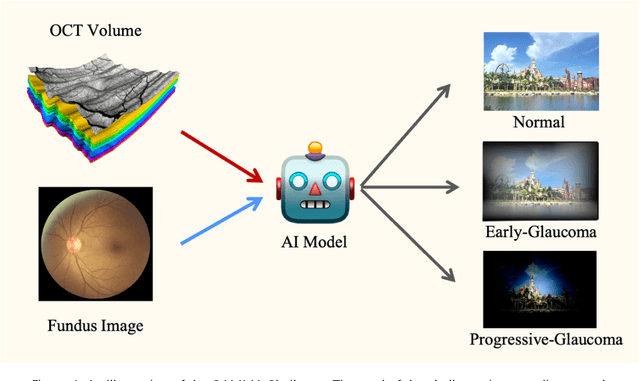
Abstract:GAMMA Challenge is organized to encourage the AI models to screen the glaucoma from a combination of 2D fundus image and 3D optical coherence tomography volume, like the ophthalmologists.
 Add to Chrome
 Add to Firefox
 Add to Edge