 Add to Chrome
 Add to Firefox
 Add to Edge
Routine laboratory trajectories encode the onset of organ-level complications in cancer
Jun 07, 2026Routine laboratory panels drawn during cancer treatment constitute longitudinal physiological recordings of organ function, yet their temporal structure is discarded by single-timepoint prognostic tools. A transformer trained on 2,777,595 laboratory measurements from 3,905 patients with multiple myeloma or ovarian cancer predicted the two-year onset of 162 treatment-associated complications, including therapy-related myelodysplastic syndromes, spanning eight clinical categories, achieving 1.5- to 6.1-fold enrichment above prevalence at the group level. It matched or outperformed non-sequential baselines across grouped endpoints (AUROC gains up to +0.11), demonstrating that longitudinal laboratory trajectories capture evolving complication-specific physiology inaccessible from isolated measurements. Predictions generalised across both cancers, divergence concentrating in disease-specific complications, and biomarker masking recovered signatures consistent with established pathophysiology. External validation on MIMIC-IV and MMRF CoMMpass confirmed transferability across independent healthcare systems (AUROC up to 0.85). Routine oncological laboratory data encode organ deterioration weeks to months before clinical onset, enabling complication-specific surveillance without additional testing infrastructure.
Redefining Instance Matching: A Unified Framework for Part-Aware Matching in Panoptic Segmentation Evaluation
May 29, 2026The Panoptic Quality (PQ) metric is the standard for jointly evaluating instance and semantic segmentation. However, its original definition relies on a One-to-One matching between predicted and ground truth segments, which is only straightforward when the IoU threshold exceeds 0.5. Below 0.5, multiple matching strategies emerge in a poorly explored problem space. We systematically elucidate this space by recasting segment matching as a constrained bipartite assignment problem. Independently bounding the prediction- and ground-truth-side degrees yields four matching strategies: One-to-One, Many-to-One, One-to-Many, and Many-to-Many. We show that the first three are well-defined within the PQ framework, while Many-to-Many falls outside it. These strategies become relevant when instances are fragmented, adjacent objects are difficult to delineate, or annotations are noisy. Central to our framework is a vertex-based accounting of TP, FN, and FP, anchored to ground truth and predicted segments rather than to matching edges. We further show that the framework extends naturally to part-aware panoptic segmentation, and we explore part-aware evaluation on biomedical data. Across configurable case studies we report how different combinations of thresholds and matching strategies behave in practice. We release a unified open-source package built on Panoptica. It exposes Voronoi-based region-wise analysis, part-aware evaluation, and Area Under Threshold Curve computations as configurable options.
Whole-body CT attenuation and volume charts from routine clinical scans via evidence-grounded LLM report filtering
May 07, 2026Interpreting quantitative CT biomarkers, such as organ volume and tissue attenuation, requires large-scale healthy reference distributions. However, creating these is challenging because clinical datasets are often heavily enriched with pathology. Here, we develop an evidence-grounded, cross-verified large language model (LLM) ensemble to filter pathological findings from radiology reports, enabling the construction of pathology-reduced cohorts from over 350,000 CT examinations. Five LLMs, first, flag structure-level abnormality candidates grounded in verbatim report evidence and, second, resolve disagreements via cross-verification. Using distribution-aware generalized additive models for location, scale, and shape, we establish comprehensive whole-body reference charts for 106 anatomical structures (volumes and attenuation) across adulthood, accounting for age, sex, contrast enhancement, and acquisition parameters. Longitudinal analyses reveal structure- and contrast-dependent changes distinct from cross-sectional trends. These resources facilitate covariate-adjusted centile scoring from routine CT, supporting standardized quantitative phenotyping, multi-site imaging studies, and scalable opportunistic screening research.
MAGO-SP: Detection and Correction of Water-Fat Swaps in Magnitude-Only VIBE MRI
Feb 20, 2025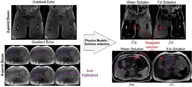
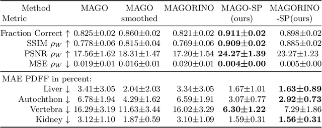
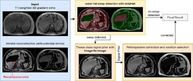
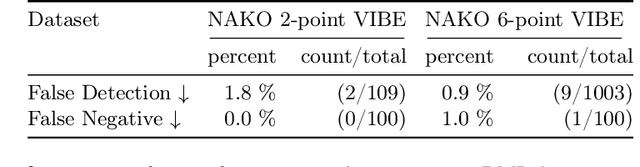
Volume Interpolated Breath-Hold Examination (VIBE) MRI generates images suitable for water and fat signal composition estimation. While the two-point VIBE provides water-fat-separated images, the six-point VIBE allows estimation of the effective transversal relaxation rate R2* and the proton density fat fraction (PDFF), which are imaging markers for health and disease. Ambiguity during signal reconstruction can lead to water-fat swaps. This shortcoming challenges the application of VIBE-MRI for automated PDFF analyses of large-scale clinical data and of population studies. This study develops an automated pipeline to detect and correct water-fat swaps in non-contrast-enhanced VIBE images. Our three-step pipeline begins with training a segmentation network to classify volumes as "fat-like" or "water-like," using synthetic water-fat swaps generated by merging fat and water volumes with Perlin noise. Next, a denoising diffusion image-to-image network predicts water volumes as signal priors for correction. Finally, we integrate this prior into a physics-constrained model to recover accurate water and fat signals. Our approach achieves a < 1% error rate in water-fat swap detection for a 6-point VIBE. Notably, swaps disproportionately affect individuals in the Underweight and Class 3 Obesity BMI categories. Our correction algorithm ensures accurate solution selection in chemical phase MRIs, enabling reliable PDFF estimation. This forms a solid technical foundation for automated large-scale population imaging analysis.
PARASIDE: An Automatic Paranasal Sinus Segmentation and Structure Analysis Tool for MRI
Jan 24, 2025


Chronic rhinosinusitis (CRS) is a common and persistent sinus imflammation that affects 5 - 12\% of the general population. It significantly impacts quality of life and is often difficult to assess due to its subjective nature in clinical evaluation. We introduce PARASIDE, an automatic tool for segmenting air and soft tissue volumes of the structures of the sinus maxillaris, frontalis, sphenodalis and ethmoidalis in T1 MRI. By utilizing that segmentation, we can quantify feature relations that have been observed only manually and subjectively before. We performed an exemplary study and showed both volume and intensity relations between structures and radiology reports. While the soft tissue segmentation is good, the automated annotations of the air volumes are excellent. The average intensity over air structures are consistently below those of the soft tissues, close to perfect separability. Healthy subjects exhibit lower soft tissue volumes and lower intensities. Our developed system is the first automated whole nasal segmentation of 16 structures, and capable of calculating medical relevant features such as the Lund-Mackay score.
Enhancing Interpretability of Vertebrae Fracture Grading using Human-interpretable Prototypes
Apr 03, 2024


Vertebral fracture grading classifies the severity of vertebral fractures, which is a challenging task in medical imaging and has recently attracted Deep Learning (DL) models. Only a few works attempted to make such models human-interpretable despite the need for transparency and trustworthiness in critical use cases like DL-assisted medical diagnosis. Moreover, such models either rely on post-hoc methods or additional annotations. In this work, we propose a novel interpretable-by-design method, ProtoVerse, to find relevant sub-parts of vertebral fractures (prototypes) that reliably explain the model's decision in a human-understandable way. Specifically, we introduce a novel diversity-promoting loss to mitigate prototype repetitions in small datasets with intricate semantics. We have experimented with the VerSe'19 dataset and outperformed the existing prototype-based method. Further, our model provides superior interpretability against the post-hoc method. Importantly, expert radiologists validated the visual interpretability of our results, showing clinical applicability.
The Brain Tumor Segmentation Challenge 2023: Brain MR Image Synthesis for Tumor Segmentation
May 20, 2023
Automated brain tumor segmentation methods are well established, reaching performance levels with clear clinical utility. Most algorithms require four input magnetic resonance imaging (MRI) modalities, typically T1-weighted images with and without contrast enhancement, T2-weighted images, and FLAIR images. However, some of these sequences are often missing in clinical practice, e.g., because of time constraints and/or image artifacts (such as patient motion). Therefore, substituting missing modalities to recover segmentation performance in these scenarios is highly desirable and necessary for the more widespread adoption of such algorithms in clinical routine. In this work, we report the set-up of the Brain MR Image Synthesis Benchmark (BraSyn), organized in conjunction with the Medical Image Computing and Computer-Assisted Intervention (MICCAI) 2023. The objective of the challenge is to benchmark image synthesis methods that realistically synthesize missing MRI modalities given multiple available images to facilitate automated brain tumor segmentation pipelines. The image dataset is multi-modal and diverse, created in collaboration with various hospitals and research institutions.
The Brain Tumor Segmentation Challenge 2023: Local Synthesis of Healthy Brain Tissue via Inpainting
May 15, 2023


A myriad of algorithms for the automatic analysis of brain MR images is available to support clinicians in their decision-making. For brain tumor patients, the image acquisition time series typically starts with a scan that is already pathological. This poses problems, as many algorithms are designed to analyze healthy brains and provide no guarantees for images featuring lesions. Examples include but are not limited to algorithms for brain anatomy parcellation, tissue segmentation, and brain extraction. To solve this dilemma, we introduce the BraTS 2023 inpainting challenge. Here, the participants' task is to explore inpainting techniques to synthesize healthy brain scans from lesioned ones. The following manuscript contains the task formulation, dataset, and submission procedure. Later it will be updated to summarize the findings of the challenge. The challenge is organized as part of the BraTS 2023 challenge hosted at the MICCAI 2023 conference in Vancouver, Canada.
Approaching Peak Ground Truth
Dec 31, 2022

Machine learning models are typically evaluated by computing similarity with reference annotations and trained by maximizing similarity with such. Especially in the bio-medical domain, annotations are subjective and suffer from low inter- and intra-rater reliability. Since annotations only reflect the annotation entity's interpretation of the real world, this can lead to sub-optimal predictions even though the model achieves high similarity scores. Here, the theoretical concept of Peak Ground Truth (PGT) is introduced. PGT marks the point beyond which an increase in similarity with the reference annotation stops translating to better Real World Model Performance (RWMP). Additionally, a quantitative technique to approximate PGT by computing inter- and intra-rater reliability is proposed. Finally, three categories of PGT-aware strategies to evaluate and improve model performance are reviewed.
CheXplaining in Style: Counterfactual Explanations for Chest X-rays using StyleGAN
Jul 15, 2022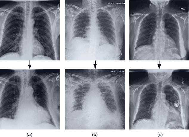
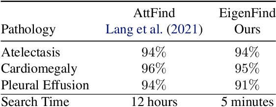
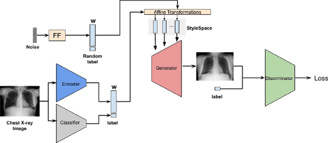
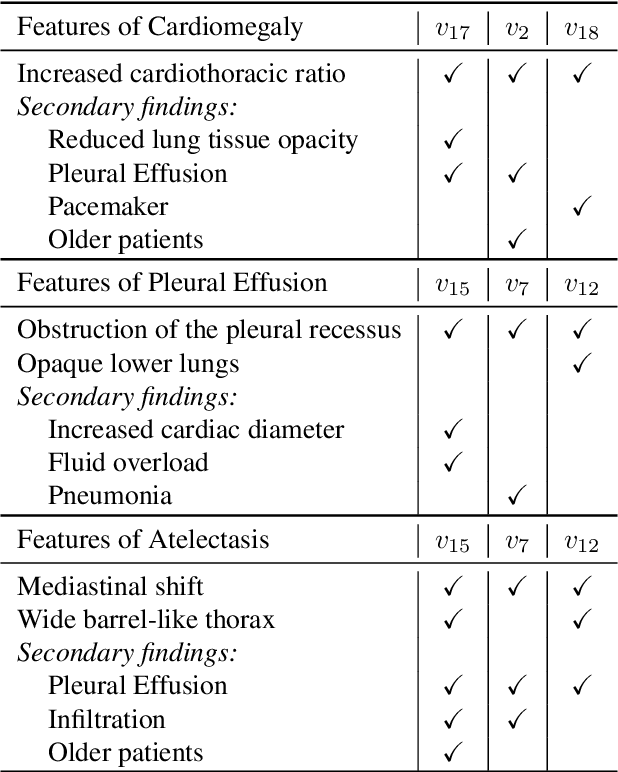
Deep learning models used in medical image analysis are prone to raising reliability concerns due to their black-box nature. To shed light on these black-box models, previous works predominantly focus on identifying the contribution of input features to the diagnosis, i.e., feature attribution. In this work, we explore counterfactual explanations to identify what patterns the models rely on for diagnosis. Specifically, we investigate the effect of changing features within chest X-rays on the classifier's output to understand its decision mechanism. We leverage a StyleGAN-based approach (StyleEx) to create counterfactual explanations for chest X-rays by manipulating specific latent directions in their latent space. In addition, we propose EigenFind to significantly reduce the computation time of generated explanations. We clinically evaluate the relevancy of our counterfactual explanations with the help of radiologists. Our code is publicly available.