 Add to Chrome
 Add to Firefox
 Add to Edge
SurgNarrator: A Generative Retrieval Framework for Surgical Video Understanding
Aug 05, 2026
Surgical procedures unfold as structured and recurring clinical events, whose real-time understanding via intraoperative surgical videos is critical for intraoperative decision-making and support. However, existing video understanding methods force a trade-off: autoregressive video-language models support comprehensive reasoning but are not practical for time-sensitive clinical applications, whereas contrastive models offer low latency but struggle with complex scene understanding. Recently, generative retrieval has been explored for general-domain video understanding, but transferring it to surgery is not trivial because near-identical visual appearances may indicate semantically distinct events, and the terminology involved is highly surgery-specific. To this end, we propose SurgNarrator, a new generative retrieval framework tailored for surgical video understanding. We construct a well-curated surgery-centric vocabulary from surgical captions to define a clinically meaningful retrieval space. We then adapt the pre-trained Qwen3-VL-Embedding-8B to learn discriminative clinical representations with a temporally-aware contrastive objective. During inference, a hierarchical, procedure-aware retrieval strategy narrows the search space to the relevant procedure type, delivering fast and effective responses. Our method is comprehensively evaluated on twelve benchmarks in a zero-shot setting and achieves consistent performance gains over state-of-the-art baselines, while reducing output-stage latency by more than two orders of magnitude compared with the generative baseline.

Overcoming Imperfect Kinematics in Surgical Robotics Through Sim-to-Real Visuomotor Learning
Jun 19, 2026Robot-Assisted Surgery is integral to modern minimally invasive procedures, with automation emerging as the next frontier to enhance precision and reduce surgeon fatigue. This evolution is largely impeded by the inherent kinematic inaccuracies of surgical robots, where unreliable internal sensors lead to significant control errors. While previous methods attempted to mitigate these issues through complex model-based calibration, they often suffer from high cost and limited effectiveness. This work utilises a learning-policy to actively compensate for hardware inaccuracies using closed-loop visual feedback that was trained from a teacher-student learning framework. The policy can fuse unreliable internal readings with precise external visual data, allowing it to correct for kinematic errors in real time without needing a perfect physical model. The learned policy was successfully deployed on the da Vinci Research Kit, where experiments validated the fundamental feasibility of using external vision to overcome internal sensor deficits. This research provides a foundational and reliable control methodology, paving the way for more advanced and robust surgical automation.
MuellerPT: Decomposition Driven Pretraining for Dense Learning in Mueller Polarimetry
May 22, 2026Mueller matrix imaging provides rich, physically meaningful contrast for biomedical tissue analysis, but supervised learning is hindered by scarce dense annotations and strong domain shifts across specimens and acquisition settings. We introduce MuellerPT, a physics guided pre-training approach that learns transferable dense representations by predicting Lu-Chipman decomposition maps from per-pixel 4x4 Mueller matrices. To scale pre-training, we collected a new large Multispectral Animal Polarimetric Organ dataset (MAP-Org). The pre-trained encoder is adapted with a segmentation head for grey vs. white matter segmentation in lamb brain. A classification head is used for colorectal cancer vs. non-cancer classification. Both segmentation and classification are evaluated across few-shot learning scenarios. In segmentation, MuellerPT improves label efficiency and cross specimen transfer compared to models without pre-training, achieving an absolute DICE gain of over 20% compared to the baseline trained from scratch when using 5% of the training data. In classification, MuellerPT also enhances label efficiency, improving overall accuracy by 8% compared to the baseline when using 1% of the training data. We demonstrate MuellerPT's robustness to domain shift with a qualitative evaluation of its predicted Lu-Chipman maps on an ex vivo human oesophagus sample. These results suggest that predicting Lu-Chipman decomposition is an effective and practical pretext task for robust biomedical inference from Mueller polarimetry and can pave the way for future work on label efficient Mueller imaging.
SAMSA: Segment Anything Model Enhanced with Spectral Angles for Hyperspectral Interactive Medical Image Segmentation
Jul 31, 2025Hyperspectral imaging (HSI) provides rich spectral information for medical imaging, yet encounters significant challenges due to data limitations and hardware variations. We introduce SAMSA, a novel interactive segmentation framework that combines an RGB foundation model with spectral analysis. SAMSA efficiently utilizes user clicks to guide both RGB segmentation and spectral similarity computations. The method addresses key limitations in HSI segmentation through a unique spectral feature fusion strategy that operates independently of spectral band count and resolution. Performance evaluation on publicly available datasets has shown 81.0% 1-click and 93.4% 5-click DICE on a neurosurgical and 81.1% 1-click and 89.2% 5-click DICE on an intraoperative porcine hyperspectral dataset. Experimental results demonstrate SAMSA's effectiveness in few-shot and zero-shot learning scenarios and using minimal training examples. Our approach enables seamless integration of datasets with different spectral characteristics, providing a flexible framework for hyperspectral medical image analysis.
Automatic Robotic-Assisted Diffuse Reflectance Spectroscopy Scanning System
Mar 11, 2025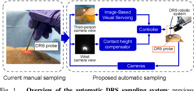
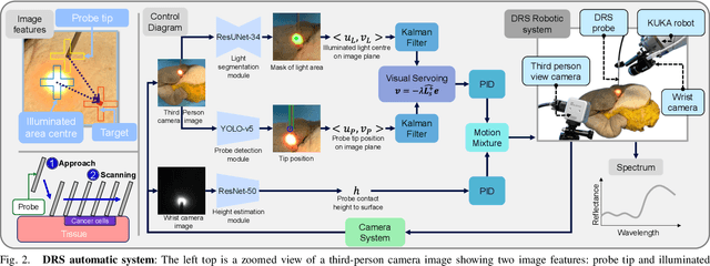
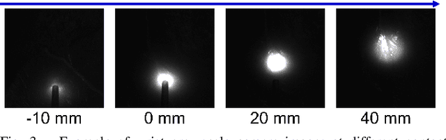
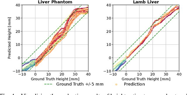
Diffuse Reflectance Spectroscopy (DRS) is a well-established optical technique for tissue composition assessment which has been clinically evaluated for tumour detection to ensure the complete removal of cancerous tissue. While point-wise assessment has many potential applications, incorporating automated large-area scanning would enable holistic tissue sampling with higher consistency. We propose a robotic system to facilitate autonomous DRS scanning with hybrid visual servoing control. A specially designed height compensation module enables precise contact condition control. The evaluation results show that the system can accurately execute the scanning command and acquire consistent DRS spectra with comparable results to the manual collection, which is the current gold standard protocol. Integrating the proposed system into surgery lays the groundwork for autonomous intra-operative DRS tissue assessment with high reliability and repeatability. This could reduce the need for manual scanning by the surgeon while ensuring complete tumor removal in clinical practice.
Hybrid Deep Reinforcement Learning for Radio Tracer Localisation in Robotic-assisted Radioguided Surgery
Mar 11, 2025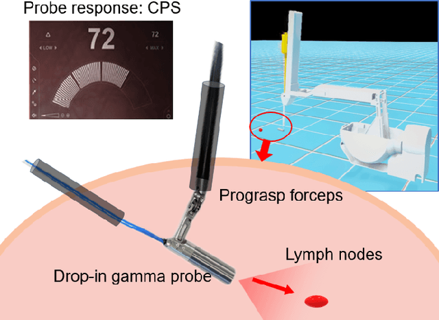
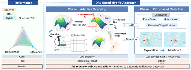
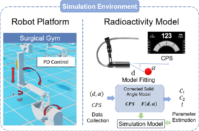
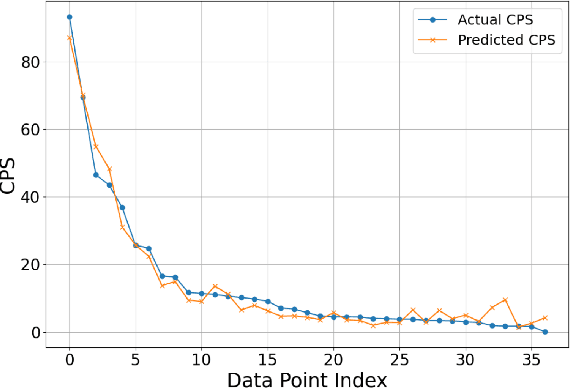
Radioguided surgery, such as sentinel lymph node biopsy, relies on the precise localization of radioactive targets by non-imaging gamma/beta detectors. Manual radioactive target detection based on visual display or audible indication of gamma level is highly dependent on the ability of the surgeon to track and interpret the spatial information. This paper presents a learning-based method to realize the autonomous radiotracer detection in robot-assisted surgeries by navigating the probe to the radioactive target. We proposed novel hybrid approach that combines deep reinforcement learning (DRL) with adaptive robotic scanning. The adaptive grid-based scanning could provide initial direction estimation while the DRL-based agent could efficiently navigate to the target utilising historical data. Simulation experiments demonstrate a 95% success rate, and improved efficiency and robustness compared to conventional techniques. Real-world evaluation on the da Vinci Research Kit (dVRK) further confirms the feasibility of the approach, achieving an 80% success rate in radiotracer detection. This method has the potential to enhance consistency, reduce operator dependency, and improve procedural accuracy in radioguided surgeries.
Memorized action chunking with Transformers: Imitation learning for vision-based tissue surface scanning
Nov 06, 2024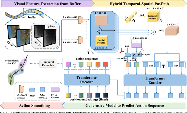
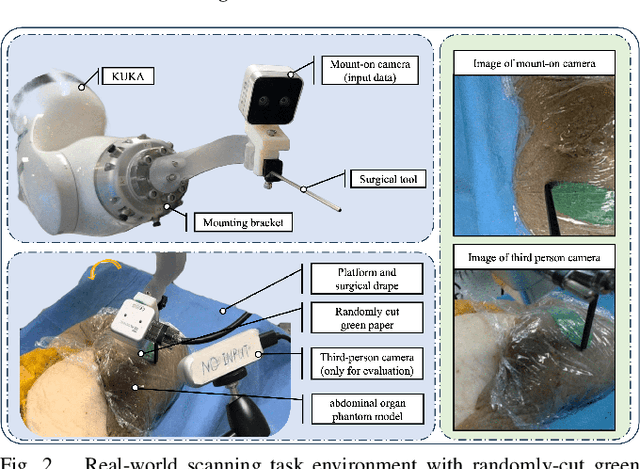
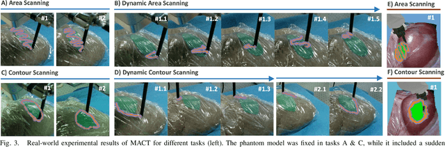
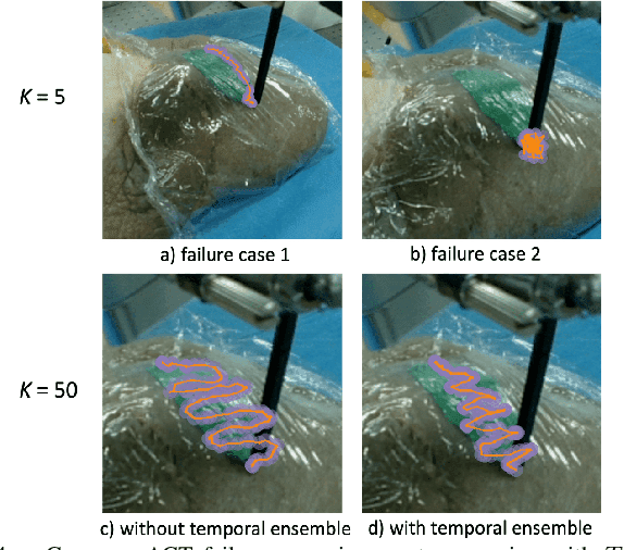
Optical sensing technologies are emerging technologies used in cancer surgeries to ensure the complete removal of cancerous tissue. While point-wise assessment has many potential applications, incorporating automated large area scanning would enable holistic tissue sampling. However, such scanning tasks are challenging due to their long-horizon dependency and the requirement for fine-grained motion. To address these issues, we introduce Memorized Action Chunking with Transformers (MACT), an intuitive yet efficient imitation learning method for tissue surface scanning tasks. It utilizes a sequence of past images as historical information to predict near-future action sequences. In addition, hybrid temporal-spatial positional embeddings were employed to facilitate learning. In various simulation settings, MACT demonstrated significant improvements in contour scanning and area scanning over the baseline model. In real-world testing, with only 50 demonstration trajectories, MACT surpassed the baseline model by achieving a 60-80% success rate on all scanning tasks. Our findings suggest that MACT is a promising model for adaptive scanning in surgical settings.
SurgicalGS: Dynamic 3D Gaussian Splatting for Accurate Robotic-Assisted Surgical Scene Reconstruction
Oct 11, 2024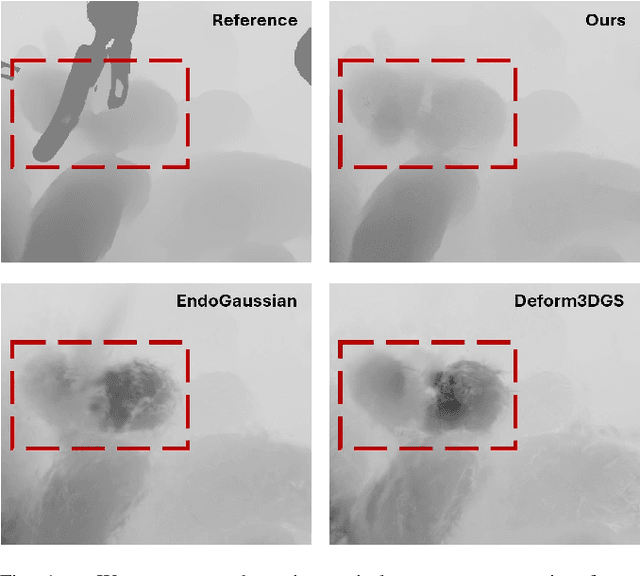


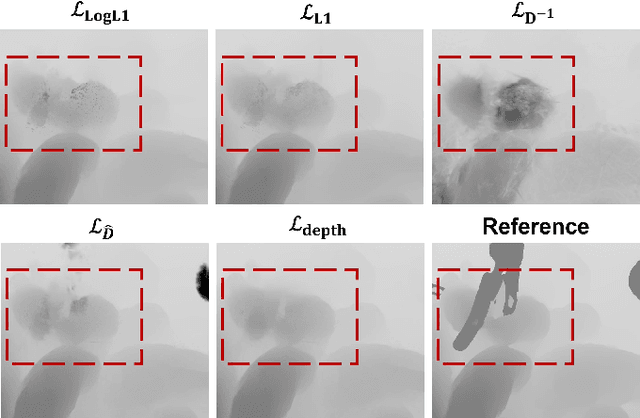
Accurate 3D reconstruction of dynamic surgical scenes from endoscopic video is essential for robotic-assisted surgery. While recent 3D Gaussian Splatting methods have shown promise in achieving high-quality reconstructions with fast rendering speeds, their use of inverse depth loss functions compresses depth variations. This can lead to a loss of fine geometric details, limiting their ability to capture precise 3D geometry and effectiveness in intraoperative application. To address these challenges, we present SurgicalGS, a dynamic 3D Gaussian Splatting framework specifically designed for surgical scene reconstruction with improved geometric accuracy. Our approach first initialises a Gaussian point cloud using depth priors, employing binary motion masks to identify pixels with significant depth variations and fusing point clouds from depth maps across frames for initialisation. We use the Flexible Deformation Model to represent dynamic scene and introduce a normalised depth regularisation loss along with an unsupervised depth smoothness constraint to ensure more accurate geometric reconstruction. Extensive experiments on two real surgical datasets demonstrate that SurgicalGS achieves state-of-the-art reconstruction quality, especially in terms of accurate geometry, advancing the usability of 3D Gaussian Splatting in robotic-assisted surgery.
Tracking Everything in Robotic-Assisted Surgery
Sep 29, 2024Accurate tracking of tissues and instruments in videos is crucial for Robotic-Assisted Minimally Invasive Surgery (RAMIS), as it enables the robot to comprehend the surgical scene with precise locations and interactions of tissues and tools. Traditional keypoint-based sparse tracking is limited by featured points, while flow-based dense two-view matching suffers from long-term drifts. Recently, the Tracking Any Point (TAP) algorithm was proposed to overcome these limitations and achieve dense accurate long-term tracking. However, its efficacy in surgical scenarios remains untested, largely due to the lack of a comprehensive surgical tracking dataset for evaluation. To address this gap, we introduce a new annotated surgical tracking dataset for benchmarking tracking methods for surgical scenarios, comprising real-world surgical videos with complex tissue and instrument motions. We extensively evaluate state-of-the-art (SOTA) TAP-based algorithms on this dataset and reveal their limitations in challenging surgical scenarios, including fast instrument motion, severe occlusions, and motion blur, etc. Furthermore, we propose a new tracking method, namely SurgMotion, to solve the challenges and further improve the tracking performance. Our proposed method outperforms most TAP-based algorithms in surgical instruments tracking, and especially demonstrates significant improvements over baselines in challenging medical videos.
Detecting the Sensing Area of A Laparoscopic Probe in Minimally Invasive Cancer Surgery
Jul 07, 2023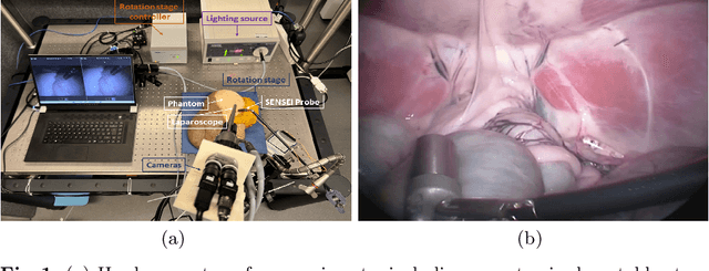
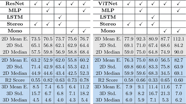

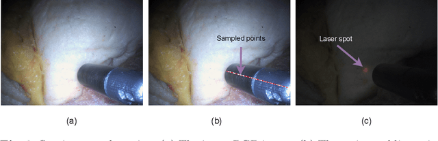
In surgical oncology, it is challenging for surgeons to identify lymph nodes and completely resect cancer even with pre-operative imaging systems like PET and CT, because of the lack of reliable intraoperative visualization tools. Endoscopic radio-guided cancer detection and resection has recently been evaluated whereby a novel tethered laparoscopic gamma detector is used to localize a preoperatively injected radiotracer. This can both enhance the endoscopic imaging and complement preoperative nuclear imaging data. However, gamma activity visualization is challenging to present to the operator because the probe is non-imaging and it does not visibly indicate the activity origination on the tissue surface. Initial failed attempts used segmentation or geometric methods, but led to the discovery that it could be resolved by leveraging high-dimensional image features and probe position information. To demonstrate the effectiveness of this solution, we designed and implemented a simple regression network that successfully addressed the problem. To further validate the proposed solution, we acquired and publicly released two datasets captured using a custom-designed, portable stereo laparoscope system. Through intensive experimentation, we demonstrated that our method can successfully and effectively detect the sensing area, establishing a new performance benchmark. Code and data are available at https://github.com/br0202/Sensing_area_detection.git