 Add to Chrome
 Add to Firefox
 Add to Edge
Deep Learning Methods for Lung Cancer Segmentation in Whole-slide Histopathology Images -- the ACDC@LungHP Challenge 2019
Aug 21, 2020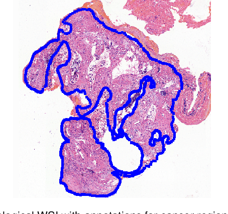
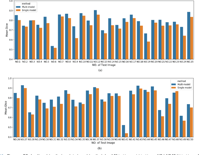
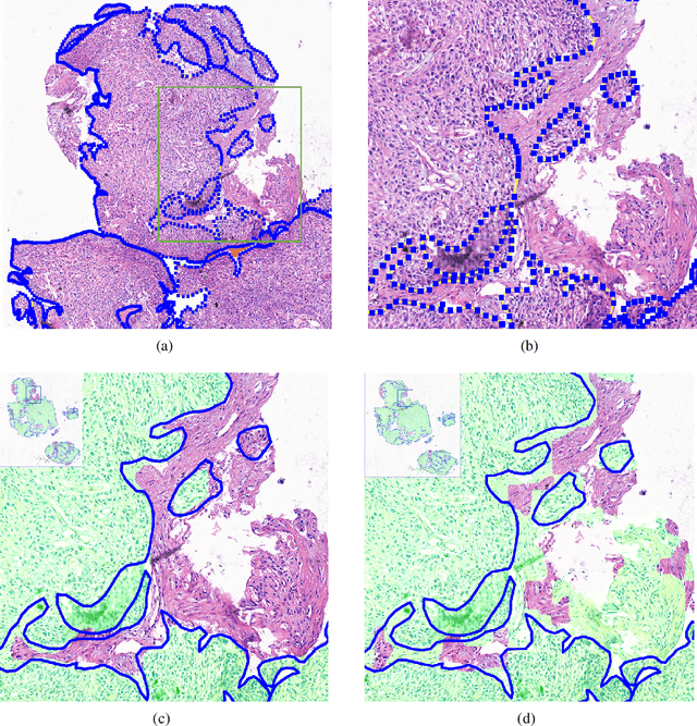
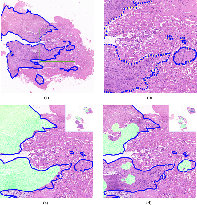
Accurate segmentation of lung cancer in pathology slides is a critical step in improving patient care. We proposed the ACDC@LungHP (Automatic Cancer Detection and Classification in Whole-slide Lung Histopathology) challenge for evaluating different computer-aided diagnosis (CADs) methods on the automatic diagnosis of lung cancer. The ACDC@LungHP 2019 focused on segmentation (pixel-wise detection) of cancer tissue in whole slide imaging (WSI), using an annotated dataset of 150 training images and 50 test images from 200 patients. This paper reviews this challenge and summarizes the top 10 submitted methods for lung cancer segmentation. All methods were evaluated using the false positive rate, false negative rate, and DICE coefficient (DC). The DC ranged from 0.7354$\pm$0.1149 to 0.8372$\pm$0.0858. The DC of the best method was close to the inter-observer agreement (0.8398$\pm$0.0890). All methods were based on deep learning and categorized into two groups: multi-model method and single model method. In general, multi-model methods were significantly better ($\textit{p}$<$0.01$) than single model methods, with mean DC of 0.7966 and 0.7544, respectively. Deep learning based methods could potentially help pathologists find suspicious regions for further analysis of lung cancer in WSI.
Computer-aided diagnosis of lung carcinoma using deep learning - a pilot study
Mar 14, 2018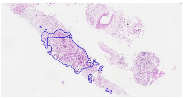
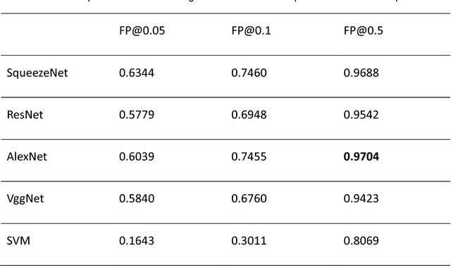
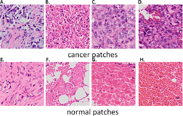
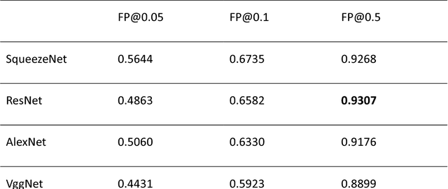
Aim: Early detection and correct diagnosis of lung cancer are the most important steps in improving patient outcome. This study aims to assess which deep learning models perform best in lung cancer diagnosis. Methods: Non-small cell lung carcinoma and small cell lung carcinoma biopsy specimens were consecutively obtained and stained. The specimen slides were diagnosed by two experienced pathologists (over 20 years). Several deep learning models were trained to discriminate cancer and non-cancer biopsies. Result: Deep learning models give reasonable AUC from 0.8810 to 0.9119. Conclusion: The deep learning analysis could help to speed up the detection process for the whole-slide image (WSI) and keep the comparable detection rate with human observer.
Optimize transfer learning for lung diseases in bronchoscopy using a new concept: sequential fine-tuning
Feb 10, 2018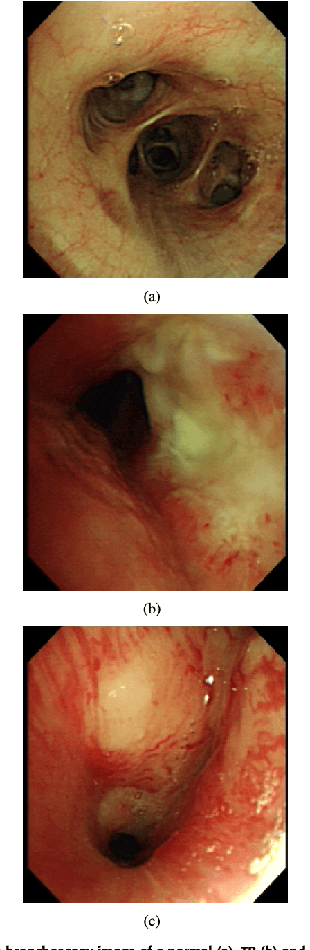
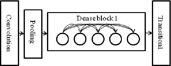
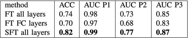
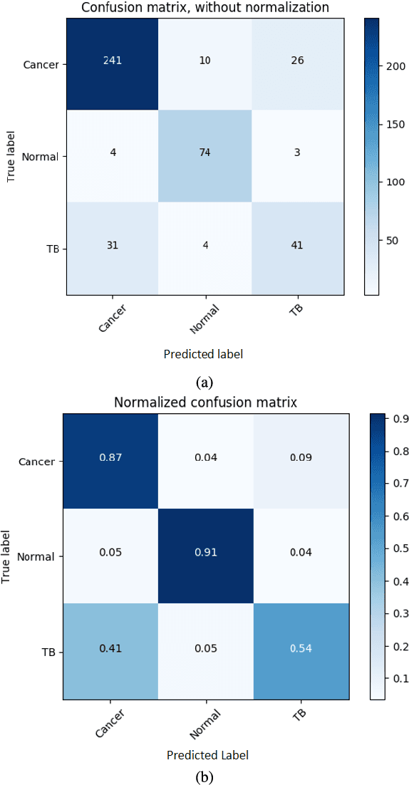
Bronchoscopy inspection as a follow-up procedure from the radiological imaging plays a key role in lung disease diagnosis and determining treatment plans for the patients. Doctors needs to make a decision whether to biopsy the patients timely when performing bronchoscopy. However, the doctors also needs to be very selective with biopsies as biopsies may cause uncontrollable bleeding of the lung tissue which is life-threaten. To help doctors to be more selective on biopsies and provide a second opinion on diagnosis, in this work, we propose a computer-aided diagnosis (CAD) system for lung diseases including cancers and tuberculosis (TB). The system is developed based on transfer learning. We propose a novel transfer learning method: sentential fine-tuning . Compared to traditional fine-tuning methods, our methods achieves the best performance. We obtained a overall accuracy of 77.0% a dataset of 81 normal cases, 76 tuberculosis cases and 277 lung cancer cases while the other traditional transfer learning methods achieve an accuracy of 73% and 68%. . The detection accuracy of our method for cancers, TB and normal cases are 87%, 54% and 91% respectively. This indicates that the CAD system has potential to improve lung disease diagnosis accuracy in bronchoscopy and it also might be used to be more selective with biopsies.