 Add to Chrome
 Add to Firefox
 Add to Edge
Weakly Supervised Estimation of Shadow Confidence Maps in Ultrasound Imaging
Nov 21, 2018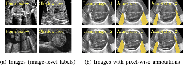
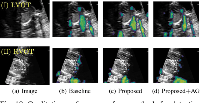
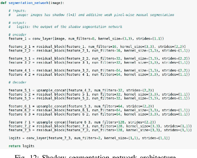
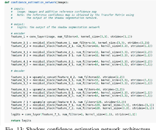
Detecting acoustic shadows in ultrasound images is important in many clinical and engineering applications. Real-time feedback of acoustic shadows can guide sonographers to a standardized diagnostic viewing plane with minimal artifacts and can provide additional information for other automatic image analysis algorithms. However, automatically detecting shadow regions is challenging because pixel-wise annotation of acoustic shadows is subjective and time consuming. In this paper we propose a weakly supervised method for automatic confidence estimation of acoustic shadow regions, which is able to generate a dense shadow-focused confidence map. During training, a multi-task module for shadow segmentation is built to learn general shadow features according based image-level annotations as well as a small number of coarse pixel-wise shadow annotations. A transfer function is then established to extend the binary shadow segmentation to a reference confidence map. In addition, a confidence estimation network is proposed to learn the mapping between input images and the reference confidence maps. This confidence estimation network is able to predict shadow confidence maps directly from input images during inference. We evaluate DICE, soft DICE, recall, precision, mean squared error and inter-class correlation to verify the effectiveness of our method. Our method outperforms the state-of-the-art qualitatively and quantitatively. We further demonstrate the applicability of our method by integrating shadow confidence maps into tasks such as ultrasound image classification, multi-view image fusion and automated biometric measurements.
Standard Plane Detection in 3D Fetal Ultrasound Using an Iterative Transformation Network
Oct 07, 2018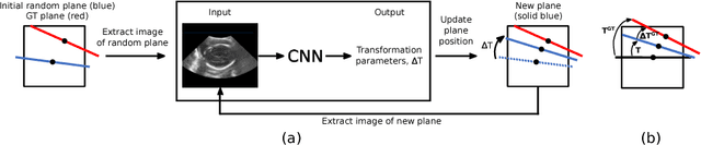
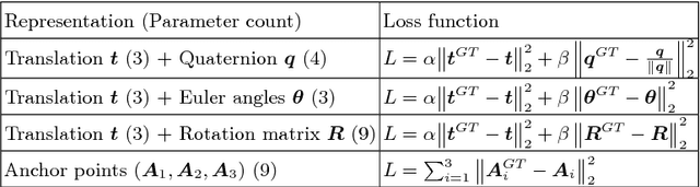
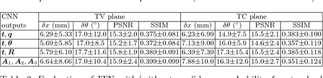
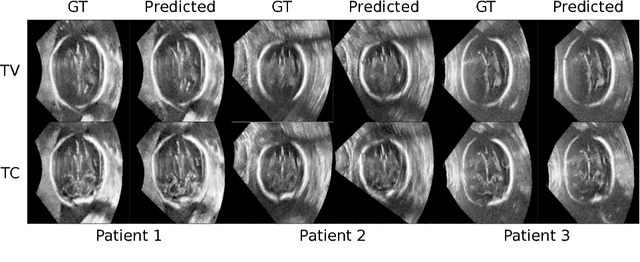
Standard scan plane detection in fetal brain ultrasound (US) forms a crucial step in the assessment of fetal development. In clinical settings, this is done by manually manoeuvring a 2D probe to the desired scan plane. With the advent of 3D US, the entire fetal brain volume containing these standard planes can be easily acquired. However, manual standard plane identification in 3D volume is labour-intensive and requires expert knowledge of fetal anatomy. We propose a new Iterative Transformation Network (ITN) for the automatic detection of standard planes in 3D volumes. ITN uses a convolutional neural network to learn the relationship between a 2D plane image and the transformation parameters required to move that plane towards the location/orientation of the standard plane in the 3D volume. During inference, the current plane image is passed iteratively to the network until it converges to the standard plane location. We explore the effect of using different transformation representations as regression outputs of ITN. Under a multi-task learning framework, we introduce additional classification probability outputs to the network to act as confidence measures for the regressed transformation parameters in order to further improve the localisation accuracy. When evaluated on 72 US volumes of fetal brain, our method achieves an error of 3.83mm/12.7 degrees and 3.80mm/12.6 degrees for the transventricular and transcerebellar planes respectively and takes 0.46s per plane. Source code is publicly available at https://github.com/yuanwei1989/plane-detection.
* 8 pages, 2 figures, accepted for MICCAI 2018; Added link to source code
Fast Multiple Landmark Localisation Using a Patch-based Iterative Network
Oct 07, 2018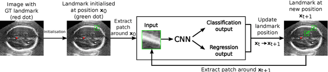
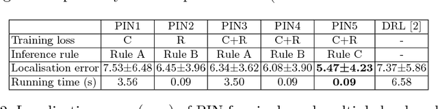
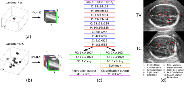
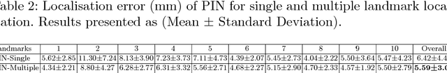
We propose a new Patch-based Iterative Network (PIN) for fast and accurate landmark localisation in 3D medical volumes. PIN utilises a Convolutional Neural Network (CNN) to learn the spatial relationship between an image patch and anatomical landmark positions. During inference, patches are repeatedly passed to the CNN until the estimated landmark position converges to the true landmark location. PIN is computationally efficient since the inference stage only selectively samples a small number of patches in an iterative fashion rather than a dense sampling at every location in the volume. Our approach adopts a multi-task learning framework that combines regression and classification to improve localisation accuracy. We extend PIN to localise multiple landmarks by using principal component analysis, which models the global anatomical relationships between landmarks. We have evaluated PIN using 72 3D ultrasound images from fetal screening examinations. PIN achieves quantitatively an average landmark localisation error of 5.59mm and a runtime of 0.44s to predict 10 landmarks per volume. Qualitatively, anatomical 2D standard scan planes derived from the predicted landmark locations are visually similar to the clinical ground truth. Source code is publicly available at https://github.com/yuanwei1989/landmark-detection.
* 8 pages, 4 figures, Accepted for MICCAI 2018
Weakly Supervised Localisation for Fetal Ultrasound Images
Aug 02, 2018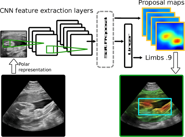
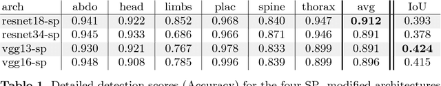
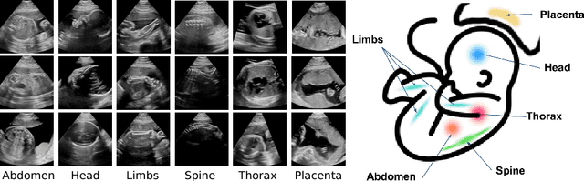
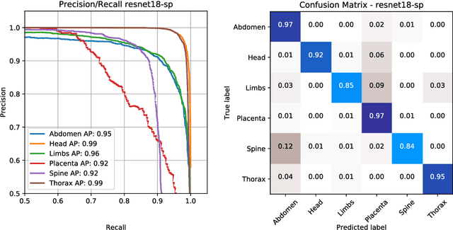
This paper addresses the task of detecting and localising fetal anatomical regions in 2D ultrasound images, where only image-level labels are present at training, i.e. without any localisation or segmentation information. We examine the use of convolutional neural network architectures coupled with soft proposal layers. The resulting network simultaneously performs anatomical region detection (classification) and localisation tasks. We generate a proposal map describing the attention of the network for a particular class. The network is trained on 85,500 2D fetal Ultrasound images and their associated labels. Labels correspond to six anatomical regions: head, spine, thorax, abdomen, limbs, and placenta. Detection achieves an average accuracy of 90\% on individual regions, and show that the proposal maps correlate well with relevant anatomical structures. This work presents itself as a powerful and essential step towards subsequent tasks such as fetal position and pose estimation, organ-specific segmentation, or image-guided navigation. Code and additional material is available at https://ntoussaint.github.io/fetalnav
EchoFusion: Tracking and Reconstruction of Objects in 4D Freehand Ultrasound Imaging without External Trackers
Jul 19, 2018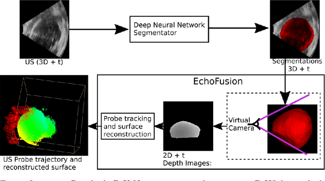
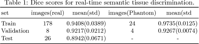
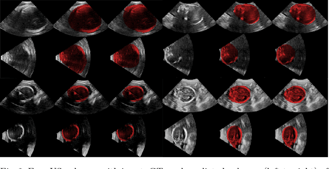
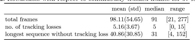
Ultrasound (US) is the most widely used fetal imaging technique. However, US images have limited capture range, and suffer from view dependent artefacts such as acoustic shadows. Compounding of overlapping 3D US acquisitions into a high-resolution volume can extend the field of view and remove image artefacts, which is useful for retrospective analysis including population based studies. However, such volume reconstructions require information about relative transformations between probe positions from which the individual volumes were acquired. In prenatal US scans, the fetus can move independently from the mother, making external trackers such as electromagnetic or optical tracking unable to track the motion between probe position and the moving fetus. We provide a novel methodology for image-based tracking and volume reconstruction by combining recent advances in deep learning and simultaneous localisation and mapping (SLAM). Tracking semantics are established through the use of a Residual 3D U-Net and the output is fed to the SLAM algorithm. As a proof of concept, experiments are conducted on US volumes taken from a whole body fetal phantom, and from the heads of real fetuses. For the fetal head segmentation, we also introduce a novel weak annotation approach to minimise the required manual effort for ground truth annotation. We evaluate our method qualitatively, and quantitatively with respect to tissue discrimination accuracy and tracking robustness.
Human-level Performance On Automatic Head Biometrics In Fetal Ultrasound Using Fully Convolutional Neural Networks
Apr 24, 2018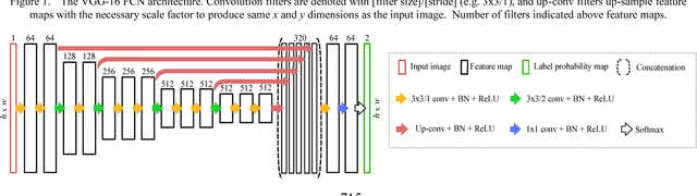
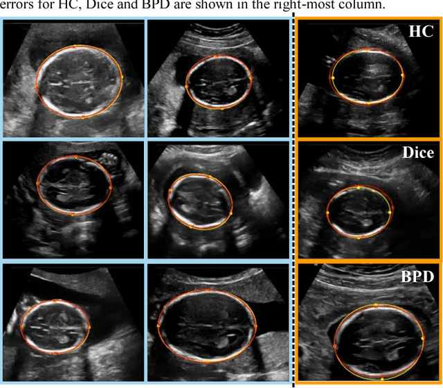
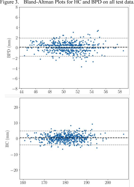
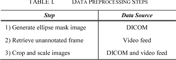
Measurement of head biometrics from fetal ultrasonography images is of key importance in monitoring the healthy development of fetuses. However, the accurate measurement of relevant anatomical structures is subject to large inter-observer variability in the clinic. To address this issue, an automated method utilizing Fully Convolutional Networks (FCN) is proposed to determine measurements of fetal head circumference (HC) and biparietal diameter (BPD). An FCN was trained on approximately 2000 2D ultrasound images of the head with annotations provided by 45 different sonographers during routine screening examinations to perform semantic segmentation of the head. An ellipse is fitted to the resulting segmentation contours to mimic the annotation typically produced by a sonographer. The model's performance was compared with inter-observer variability, where two experts manually annotated 100 test images. Mean absolute model-expert error was slightly better than inter-observer error for HC (1.99mm vs 2.16mm), and comparable for BPD (0.61mm vs 0.59mm), as well as Dice coefficient (0.980 vs 0.980). Our results demonstrate that the model performs at a level similar to a human expert, and learns to produce accurate predictions from a large dataset annotated by many sonographers. Additionally, measurements are generated in near real-time at 15fps on a GPU, which could speed up clinical workflow for both skilled and trainee sonographers.
Attention-Gated Networks for Improving Ultrasound Scan Plane Detection
Apr 15, 2018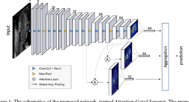
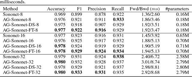
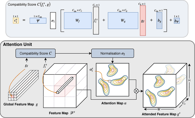
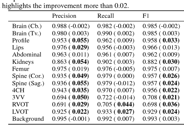
In this work, we apply an attention-gated network to real-time automated scan plane detection for fetal ultrasound screening. Scan plane detection in fetal ultrasound is a challenging problem due the poor image quality resulting in low interpretability for both clinicians and automated algorithms. To solve this, we propose incorporating self-gated soft-attention mechanisms. A soft-attention mechanism generates a gating signal that is end-to-end trainable, which allows the network to contextualise local information useful for prediction. The proposed attention mechanism is generic and it can be easily incorporated into any existing classification architectures, while only requiring a few additional parameters. We show that, when the base network has a high capacity, the incorporated attention mechanism can provide efficient object localisation while improving the overall performance. When the base network has a low capacity, the method greatly outperforms the baseline approach and significantly reduces false positives. Lastly, the generated attention maps allow us to understand the model's reasoning process, which can also be used for weakly supervised object localisation.
SonoNet: Real-Time Detection and Localisation of Fetal Standard Scan Planes in Freehand Ultrasound
Jul 25, 2017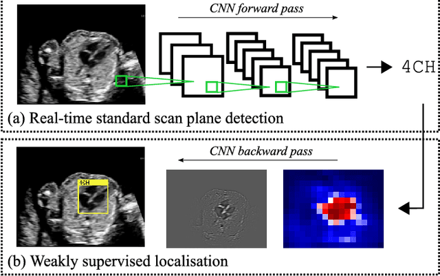
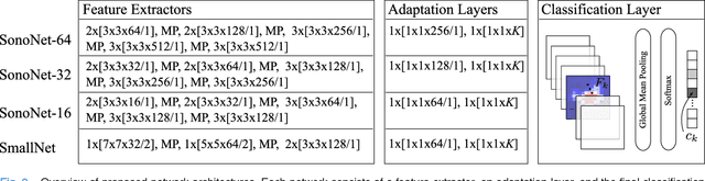
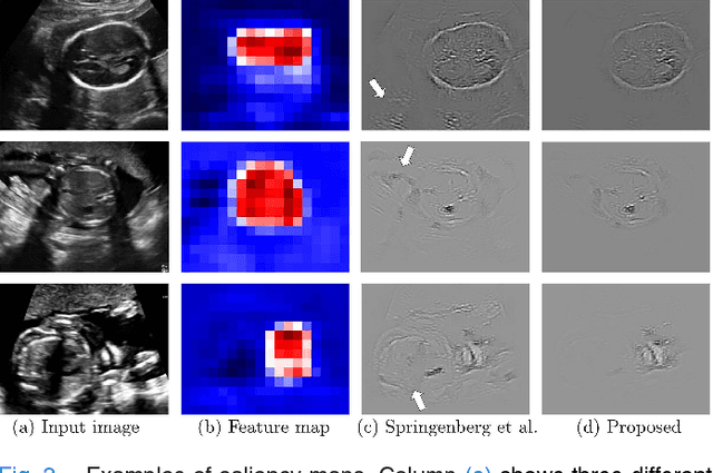
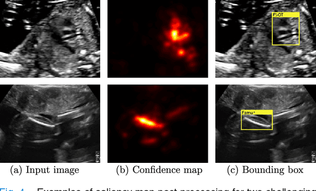
Identifying and interpreting fetal standard scan planes during 2D ultrasound mid-pregnancy examinations are highly complex tasks which require years of training. Apart from guiding the probe to the correct location, it can be equally difficult for a non-expert to identify relevant structures within the image. Automatic image processing can provide tools to help experienced as well as inexperienced operators with these tasks. In this paper, we propose a novel method based on convolutional neural networks which can automatically detect 13 fetal standard views in freehand 2D ultrasound data as well as provide a localisation of the fetal structures via a bounding box. An important contribution is that the network learns to localise the target anatomy using weak supervision based on image-level labels only. The network architecture is designed to operate in real-time while providing optimal output for the localisation task. We present results for real-time annotation, retrospective frame retrieval from saved videos, and localisation on a very large and challenging dataset consisting of images and video recordings of full clinical anomaly screenings. We found that the proposed method achieved an average F1-score of 0.798 in a realistic classification experiment modelling real-time detection, and obtained a 90.09% accuracy for retrospective frame retrieval. Moreover, an accuracy of 77.8% was achieved on the localisation task.