 Add to Chrome
 Add to Firefox
 Add to Edge
Agentic Visual Reasoning in Whole-Slide Pathology Images via Active Perception
Aug 09, 2026
Whole-slide visual reasoning requires identifying sparse diagnostic evidence in gigapixel pathology slides and integrating observations across spatial scales. Existing WSI methods either compress densely sampled patches into global representations or use pretrained vision-language models with heuristic region selection, weakening links between predictions and morphology or lacking pathology-trained observation policies. We present AdaptivePath, an active-perception framework that formulates WSI evidence acquisition as sequential decision making. The Navigator learns question-agnostic abnormality-driven navigation from pathologist-reviewed labels to select observation locations and spatial extents, avoiding costly question-specific trajectory annotations. We train this policy through alternating representation learning and proximal policy optimization, followed by fine-tuning with geometric and appearance consistency objectives to stabilize focus trajectories. During inference, the Navigator hierarchically acquires sparse observations from low to high magnification under a limited ROI budget. A Morphology Interpreter converts observations into question-conditioned evidence, while the Deliberator evaluates evidence and revises intermediate answers across magnifications. The Arbiter integrates deliberation history to produce final answers. AdaptivePath achieves state-of-the-art zero-shot performance on WSI and region pathology VQA benchmarks and reaches 80.14% accuracy for cancer subtype classification across six TCGA cohorts. In a blinded diagnostic-utility study, pathologists using AdaptivePath-selected observation sequences achieve 82.9% accuracy. These results demonstrate that learned active perception enables effective and traceable visual reasoning over gigapixel pathology slides.

PathScale-R1: Cross-scale Reasoning for Pathological Image Analysis
Jul 26, 2026Pathological diagnosis is inherently multi-scale, requiring the integration of global tissue architecture at low magnification with cellular morphology at higher magnification. However, existing pathology benchmarks and vision-language models (VLMs) are still largely developed under single-scale settings, limiting their ability to learn clinically meaningful multi-magnification reasoning. Moreover, naively constructed visual question answering (VQA) tasks may be susceptible to text-only or superficial visual shortcuts, leading to unreliable assessments of visual understanding. To address these limitations, we introduce a benchmark and training framework for shortcut-resistant cross-scale pathology reasoning. We design an Adversarial Text-only Screening strategy for semantic reasoning questions and a Structure-controlled Distractor Sampling strategy for visual grounding questions, encouraging models to rely on cross-scale visual evidence. Based on this pipeline, we construct PathScale-VQA, a high-quality cross-scale pathology VQA benchmark with 10,373 multiple-choice questions grounded in 1,368 diagnostic paths across multiple magnification levels. Building on the semantic reasoning set, PathScale-R1 is optimized through Difficulty-driven Reasoning Distillation supervised fine-tuning followed by reinforcement learning with a Scale-aware Reasoning Structure reward, which encourages the use of evidence across magnifications. Extensive experiments demonstrate state-of-the-art performance of PathScale-R1 on cross-scale reasoning tasks and effective transfer to conventional single-scale pathology VQA. Our code is available at https://github.com/iMVR-PL/PathScale-R1.
PathAgentBench: Benchmarking Evidence-Seeking Vision-Language Models on Whole-Slide Pathology Image
Jul 21, 2026Whole-slide image (WSI) diagnosis requires identifying diagnostically relevant regions, examining them across magnifications, and integrating multi-scale evidence. However, most existing pathology benchmarks evaluate models on pre-cropped patches or pre-extracted slide features, leaving their ability to acquire evidence directly from gigapixel WSIs largely untested. We introduce PathAgentBench, a benchmark for evaluating evidence-seeking vision-language models (VLMs) across four complementary capabilities: image-to-text matching for evidence interpretation, text-to-image retrieval for evidence verification, diagnostic-region localization for evidence acquisition, and multi-scale reasoning for evidence integration. The benchmark is organized as a diagnostic tree that links nested regions across magnifications with scale-specific findings and path-level diagnoses. It contains 1,822 TCGA WSIs and 17,135 diagnostic paths annotated by ten board-certified pathologists. An additional private cohort of 190 breast cancer WSIs with detailed annotations is used to evaluate autonomous whole-slide exploration. We evaluate 20 general-purpose, medical, and pathology-specialized models. Leading open-weight models achieve over 93% accuracy in multi-scale reasoning and over 50% accuracy in both cross-modal matching tasks. In contrast, diagnostic-region localization remains challenging: the best text-guided mean intersection-over-union is below 0.09, underperforming a simple center-based heuristic. During autonomous exploration, the unconditional hit rate decreases from 0.522 at low magnification to 0.185 at intermediate magnification and 0.020 at high magnification. These results reveal a pronounced gap between reasoning over curated evidence and acquiring that evidence directly from WSIs. PathAgentBench provides a unified framework for measuring and improving evidence-seeking pathology models.
Current World Models Lack a Persistent State Core
Jun 18, 2026World models are increasingly regarded as a decisive step toward artificial general intelligence, yet modeling the physical world demands more than rendering convincing frames on demand: it requires an internal world state that keeps evolving over time, decoupled from observation, so that objects endure and events run to their conclusions whether or not a camera is watching, much as the moon holds to its orbit when no one is looking. This requirement is a blind spot of existing benchmarks, which reward surface properties such as fidelity, motion, and camera controllability while never asking whether a generated world keeps evolving once it is unobserved. We introduce \textbf{WRBench}, the first systematic diagnostic benchmark that treats camera motion as an intervention on observability and resolves evaluation into a human-calibrated chain that asks whether the camera executes the requested interaction, whether the scene stays continuous and identifiable while in view, and whether a returning target remains consistent with the event that was set in motion. Across 9{,}600 videos from 23 models spanning four control paradigms, one finding proves stubborn: current systems maintain the observed world as a tracking shot, resuming a returning target in the state at which it was abandoned rather than advancing the event while it went unseen. Because this failure recurs across control paradigms, model families, and increments of scale, robust world-state evolution does not follow from cleaner imagery, tighter control, richer geometric priors, or sheer parameter count We therefore argue that the stability of the physical state kernel and the consistency of worldlines under viewpoint intervention should become first-class objectives of world-model design, so that a world model captures how the world will unfold rather than how the next frame appears.
PathReasoner-R1: Instilling Structured Reasoning into Pathology Vision-Language Model via Knowledge-Guided Policy Optimization
Jan 29, 2026Vision-Language Models (VLMs) are advancing computational pathology with superior visual understanding capabilities. However, current systems often reduce diagnosis to directly output conclusions without verifiable evidence-linked reasoning, which severely limits clinical trust and hinders expert error rectification. To address these barriers, we construct PathReasoner, the first large-scale dataset of whole-slide image (WSI) reasoning. Unlike previous work reliant on unverified distillation, we develop a rigorous knowledge-guided generation pipeline. By leveraging medical knowledge graphs, we explicitly align structured pathological findings and clinical reasoning with diagnoses, generating over 20K high-quality instructional samples. Based on the database, we propose PathReasoner-R1, which synergizes trajectory-masked supervised fine-tuning with reasoning-oriented reinforcement learning to instill structured chain-of-thought capabilities. To ensure medical rigor, we engineer a knowledge-aware multi-granular reward function incorporating an Entity Reward mechanism strictly aligned with knowledge graphs. This effectively guides the model to optimize for logical consistency rather than mere outcome matching, thereby enhancing robustness. Extensive experiments demonstrate that PathReasoner-R1 achieves state-of-the-art performance on both PathReasoner and public benchmarks across various image scales, equipping pathology models with transparent, clinically grounded reasoning capabilities. Dataset and code are available at https://github.com/cyclexfy/PathReasoner-R1.
PathFLIP: Fine-grained Language-Image Pretraining for Versatile Computational Pathology
Dec 19, 2025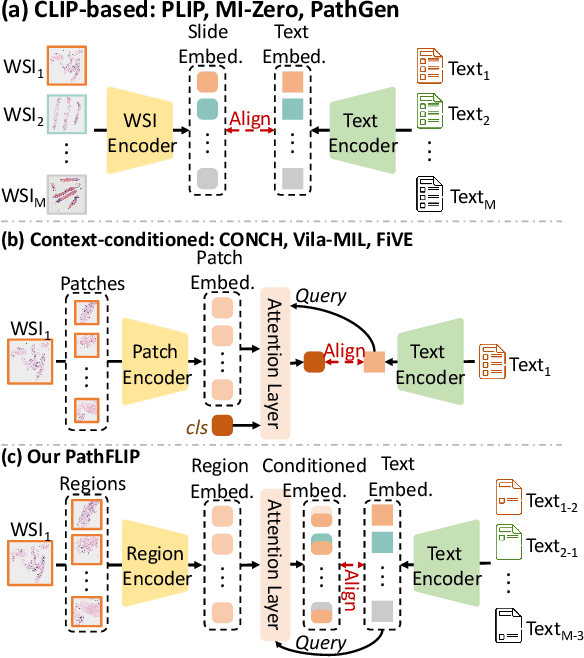

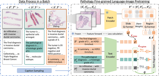
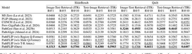
While Vision-Language Models (VLMs) have achieved notable progress in computational pathology (CPath), the gigapixel scale and spatial heterogeneity of Whole Slide Images (WSIs) continue to pose challenges for multimodal understanding. Existing alignment methods struggle to capture fine-grained correspondences between textual descriptions and visual cues across thousands of patches from a slide, compromising their performance on downstream tasks. In this paper, we propose PathFLIP (Pathology Fine-grained Language-Image Pretraining), a novel framework for holistic WSI interpretation. PathFLIP decomposes slide-level captions into region-level subcaptions and generates text-conditioned region embeddings to facilitate precise visual-language grounding. By harnessing Large Language Models (LLMs), PathFLIP can seamlessly follow diverse clinical instructions and adapt to varied diagnostic contexts. Furthermore, it exhibits versatile capabilities across multiple paradigms, efficiently handling slide-level classification and retrieval, fine-grained lesion localization, and instruction following. Extensive experiments demonstrate that PathFLIP outperforms existing large-scale pathological VLMs on four representative benchmarks while requiring significantly less training data, paving the way for fine-grained, instruction-aware WSI interpretation in clinical practice.
Johnson-Lindenstrauss Lemma Guided Network for Efficient 3D Medical Segmentation
Sep 26, 2025Lightweight 3D medical image segmentation remains constrained by a fundamental "efficiency / robustness conflict", particularly when processing complex anatomical structures and heterogeneous modalities. In this paper, we study how to redesign the framework based on the characteristics of high-dimensional 3D images, and explore data synergy to overcome the fragile representation of lightweight methods. Our approach, VeloxSeg, begins with a deployable and extensible dual-stream CNN-Transformer architecture composed of Paired Window Attention (PWA) and Johnson-Lindenstrauss lemma-guided convolution (JLC). For each 3D image, we invoke a "glance-and-focus" principle, where PWA rapidly retrieves multi-scale information, and JLC ensures robust local feature extraction with minimal parameters, significantly enhancing the model's ability to operate with low computational budget. Followed by an extension of the dual-stream architecture that incorporates modal interaction into the multi-scale image-retrieval process, VeloxSeg efficiently models heterogeneous modalities. Finally, Spatially Decoupled Knowledge Transfer (SDKT) via Gram matrices injects the texture prior extracted by a self-supervised network into the segmentation network, yielding stronger representations than baselines at no extra inference cost. Experimental results on multimodal benchmarks show that VeloxSeg achieves a 26% Dice improvement, alongside increasing GPU throughput by 11x and CPU by 48x. Codes are available at https://github.com/JinPLu/VeloxSeg.
IPGPhormer: Interpretable Pathology Graph-Transformer for Survival Analysis
Aug 17, 2025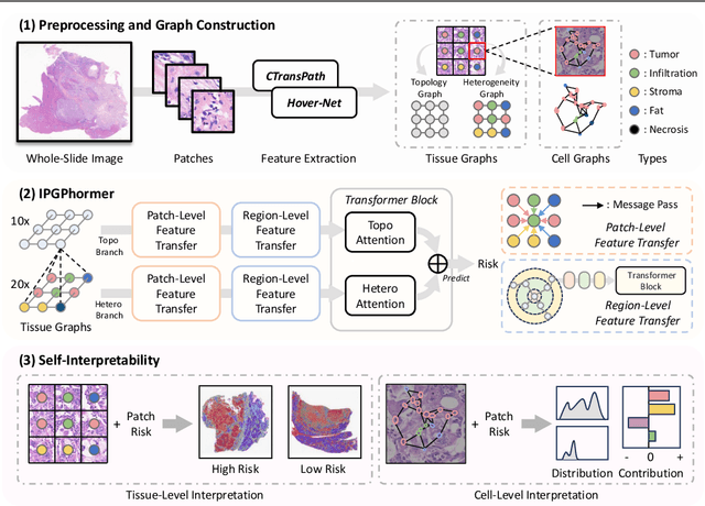
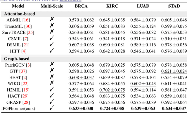

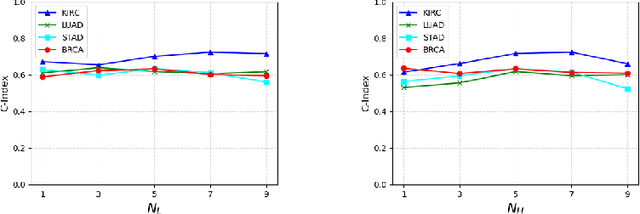
Pathological images play an essential role in cancer prognosis, while survival analysis, which integrates computational techniques, can predict critical clinical events such as patient mortality or disease recurrence from whole-slide images (WSIs). Recent advancements in multiple instance learning have significantly improved the efficiency of survival analysis. However, existing methods often struggle to balance the modeling of long-range spatial relationships with local contextual dependencies and typically lack inherent interpretability, limiting their clinical utility. To address these challenges, we propose the Interpretable Pathology Graph-Transformer (IPGPhormer), a novel framework that captures the characteristics of the tumor microenvironment and models their spatial dependencies across the tissue. IPGPhormer uniquely provides interpretability at both tissue and cellular levels without requiring post-hoc manual annotations, enabling detailed analyses of individual WSIs and cross-cohort assessments. Comprehensive evaluations on four public benchmark datasets demonstrate that IPGPhormer outperforms state-of-the-art methods in both predictive accuracy and interpretability. In summary, our method, IPGPhormer, offers a promising tool for cancer prognosis assessment, paving the way for more reliable and interpretable decision-support systems in pathology. The code is publicly available at https://anonymous.4open.science/r/IPGPhormer-6EEB.
Prototype-Guided Cross-Modal Knowledge Enhancement for Adaptive Survival Prediction
Mar 13, 2025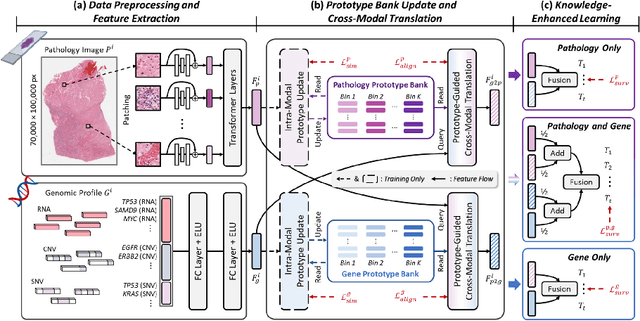
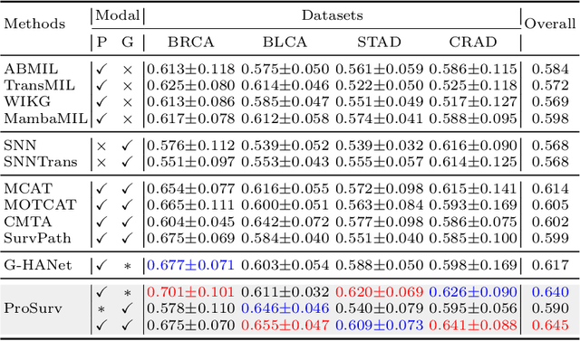
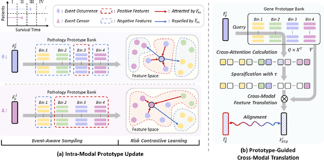
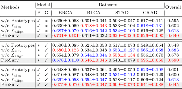
Histo-genomic multimodal survival prediction has garnered growing attention for its remarkable model performance and potential contributions to precision medicine. However, a significant challenge in clinical practice arises when only unimodal data is available, limiting the usability of these advanced multimodal methods. To address this issue, this study proposes a prototype-guided cross-modal knowledge enhancement (ProSurv) framework, which eliminates the dependency on paired data and enables robust learning and adaptive survival prediction. Specifically, we first introduce an intra-modal updating mechanism to construct modality-specific prototype banks that encapsulate the statistics of the whole training set and preserve the modality-specific risk-relevant features/prototypes across intervals. Subsequently, the proposed cross-modal translation module utilizes the learned prototypes to enhance knowledge representation for multimodal inputs and generate features for missing modalities, ensuring robust and adaptive survival prediction across diverse scenarios. Extensive experiments on four public datasets demonstrate the superiority of ProSurv over state-of-the-art methods using either unimodal or multimodal input, and the ablation study underscores its feasibility for broad applicability. Overall, this study addresses a critical practical challenge in computational pathology, offering substantial significance and potential impact in the field.
Multimodal Cross-Task Interaction for Survival Analysis in Whole Slide Pathological Images
Jun 25, 2024

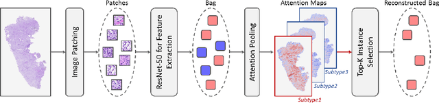

Survival prediction, utilizing pathological images and genomic profiles, is increasingly important in cancer analysis and prognosis. Despite significant progress, precise survival analysis still faces two main challenges: (1) The massive pixels contained in whole slide images (WSIs) complicate the process of pathological images, making it difficult to generate an effective representation of the tumor microenvironment (TME). (2) Existing multimodal methods often rely on alignment strategies to integrate complementary information, which may lead to information loss due to the inherent heterogeneity between pathology and genes. In this paper, we propose a Multimodal Cross-Task Interaction (MCTI) framework to explore the intrinsic correlations between subtype classification and survival analysis tasks. Specifically, to capture TME-related features in WSIs, we leverage the subtype classification task to mine tumor regions. Simultaneously, multi-head attention mechanisms are applied in genomic feature extraction, adaptively performing genes grouping to obtain task-related genomic embedding. With the joint representation of pathological images and genomic data, we further introduce a Transport-Guided Attention (TGA) module that uses optimal transport theory to model the correlation between subtype classification and survival analysis tasks, effectively transferring potential information. Extensive experiments demonstrate the superiority of our approaches, with MCTI outperforming state-of-the-art frameworks on three public benchmarks. \href{https://github.com/jsh0792/MCTI}{https://github.com/jsh0792/MCTI}.