 Add to Chrome
 Add to Firefox
 Add to Edge
HiMatch-AD: DINOv3-driven Hierarchical Matching for Training-free Medical Anomaly Detection
Jun 21, 2026
Anomaly detection is essential for medical image analysis, where pathological regions often appear as rare deviations from normal anatomical structures. While training-based methods have achieved promising performance, they require task-specific optimization and extensive normal data, which limits scalability across modalities and institutions. Training-free approaches offer greater flexibility by leveraging pretrained visual representations, yet existing methods typically rely on simple nearest-neighbor retrieval and naive aggregation strategies, which may fail to capture hierarchical semantics and ignore the reliability of multiple anomaly responses. In this work, we propose HiMatch-AD, a DINOv3-driven hierarchical matching framework for training-free medical anomaly detection. Our method first retrieves semantically relevant normal references via dual-branch matching that jointly considers global CLS-token similarity and patch-level representations. Hierarchical anomaly maps are then generated across multiple transformer stages by comparing clustered normal features with query representations. To robustly aggregate anomaly responses, we introduce a unified uncertainty-based fusion mechanism that adaptively weights maps according to their reliability. The entire framework operates without any task-specific training. Extensive experiments on the BMAD benchmark, including brain MRI, liver CT, and retinal OCT datasets, demonstrate that HiMatch-AD consistently outperforms both training-based and DINO-based state-of-the-art methods, which highlights the effectiveness of multi-level matching and uncertainty-aware fusion for scalable medical anomaly detection.

A Signal-Language Foundation Model for Broad-Spectrum Cardiovascular Assessment from Routine Electrocardiography
May 25, 2026Electrocardiography (ECG) is central to cardiovascular care, but conventional AI models are often restricted to common arrhythmias and may generalize poorly across populations or clinically subtle diseases. We developed ECG Contrastive Language-Image Pre-training (ECGCLIP), a signal-language contrastive learning framework that aligns ECG waveforms with expert diagnostic reports. ECGCLIP was pre-trained on 2,837,962 ECG studies from 1,324,856 patients and evaluated on a held-out internal test set plus nine independent external cohorts comprising about 1.5 million ECGs. Evaluation covered 89 downstream tasks, including 45 ECG diagnoses, 39 echocardiographic targets, and 5 rare cardiac diseases, using PRAUC as the primary metric. ECGCLIP consistently improved performance over random initialization and Merl-R18 baselines. On the internal test set, ECGCLIP-R34 achieved strong performance for atrial fibrillation (PRAUC 0.900) and ST-segment elevation myocardial infarction (PRAUC 0.383), with robust generalization across all external cohorts. It also improved low-prevalence and diagnostically elusive diseases, including Ebstein anomaly, constrictive pericarditis, dextrocardia, and cardiac amyloidosis, with internal PRAUC values of 0.253, 0.175, 0.121, and 0.201, respectively. ECGCLIP was data efficient, matching or exceeding full-dataset baseline performance with only 10% of training data. Feature visualization and saliency analysis suggested clinically meaningful representations aligned with established electrocardiographic criteria. These findings indicate that large-scale ECG-report contrastive pre-training can expand routine ECG interpretation beyond common arrhythmias toward broad cardiovascular assessment and opportunistic screening of echocardiographic and rare conditions.
DINO-AD: Unsupervised Anomaly Detection with Frozen DINO-V3 Features
Jan 31, 2026Unsupervised anomaly detection (AD) in medical images aims to identify abnormal regions without relying on pixel-level annotations, which is crucial for scalable and label-efficient diagnostic systems. In this paper, we propose a novel anomaly detection framework based on DINO-V3 representations, termed DINO-AD, which leverages self-supervised visual features for precise and interpretable anomaly localization. Specifically, we introduce an embedding similarity matching strategy to select a semantically aligned support image and a foreground-aware K-means clustering module to model the distribution of normal features. Anomaly maps are then computed by comparing the query features with clustered normal embeddings through cosine similarity. Experimental results on both the Brain and Liver datasets demonstrate that our method achieves superior quantitative performance compared with state-of-the-art approaches, achieving AUROC scores of up to 98.71. Qualitative results further confirm that our framework produces clearer and more accurate anomaly localization. Extensive ablation studies validate the effectiveness of each proposed component, highlighting the robustness and generalizability of our approach.
SAM-I2I: Unleash the Power of Segment Anything Model for Medical Image Translation
Nov 13, 2024



Medical image translation is crucial for reducing the need for redundant and expensive multi-modal imaging in clinical field. However, current approaches based on Convolutional Neural Networks (CNNs) and Transformers often fail to capture fine-grain semantic features, resulting in suboptimal image quality. To address this challenge, we propose SAM-I2I, a novel image-to-image translation framework based on the Segment Anything Model 2 (SAM2). SAM-I2I utilizes a pre-trained image encoder to extract multiscale semantic features from the source image and a decoder, based on the mask unit attention module, to synthesize target modality images. Our experiments on multi-contrast MRI datasets demonstrate that SAM-I2I outperforms state-of-the-art methods, offering more efficient and accurate medical image translation.
Self-supervised Brain Lesion Generation for Effective Data Augmentation of Medical Images
Jun 21, 2024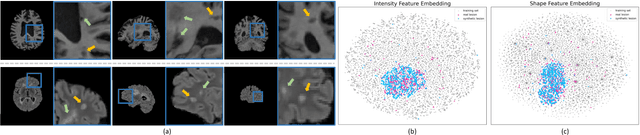
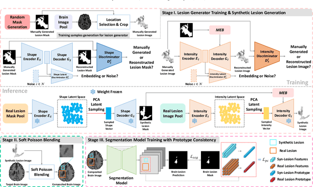
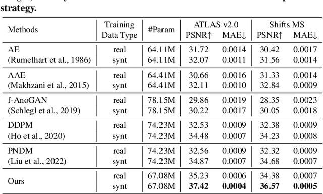
Accurate brain lesion delineation is important for planning neurosurgical treatment. Automatic brain lesion segmentation methods based on convolutional neural networks have demonstrated remarkable performance. However, neural network performance is constrained by the lack of large-scale well-annotated training datasets. In this manuscript, we propose a comprehensive framework to efficiently generate new, realistic samples for training a brain lesion segmentation model. We first train a lesion generator, based on an adversarial autoencoder, in a self-supervised manner. Next, we utilize a novel image composition algorithm, Soft Poisson Blending, to seamlessly combine synthetic lesions and brain images to obtain training samples. Finally, to effectively train the brain lesion segmentation model with augmented images we introduce a new prototype consistence regularization to align real and synthetic features. Our framework is validated by extensive experiments on two public brain lesion segmentation datasets: ATLAS v2.0 and Shift MS. Our method outperforms existing brain image data augmentation schemes. For instance, our method improves the Dice from 50.36% to 60.23% compared to the U-Net with conventional data augmentation techniques for the ATLAS v2.0 dataset.
A Robust Ensemble Algorithm for Ischemic Stroke Lesion Segmentation: Generalizability and Clinical Utility Beyond the ISLES Challenge
Apr 03, 2024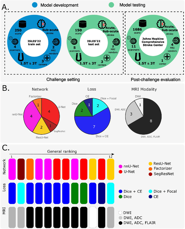
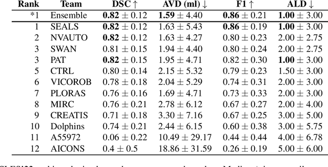
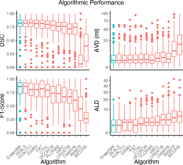
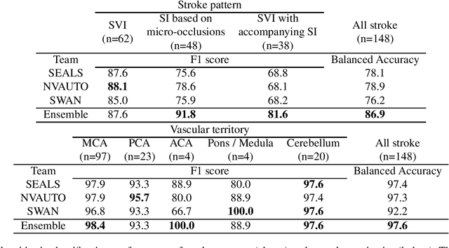
Diffusion-weighted MRI (DWI) is essential for stroke diagnosis, treatment decisions, and prognosis. However, image and disease variability hinder the development of generalizable AI algorithms with clinical value. We address this gap by presenting a novel ensemble algorithm derived from the 2022 Ischemic Stroke Lesion Segmentation (ISLES) challenge. ISLES'22 provided 400 patient scans with ischemic stroke from various medical centers, facilitating the development of a wide range of cutting-edge segmentation algorithms by the research community. Through collaboration with leading teams, we combined top-performing algorithms into an ensemble model that overcomes the limitations of individual solutions. Our ensemble model achieved superior ischemic lesion detection and segmentation accuracy on our internal test set compared to individual algorithms. This accuracy generalized well across diverse image and disease variables. Furthermore, the model excelled in extracting clinical biomarkers. Notably, in a Turing-like test, neuroradiologists consistently preferred the algorithm's segmentations over manual expert efforts, highlighting increased comprehensiveness and precision. Validation using a real-world external dataset (N=1686) confirmed the model's generalizability. The algorithm's outputs also demonstrated strong correlations with clinical scores (admission NIHSS and 90-day mRS) on par with or exceeding expert-derived results, underlining its clinical relevance. This study offers two key findings. First, we present an ensemble algorithm (https://github.com/Tabrisrei/ISLES22_Ensemble) that detects and segments ischemic stroke lesions on DWI across diverse scenarios on par with expert (neuro)radiologists. Second, we show the potential for biomedical challenge outputs to extend beyond the challenge's initial objectives, demonstrating their real-world clinical applicability.
Generative Medical Segmentation
Mar 27, 2024

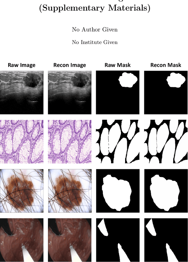
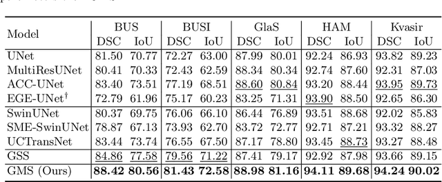
Rapid advancements in medical image segmentation performance have been significantly driven by the development of Convolutional Neural Networks (CNNs) and Vision Transformers (ViTs). However, these models introduce high computational demands and often have limited ability to generalize across diverse medical imaging datasets. In this manuscript, we introduce Generative Medical Segmentation (GMS), a novel approach leveraging a generative model for image segmentation. Concretely, GMS employs a robust pre-trained Variational Autoencoder (VAE) to derive latent representations of both images and masks, followed by a mapping model that learns the transition from image to mask in the latent space. This process culminates in generating a precise segmentation mask within the image space using the pre-trained VAE decoder. The design of GMS leads to fewer learnable parameters in the model, resulting in a reduced computational burden and enhanced generalization capability. Our extensive experimental analysis across five public datasets in different medical imaging domains demonstrates GMS outperforms existing discriminative segmentation models and has remarkable domain generalization. Our experiments suggest GMS could set a new benchmark for medical image segmentation, offering a scalable and effective solution. GMS implementation and model weights are available at https://github.com/King-HAW/GMS.
MatchSeg: Towards Better Segmentation via Reference Image Matching
Mar 23, 2024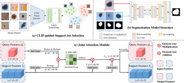
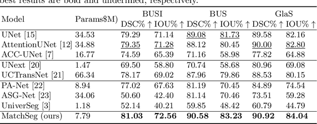
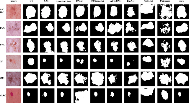
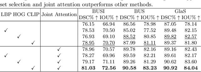
Recently, automated medical image segmentation methods based on deep learning have achieved great success. However, they heavily rely on large annotated datasets, which are costly and time-consuming to acquire. Few-shot learning aims to overcome the need for annotated data by using a small labeled dataset, known as a support set, to guide predicting labels for new, unlabeled images, known as the query set. Inspired by this paradigm, we introduce MatchSeg, a novel framework that enhances medical image segmentation through strategic reference image matching. We leverage contrastive language-image pre-training (CLIP) to select highly relevant samples when defining the support set. Additionally, we design a joint attention module to strengthen the interaction between support and query features, facilitating a more effective knowledge transfer between support and query sets. We validated our method across four public datasets. Experimental results demonstrate superior segmentation performance and powerful domain generalization ability of MatchSeg against existing methods for domain-specific and cross-domain segmentation tasks. Our code is made available at https://github.com/keeplearning-again/MatchSeg
RetiGen: A Framework for Generalized Retinal Diagnosis Using Multi-View Fundus Images
Mar 22, 2024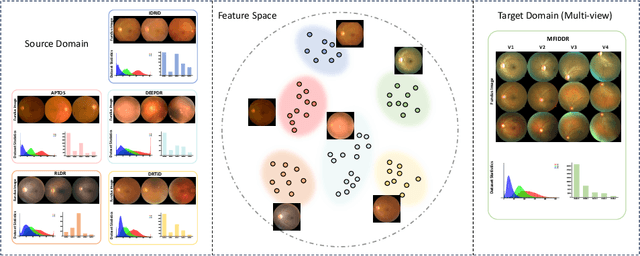

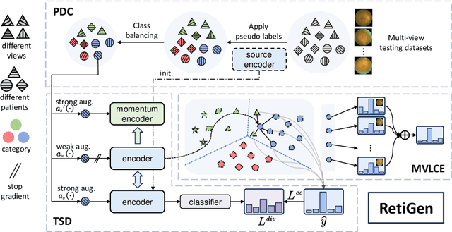

This study introduces a novel framework for enhancing domain generalization in medical imaging, specifically focusing on utilizing unlabelled multi-view colour fundus photographs. Unlike traditional approaches that rely on single-view imaging data and face challenges in generalizing across diverse clinical settings, our method leverages the rich information in the unlabelled multi-view imaging data to improve model robustness and accuracy. By incorporating a class balancing method, a test-time adaptation technique and a multi-view optimization strategy, we address the critical issue of domain shift that often hampers the performance of machine learning models in real-world applications. Experiments comparing various state-of-the-art domain generalization and test-time optimization methodologies show that our approach consistently outperforms when combined with existing baseline and state-of-the-art methods. We also show our online method improves all existing techniques. Our framework demonstrates improvements in domain generalization capabilities and offers a practical solution for real-world deployment by facilitating online adaptation to new, unseen datasets. Our code is available at https://github.com/zgy600/RetiGen .
DDSB: An Unsupervised and Training-free Method for Phase Detection in Echocardiography
Mar 19, 2024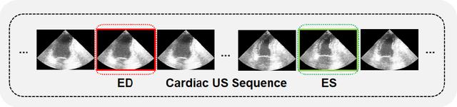
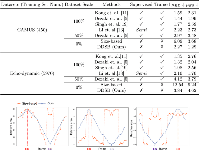
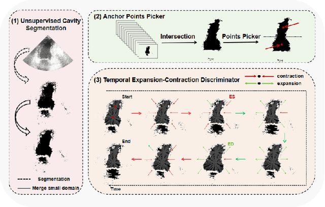
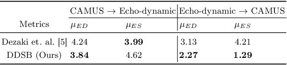
Accurate identification of End-Diastolic (ED) and End-Systolic (ES) frames is key for cardiac function assessment through echocardiography. However, traditional methods face several limitations: they require extensive amounts of data, extensive annotations by medical experts, significant training resources, and often lack robustness. Addressing these challenges, we proposed an unsupervised and training-free method, our novel approach leverages unsupervised segmentation to enhance fault tolerance against segmentation inaccuracies. By identifying anchor points and analyzing directional deformation, we effectively reduce dependence on the accuracy of initial segmentation images and enhance fault tolerance, all while improving robustness. Tested on Echo-dynamic and CAMUS datasets, our method achieves comparable accuracy to learning-based models without their associated drawbacks. The code is available at https://github.com/MRUIL/DDSB