 Add to Chrome
 Add to Firefox
 Add to Edge
HiMatch-AD: DINOv3-driven Hierarchical Matching for Training-free Medical Anomaly Detection
Jun 21, 2026
Anomaly detection is essential for medical image analysis, where pathological regions often appear as rare deviations from normal anatomical structures. While training-based methods have achieved promising performance, they require task-specific optimization and extensive normal data, which limits scalability across modalities and institutions. Training-free approaches offer greater flexibility by leveraging pretrained visual representations, yet existing methods typically rely on simple nearest-neighbor retrieval and naive aggregation strategies, which may fail to capture hierarchical semantics and ignore the reliability of multiple anomaly responses. In this work, we propose HiMatch-AD, a DINOv3-driven hierarchical matching framework for training-free medical anomaly detection. Our method first retrieves semantically relevant normal references via dual-branch matching that jointly considers global CLS-token similarity and patch-level representations. Hierarchical anomaly maps are then generated across multiple transformer stages by comparing clustered normal features with query representations. To robustly aggregate anomaly responses, we introduce a unified uncertainty-based fusion mechanism that adaptively weights maps according to their reliability. The entire framework operates without any task-specific training. Extensive experiments on the BMAD benchmark, including brain MRI, liver CT, and retinal OCT datasets, demonstrate that HiMatch-AD consistently outperforms both training-based and DINO-based state-of-the-art methods, which highlights the effectiveness of multi-level matching and uncertainty-aware fusion for scalable medical anomaly detection.

DINO-AD: Unsupervised Anomaly Detection with Frozen DINO-V3 Features
Jan 31, 2026Unsupervised anomaly detection (AD) in medical images aims to identify abnormal regions without relying on pixel-level annotations, which is crucial for scalable and label-efficient diagnostic systems. In this paper, we propose a novel anomaly detection framework based on DINO-V3 representations, termed DINO-AD, which leverages self-supervised visual features for precise and interpretable anomaly localization. Specifically, we introduce an embedding similarity matching strategy to select a semantically aligned support image and a foreground-aware K-means clustering module to model the distribution of normal features. Anomaly maps are then computed by comparing the query features with clustered normal embeddings through cosine similarity. Experimental results on both the Brain and Liver datasets demonstrate that our method achieves superior quantitative performance compared with state-of-the-art approaches, achieving AUROC scores of up to 98.71. Qualitative results further confirm that our framework produces clearer and more accurate anomaly localization. Extensive ablation studies validate the effectiveness of each proposed component, highlighting the robustness and generalizability of our approach.
Unsupervised Landmark Detection Based Spatiotemporal Motion Estimation for 4D Dynamic Medical Images
Oct 12, 2021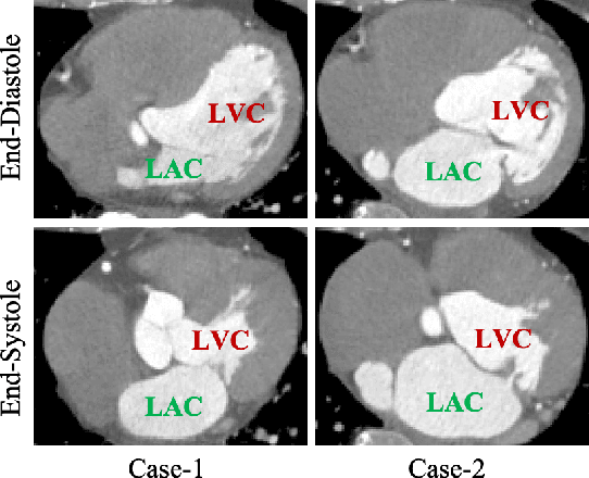
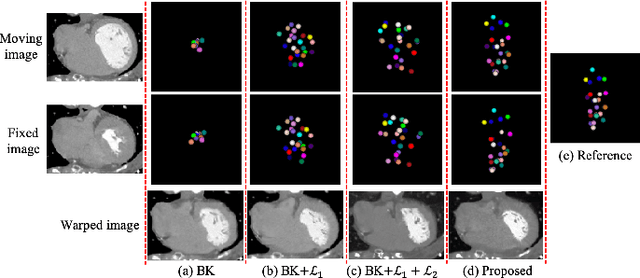
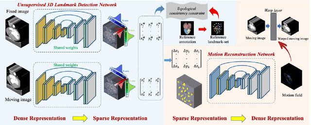
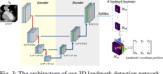
Motion estimation is a fundamental step in dynamic medical image processing for the assessment of target organ anatomy and function. However, existing image-based motion estimation methods, which optimize the motion field by evaluating the local image similarity, are prone to produce implausible estimation, especially in the presence of large motion. In this study, we provide a novel motion estimation framework of Dense-Sparse-Dense (DSD), which comprises two stages. In the first stage, we process the raw dense image to extract sparse landmarks to represent the target organ anatomical topology and discard the redundant information that is unnecessary for motion estimation. For this purpose, we introduce an unsupervised 3D landmark detection network to extract spatially sparse but representative landmarks for the target organ motion estimation. In the second stage, we derive the sparse motion displacement from the extracted sparse landmarks of two images of different time points. Then, we present a motion reconstruction network to construct the motion field by projecting the sparse landmarks displacement back into the dense image domain. Furthermore, we employ the estimated motion field from our two-stage DSD framework as initialization and boost the motion estimation quality in light-weight yet effective iterative optimization. We evaluate our method on two dynamic medical imaging tasks to model cardiac motion and lung respiratory motion, respectively. Our method has produced superior motion estimation accuracy compared to existing comparative methods. Besides, the extensive experimental results demonstrate that our solution can extract well representative anatomical landmarks without any requirement of manual annotation. Our code is publicly available online.