 Add to Chrome
 Add to Firefox
 Add to Edge
Panda: Unsupervised Pelvic Anomaly Detection for Real-Time MR Imaging
Jul 27, 2026
Female pelvic diseases remain an under researched area characterized by often delayed diagnosis. While pelvic MRI offers superior soft-tissue contrast for diagnosis and image-guided procedures, real-time anomaly detection remains challenging due to physiological motion, tissue deformation, and instrument artifacts. Existing supervised approaches are impractical, as adverse events are rare, heterogeneous, and difficult to annotate. We present a Dinomaly-based unsupervised anomaly detection framework adapted for pelvic MRI that learns normative representations from healthy cases and flags deviations without requiring labels. Our approach leverages a frozen DINOv3 Vision Transformer encoder combined with a noisy MLP bottleneck and Linear Attention decoder to prevent identity mapping while maintaining computational efficiency. Anomalies are localized via per-token cosine distance between encoder and decoder representations, yielding spatial anomaly maps that provide immediate feedback at the scanner to support radiologist decision-making and adaptive protocol adjustment. Evaluated on a curated subset of the Uterine Myoma Dataset, the framework achieves a pixel-level AUROC of 88.06% and high specificity (95.45%) at frame level at 40.5 slices/s, meeting real-time clinical deployment requirements. The spatial anomaly maps and frame-level scores provide immediate, localized feedback at the scanner to support radiologist decision-making and adaptive protocol adjustment during active procedures.

PRIME-SVR: Physics-infoRmed Implicit Multi-Echo Slice-to-Volume Reconstruction for Fetal T2 mapping
Jul 22, 2026Slice-to-volume reconstruction (SVR) is the standard method for obtaining high-resolution (HR) 3D fetal brain volumes from motion-corrupted 2D MRI slice stacks acquired in multiple orientations. Existing SVR methods are optimized and validated only for clinical-range echo times (TEs), limiting their use at non-clinical TEs and making them incompatible with quantitative T2 mapping, a protocol- and center-independent biomarker of fetal brain maturation requiring HR reconstructions across multiple TEs. We present PRIME-SVR, the first implicit neural representation (INR) framework for joint HR reconstruction from multi-echo MRI. A single fully connected network models a continuous function from spatial coordinates to signal intensities across TEs, while a second network estimates slice-specific acquisition degradations. Cross-TE coherence is enforced via a Bloch equation-derived regularization penalizing deviations from expected T2 decay, with adaptive weighting that strengthens coupling for degraded stacks. The method is fully self-supervised. We validate PRIME-SVR on 39 in vivo fetal acquisitions (13 subjects x 3 TEs) from two centers, two vendors, and two field strengths (1.5 T and 0.55 T). Compared to state-of-the-art SVR, PRIME-SVR improves reconstruction sharpness by 47%, anatomical accuracy by 30%, and cross-TE structural consistency by 14%. It enables reconstruction at late TEs previously inaccessible to SVR, yielding the first 0.8 mm isotropic T2 maps at 0.55 T and the first T2 maps derived from INR-based SVR. PRIME-SVR also accelerates quantitative imaging by reducing the data needed for multi-TE reconstruction, cutting acquisition from 15 to 10 minutes while keeping T2 accuracy within 1.7% in white and deep gray matter, or to 5 minutes with a mean T2 error of 2.3% for high-quality acquisitions.
Female-RHINO: A Real-Time Scanner-Integrated Framework for Automated Quantitative Uterine MRI Analysis and Structured Reporting
Jun 23, 2026Standardized assessment of uterine MRI remains challenging due to anatomical variability, observer dependence, and the lack of workflow-integrated automated analysis tools. This work presents Female-RHINO: (R)eproductive (H)ealth (I)maging A(N)alysis T(O)ol, a real-time AI-assisted framework for automated quantitative uterine MRI analysis and structured reporting during image acquisition. We present an end-to-end system that integrates inline communication with the MRI scanner and deep learning-based analysis to derive quantitative uterine biomarkers from sagittal T2-weighted pelvic MRI. The framework combines segmentation and anatomical landmark detection models trained and evaluated on more than 500 multi-center datasets spanning diverse protocols, vendors, and patient populations. It performs volumetry, detects and quantifies common incidental findings such as fibroids and Nabothian cysts, and extracts six anatomical landmarks for biometric assessment. Results are compiled into a structured clinician-oriented report with integrated visualizations, without manual interaction. Evaluation on independent retrospective and prospective cohorts demonstrated robust performance across varying acquisition settings. Mean Dice similarity coefficients were 0.82 for the uterus and 0.80 for fibroids, with lower but consistent agreement for Nabothian cysts. Landmark detection achieved a mean radial error of 3.7 mm. End-to-end processing was completed in under 70 seconds, enabling availability of results during the ongoing scan. Prospective deployment yielded immediate, standardized, and reproducible analyses supported by inter-observer agreement. The proposed system enables real-time scanner-integrated AI for automated uterine MRI analysis and reporting, with potential to improve standardization, efficiency, and clinical workflow in pelvic imaging.
Predicting gestational age at birth in the context of preterm birth from multi-modal fetal MRI
Jun 18, 2026Preterm birth is associated with significant mortality and a risk for lifelong morbidity. The complex multifactorial aetiology hampers accurate prediction and thus optimal care. A pipeline consisting of bespoke machine learning methods for data imputation, feature selection, and regression models to predict gestational age (GA) at birth was developed and evaluated from comprehensive multi-modal morphological and functional fetal MRI data from 333 control cases and 93 preterm birth cases. The GA at birth predictions were classified into term and preterm categories and their accuracy, sensitivity, and specificity were reported. An ablation study was performed to further validate the design of the pipeline. Performance was evaluated using stratified 10-fold cross-validation. The pipeline achieves an R2 score of 0.13 and a mean absolute error of 2.74 weeks. It also achieves a 0.77 accuracy, 0.59 sensitivity, and 0.82 specificity across folds. The predominant features selected by the pipeline include cervical length and statistics derived from placental T2* values. The confluence of fast, motion-robust and multi-modal fetal MRI techniques and machine learning prediction allowed the prediction of the gestation at birth. This information is essential for any pregnancy. To the best of our knowledge, preterm birth had only been addressed as a classification problem in the literature. Therefore, this work provides a proof of concept. Future work will increase the cohort size to allow for finer stratification within the preterm birth cohort. Our code is available at https://github.com/dfajardorojas/ml-for-preterm-birth-.
* Accepted for publication at the Journal of Machine Learning for Biomedical Imaging (MELBA) https://melba-journal.org/2026:013
SIREM: Speech-Informed MRI Reconstruction with Learned Sampling
May 18, 2026Real-time magnetic resonance imaging (rtMRI) of speech production enables non-invasive visualization of dynamic vocal-tract motion and is valuable for speech science and clinical assessment. However, rtMRI is fundamentally constrained by trade-offs among spatial resolution, temporal resolution, and acquisition speed, often leading to undersampled k-space measurements and degraded reconstructions. We propose SIREM, a speech-informed MRI reconstruction framework that uses synchronized speech as a cross-modal prior. The central idea is that vocal-tract configurations during speech are correlated with the produced acoustics, making part of the image content predictable from audio. SIREM models each frame as a fusion of an audio-driven component and an MRI-driven component through a spatial weighting map. The audio branch predicts articulator-related structure from speech, while the MRI branch reconstructs complementary content from measured k-space data. We further introduce a learnable soft weighting profile over spiral arms, enabling a differentiable study of how k-space arm usage interacts with speech-informed fusion. This yields a unified multimodal formulation that combines audio-driven prediction, MRI reconstruction, and sampling adaptation. We evaluate SIREM on the USC speech rtMRI benchmark against standard baselines, including gridding, wavelet-based compressed sensing, and total variation. SIREM introduces a speech-informed reconstruction paradigm that operates in a substantially higher-throughput regime than iterative methods while preserving anatomically plausible vocal-tract structure. These results establish an initial benchmark for multimodal speech-informed rtMRI reconstruction and highlight the potential of synchronized speech as an auxiliary prior for fast reconstruction. The source code is available at https://github.com/mdhasanai/SIREM
Speech-Guided Multimodal Learning for Vocal Tract Segmentation in Real-Time MRI
May 18, 2026Segmenting vocal tract articulators in real-time MRI (rtMRI) is a challenging dynamic image segmentation problem characterized by low contrast, rapid motion, and limited spatial resolution. However, while rtMRI acquisitions may provide synchronized acoustic signals, existing methods discard this information, and the few multimodal approaches that incorporate audio cannot be deployed when audio is unavailable. We propose a three-stage framework that leverages acoustic and phonological supervision during training while requiring only the rtMRI image at inference: phonological representations are converted into spatial bounding-box priors for articulator localization, visual and acoustic encoders are aligned via dual-level cross-modal contrastive pretraining, and the learned representations are fused through a cross-attention decoder, effectively transferring multimodal knowledge into a single-modality inference pipeline. Evaluated on 75-Speaker~Annot-16 and USC-TIMIT datasets, our method outperforms existing unimodal and multimodal methods, demonstrating that multimodal supervision provides transferable benefits for precise and clinically deployable vocal tract segmentation.
Unsupervised Anomaly Detection of Diseases in the Female Pelvis for Real-Time MR Imaging
Feb 05, 2026Pelvic diseases in women of reproductive age represent a major global health burden, with diagnosis frequently delayed due to high anatomical variability, complicating MRI interpretation. Existing AI approaches are largely disease-specific and lack real-time compatibility, limiting generalizability and clinical integration. To address these challenges, we establish a benchmark framework for disease- and parameter-agnostic, real-time-compatible unsupervised anomaly detection in pelvic MRI. The method uses a residual variational autoencoder trained exclusively on healthy sagittal T2-weighted scans acquired across diverse imaging protocols to model normal pelvic anatomy. During inference, reconstruction error heatmaps indicate deviations from learned healthy structure, enabling detection of pathological regions without labeled abnormal data. The model is trained on 294 healthy scans and augmented with diffusion-generated synthetic data to improve robustness. Quantitative evaluation on the publicly available Uterine Myoma MRI Dataset yields an average area-under-the-curve (AUC) value of 0.736, with 0.828 sensitivity and 0.692 specificity. Additional inter-observer clinical evaluation extends analysis to endometrial cancer, endometriosis, and adenomyosis, revealing the influence of anatomical heterogeneity and inter-observer variability on performance interpretation. With a reconstruction time of approximately 92.6 frames per second, the proposed framework establishes a baseline for unsupervised anomaly detection in the female pelvis and supports future integration into real-time MRI. Code is available upon request (https://github.com/AniKnu/UADPelvis), prospective data sets are available for academic collaboration.
Diffusing the Blind Spot: Uterine MRI Synthesis with Diffusion Models
Aug 11, 2025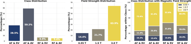
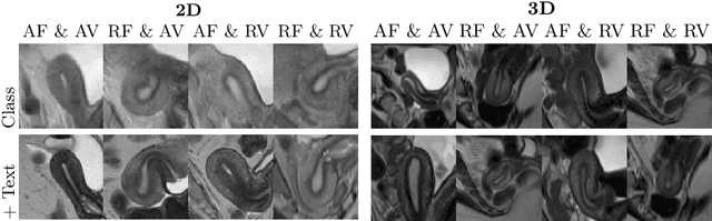
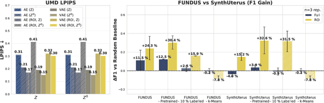
Despite significant progress in generative modelling, existing diffusion models often struggle to produce anatomically precise female pelvic images, limiting their application in gynaecological imaging, where data scarcity and patient privacy concerns are critical. To overcome these barriers, we introduce a novel diffusion-based framework for uterine MRI synthesis, integrating both unconditional and conditioned Denoising Diffusion Probabilistic Models (DDPMs) and Latent Diffusion Models (LDMs) in 2D and 3D. Our approach generates anatomically coherent, high fidelity synthetic images that closely mimic real scans and provide valuable resources for training robust diagnostic models. We evaluate generative quality using advanced perceptual and distributional metrics, benchmarking against standard reconstruction methods, and demonstrate substantial gains in diagnostic accuracy on a key classification task. A blinded expert evaluation further validates the clinical realism of our synthetic images. We release our models with privacy safeguards and a comprehensive synthetic uterine MRI dataset to support reproducible research and advance equitable AI in gynaecology.
Audio-Vision Contrastive Learning for Phonological Class Recognition
Jul 23, 2025Accurate classification of articulatory-phonological features plays a vital role in understanding human speech production and developing robust speech technologies, particularly in clinical contexts where targeted phonemic analysis and therapy can improve disease diagnosis accuracy and personalized rehabilitation. In this work, we propose a multimodal deep learning framework that combines real-time magnetic resonance imaging (rtMRI) and speech signals to classify three key articulatory dimensions: manner of articulation, place of articulation, and voicing. We perform classification on 15 phonological classes derived from the aforementioned articulatory dimensions and evaluate the system with four audio/vision configurations: unimodal rtMRI, unimodal audio signals, multimodal middle fusion, and contrastive learning-based audio-vision fusion. Experimental results on the USC-TIMIT dataset show that our contrastive learning-based approach achieves state-of-the-art performance, with an average F1-score of 0.81, representing an absolute increase of 0.23 over the unimodal baseline. The results confirm the effectiveness of contrastive representation learning for multimodal articulatory analysis. Our code and processed dataset will be made publicly available at https://github.com/DaE-plz/AC_Contrastive_Phonology to support future research.
Advances in Automated Fetal Brain MRI Segmentation and Biometry: Insights from the FeTA 2024 Challenge
May 05, 2025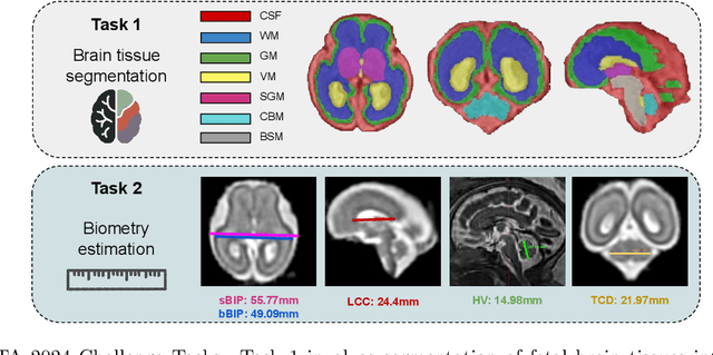
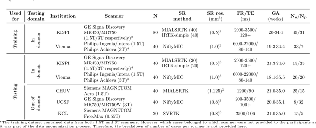
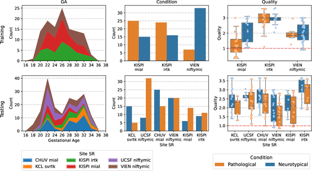
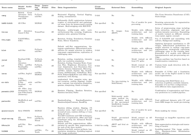
Accurate fetal brain tissue segmentation and biometric analysis are essential for studying brain development in utero. The FeTA Challenge 2024 advanced automated fetal brain MRI analysis by introducing biometry prediction as a new task alongside tissue segmentation. For the first time, our diverse multi-centric test set included data from a new low-field (0.55T) MRI dataset. Evaluation metrics were also expanded to include the topology-specific Euler characteristic difference (ED). Sixteen teams submitted segmentation methods, most of which performed consistently across both high- and low-field scans. However, longitudinal trends indicate that segmentation accuracy may be reaching a plateau, with results now approaching inter-rater variability. The ED metric uncovered topological differences that were missed by conventional metrics, while the low-field dataset achieved the highest segmentation scores, highlighting the potential of affordable imaging systems when paired with high-quality reconstruction. Seven teams participated in the biometry task, but most methods failed to outperform a simple baseline that predicted measurements based solely on gestational age, underscoring the challenge of extracting reliable biometric estimates from image data alone. Domain shift analysis identified image quality as the most significant factor affecting model generalization, with super-resolution pipelines also playing a substantial role. Other factors, such as gestational age, pathology, and acquisition site, had smaller, though still measurable, effects. Overall, FeTA 2024 offers a comprehensive benchmark for multi-class segmentation and biometry estimation in fetal brain MRI, underscoring the need for data-centric approaches, improved topological evaluation, and greater dataset diversity to enable clinically robust and generalizable AI tools.