 Add to Chrome
 Add to Firefox
 Add to Edge
Panda: Unsupervised Pelvic Anomaly Detection for Real-Time MR Imaging
Jul 27, 2026
Female pelvic diseases remain an under researched area characterized by often delayed diagnosis. While pelvic MRI offers superior soft-tissue contrast for diagnosis and image-guided procedures, real-time anomaly detection remains challenging due to physiological motion, tissue deformation, and instrument artifacts. Existing supervised approaches are impractical, as adverse events are rare, heterogeneous, and difficult to annotate. We present a Dinomaly-based unsupervised anomaly detection framework adapted for pelvic MRI that learns normative representations from healthy cases and flags deviations without requiring labels. Our approach leverages a frozen DINOv3 Vision Transformer encoder combined with a noisy MLP bottleneck and Linear Attention decoder to prevent identity mapping while maintaining computational efficiency. Anomalies are localized via per-token cosine distance between encoder and decoder representations, yielding spatial anomaly maps that provide immediate feedback at the scanner to support radiologist decision-making and adaptive protocol adjustment. Evaluated on a curated subset of the Uterine Myoma Dataset, the framework achieves a pixel-level AUROC of 88.06% and high specificity (95.45%) at frame level at 40.5 slices/s, meeting real-time clinical deployment requirements. The spatial anomaly maps and frame-level scores provide immediate, localized feedback at the scanner to support radiologist decision-making and adaptive protocol adjustment during active procedures.

Female-RHINO: A Real-Time Scanner-Integrated Framework for Automated Quantitative Uterine MRI Analysis and Structured Reporting
Jun 23, 2026Standardized assessment of uterine MRI remains challenging due to anatomical variability, observer dependence, and the lack of workflow-integrated automated analysis tools. This work presents Female-RHINO: (R)eproductive (H)ealth (I)maging A(N)alysis T(O)ol, a real-time AI-assisted framework for automated quantitative uterine MRI analysis and structured reporting during image acquisition. We present an end-to-end system that integrates inline communication with the MRI scanner and deep learning-based analysis to derive quantitative uterine biomarkers from sagittal T2-weighted pelvic MRI. The framework combines segmentation and anatomical landmark detection models trained and evaluated on more than 500 multi-center datasets spanning diverse protocols, vendors, and patient populations. It performs volumetry, detects and quantifies common incidental findings such as fibroids and Nabothian cysts, and extracts six anatomical landmarks for biometric assessment. Results are compiled into a structured clinician-oriented report with integrated visualizations, without manual interaction. Evaluation on independent retrospective and prospective cohorts demonstrated robust performance across varying acquisition settings. Mean Dice similarity coefficients were 0.82 for the uterus and 0.80 for fibroids, with lower but consistent agreement for Nabothian cysts. Landmark detection achieved a mean radial error of 3.7 mm. End-to-end processing was completed in under 70 seconds, enabling availability of results during the ongoing scan. Prospective deployment yielded immediate, standardized, and reproducible analyses supported by inter-observer agreement. The proposed system enables real-time scanner-integrated AI for automated uterine MRI analysis and reporting, with potential to improve standardization, efficiency, and clinical workflow in pelvic imaging.
Unsupervised Anomaly Detection of Diseases in the Female Pelvis for Real-Time MR Imaging
Feb 05, 2026Pelvic diseases in women of reproductive age represent a major global health burden, with diagnosis frequently delayed due to high anatomical variability, complicating MRI interpretation. Existing AI approaches are largely disease-specific and lack real-time compatibility, limiting generalizability and clinical integration. To address these challenges, we establish a benchmark framework for disease- and parameter-agnostic, real-time-compatible unsupervised anomaly detection in pelvic MRI. The method uses a residual variational autoencoder trained exclusively on healthy sagittal T2-weighted scans acquired across diverse imaging protocols to model normal pelvic anatomy. During inference, reconstruction error heatmaps indicate deviations from learned healthy structure, enabling detection of pathological regions without labeled abnormal data. The model is trained on 294 healthy scans and augmented with diffusion-generated synthetic data to improve robustness. Quantitative evaluation on the publicly available Uterine Myoma MRI Dataset yields an average area-under-the-curve (AUC) value of 0.736, with 0.828 sensitivity and 0.692 specificity. Additional inter-observer clinical evaluation extends analysis to endometrial cancer, endometriosis, and adenomyosis, revealing the influence of anatomical heterogeneity and inter-observer variability on performance interpretation. With a reconstruction time of approximately 92.6 frames per second, the proposed framework establishes a baseline for unsupervised anomaly detection in the female pelvis and supports future integration into real-time MRI. Code is available upon request (https://github.com/AniKnu/UADPelvis), prospective data sets are available for academic collaboration.
Diffusing the Blind Spot: Uterine MRI Synthesis with Diffusion Models
Aug 11, 2025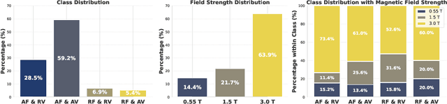
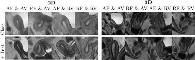
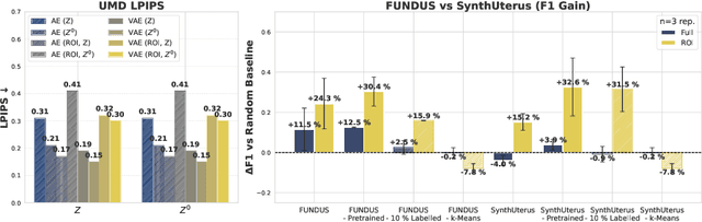
Despite significant progress in generative modelling, existing diffusion models often struggle to produce anatomically precise female pelvic images, limiting their application in gynaecological imaging, where data scarcity and patient privacy concerns are critical. To overcome these barriers, we introduce a novel diffusion-based framework for uterine MRI synthesis, integrating both unconditional and conditioned Denoising Diffusion Probabilistic Models (DDPMs) and Latent Diffusion Models (LDMs) in 2D and 3D. Our approach generates anatomically coherent, high fidelity synthetic images that closely mimic real scans and provide valuable resources for training robust diagnostic models. We evaluate generative quality using advanced perceptual and distributional metrics, benchmarking against standard reconstruction methods, and demonstrate substantial gains in diagnostic accuracy on a key classification task. A blinded expert evaluation further validates the clinical realism of our synthetic images. We release our models with privacy safeguards and a comprehensive synthetic uterine MRI dataset to support reproducible research and advance equitable AI in gynaecology.