 Add to Chrome
 Add to Firefox
 Add to Edge
PRIME-SVR: Physics-infoRmed Implicit Multi-Echo Slice-to-Volume Reconstruction for Fetal T2 mapping
Jul 22, 2026
Slice-to-volume reconstruction (SVR) is the standard method for obtaining high-resolution (HR) 3D fetal brain volumes from motion-corrupted 2D MRI slice stacks acquired in multiple orientations. Existing SVR methods are optimized and validated only for clinical-range echo times (TEs), limiting their use at non-clinical TEs and making them incompatible with quantitative T2 mapping, a protocol- and center-independent biomarker of fetal brain maturation requiring HR reconstructions across multiple TEs. We present PRIME-SVR, the first implicit neural representation (INR) framework for joint HR reconstruction from multi-echo MRI. A single fully connected network models a continuous function from spatial coordinates to signal intensities across TEs, while a second network estimates slice-specific acquisition degradations. Cross-TE coherence is enforced via a Bloch equation-derived regularization penalizing deviations from expected T2 decay, with adaptive weighting that strengthens coupling for degraded stacks. The method is fully self-supervised. We validate PRIME-SVR on 39 in vivo fetal acquisitions (13 subjects x 3 TEs) from two centers, two vendors, and two field strengths (1.5 T and 0.55 T). Compared to state-of-the-art SVR, PRIME-SVR improves reconstruction sharpness by 47%, anatomical accuracy by 30%, and cross-TE structural consistency by 14%. It enables reconstruction at late TEs previously inaccessible to SVR, yielding the first 0.8 mm isotropic T2 maps at 0.55 T and the first T2 maps derived from INR-based SVR. PRIME-SVR also accelerates quantitative imaging by reducing the data needed for multi-TE reconstruction, cutting acquisition from 15 to 10 minutes while keeping T2 accuracy within 1.7% in white and deep gray matter, or to 5 minutes with a mean T2 error of 2.3% for high-quality acquisitions.

FaBiAN: A Fetal Brain magnetic resonance Acquisition Numerical phantom
Sep 06, 2021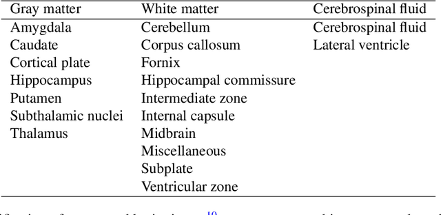
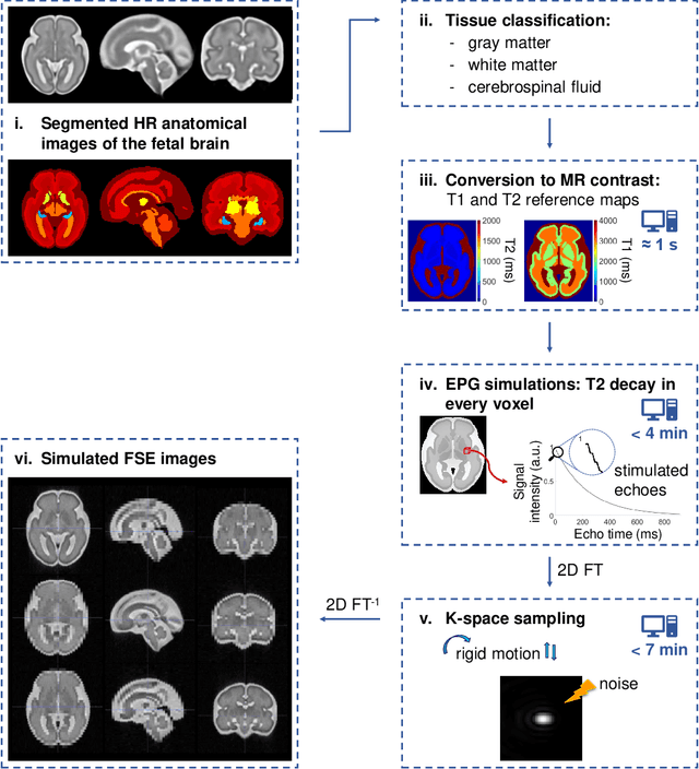
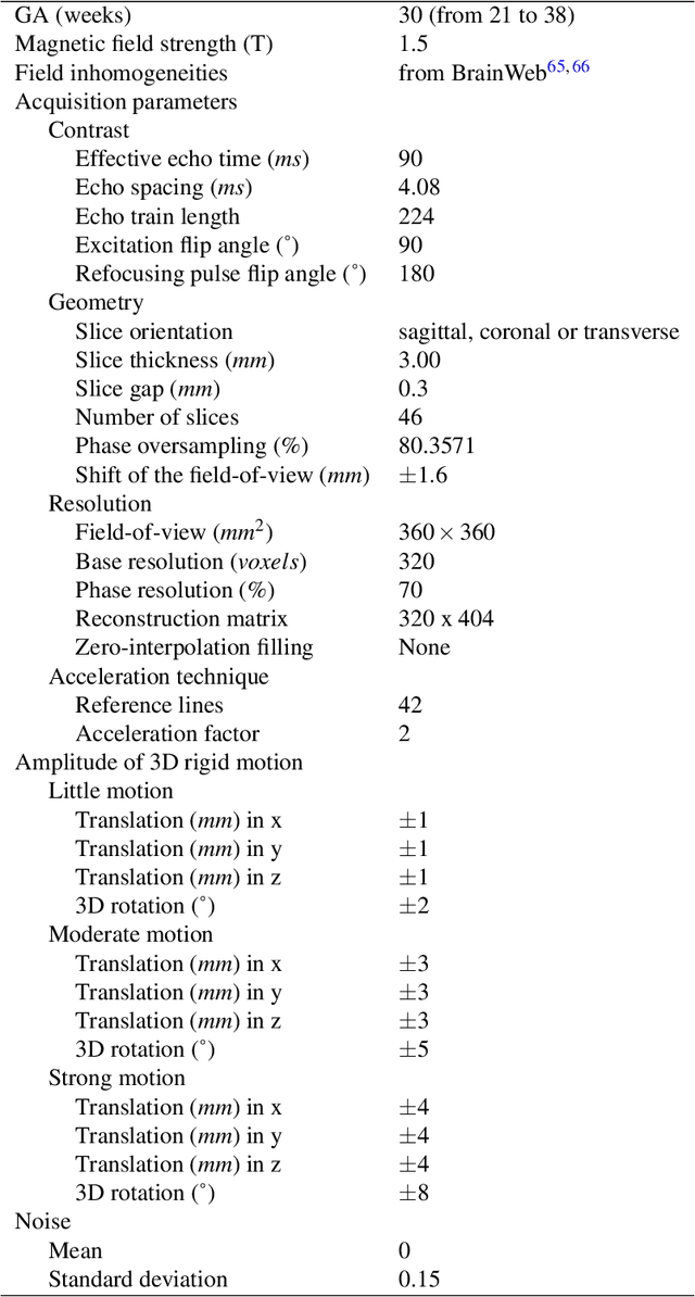
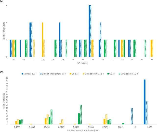
Accurate characterization of in utero human brain maturation is critical as it involves complex and interconnected structural and functional processes that may influence health later in life. Magnetic resonance imaging is a powerful tool to investigate equivocal neurological patterns during fetal development. However, the number of acquisitions of satisfactory quality available in this cohort of sensitive subjects remains scarce, thus hindering the validation of advanced image processing techniques. Numerical phantoms can mitigate these limitations by providing a controlled environment with a known ground truth. In this work, we present FaBiAN, an open-source Fetal Brain magnetic resonance Acquisition Numerical phantom that simulates clinical T2-weighted fast spin echo sequences of the fetal brain. This unique tool is based on a general, flexible and realistic setup that includes stochastic fetal movements, thus providing images of the fetal brain throughout maturation comparable to clinical acquisitions. We demonstrate its value to evaluate the robustness and optimize the accuracy of an algorithm for super-resolution fetal brain magnetic resonance imaging from simulated motion-corrupted 2D low-resolution series as compared to a synthetic high-resolution reference volume. We also show that the images generated can complement clinical datasets to support data-intensive deep learning methods for fetal brain tissue segmentation.