 Add to Chrome
 Add to Firefox
 Add to Edge
MRI2Rep: Autoregressive Structured Report Generation for 3D Liver MRI
Jun 24, 2026
Manual reporting of 3D MRI studies is time-consuming, yet end-to-end structured report generation for 3D liver MRI remains underexplored due to volumetric complexity and scarce paired data. We propose MRI2Rep, an autoregressive framework for liver MRI report generation. From 3,929 real-world MRI-report pairs acquired over a 10-year single-institution cohort, a Report-to-Label Canonicalization (RLC) module converts free-text reports into structured, closed-vocabulary diagnostic sequences without lesion-level annotations. On a held-out test set, MRI2Rep achieves 76.0% case-level sensitivity, 29.4% lesion-level F1, compared with no more than 8.3% for adapted medical vision-language baselines, and 82.4% liver-level accuracy. In a blinded reader study, two radiologists rated 75% and 70% of AI-generated reports as clinically acceptable, compared with 95% and 100% for original reports. Our automated LLM-based judge, LLM-Eval, rated 61.8% of AI-generated reports as acceptable, applying a stricter standard and supporting its use as a conservative proxy. To our knowledge, this is the first end-to-end LI-RADS-structured reporting system for 3D liver MRI.

PPDM: Pixel Puzzling Diffusion Model for Speed and Memory Efficient Volumetric Medical Image Translation
Jun 13, 2026Diffusion models have demonstrated superior fidelity for medical image-to-image translation, but their extension to high-resolution 3D volumes is severely constrained by prohibitive computational cost and GPU memory requirements. Existing memory-efficient strategies often compromise global volumetric consistency or fine anatomical detail. In this work, we propose the Pixel Puzzling Diffusion Model (PPDM), a simple and effective framework for memory- and speed-efficient 3D medical image translation. PPDM introduces a reversible pixel puzzle-unpuzzle operator that trades spatial resolution for channel dimensionality, substantially reducing activation memory while preserving global context. To further improve efficiency and stability, we adopt a direct bridge diffusion formulation that starts from the conditional input rather than pure noise, enabling the model to focus on task-relevant residuals. In addition, a puzzle-gradient loss is incorporated to enforce spatial coherence and suppress grid-like artifacts introduced by spatial rearrangement. We evaluate PPDM on multiple challenging 3D medical image translation tasks, including low-count PET denoising, joint PET denoising and attenuation correction, and cross-modal MRI translation. Across all tasks, PPDM consistently matches or outperforms full 3D diffusion models while reducing training GPU memory usage by up to an order of magnitude and significantly accelerating inference, and it outperforms existing memory-efficient diffusion approaches based on latent compression or frequency decomposition. These results demonstrate that PPDM provides a practical and scalable solution for high-fidelity 3D diffusion-based medical image translation under limited computational resources.
Learning Robust and Task-Invariant Functional Representation from fMRI through Siamese Self-Supervised Learning
May 27, 2026Functional magnetic resonance imaging (fMRI) is a powerful tool for investigating human brain function. However, the high cost of data acquisition and the inherent subjectivity of psychiatric rating scales often lead to datasets with small sample sizes and variable label quality, especially when targeting a specific neurological condition. Combined with the inherently high dimensionality of fMRI data, these limitations substantially increase the risk of model overfitting. Recent years have seen growing interest in developing fMRI foundation models by combining multiple datasets; however, the computational resources needed for pretraining and fine-tuning are often prohibitive. We show that a lightweight self-supervised framework yields representations that generalize across diverse downstream tasks, outperforming fully supervised baselines and approaching the performance of large-scale models. We introduce BrainSimSiam, a data-efficient self-supervised representation learning framework that leverages positive-only data pairs to learn robust and generalizable features. We demonstrate that the learned representations achieve strong performance across multiple downstream classification and regression tasks, highlighting the potential of BrainSimSiam for data-limited neuroimaging applications.
FM-fMRI: Event Conditioned Flow Matching for Rest-to-Task fMRI Time-Series Synthesis
May 26, 2026Task-based fMRI provides a direct readout of task-evoked neural dynamics, but it is expensive and difficult to acquire at scale, motivating rest-to-task synthesis from widely available resting-state fMRI (rsfMRI). We propose FM-fMRI, an event-conditioned flow-matching model that learns a continuous-time conditional vector field to generate task ROI time series from a subject's rsfMRI and the task event information. The formulation enables fast ODE-based sampling and flexible conditioning over heterogeneous event schedules. Rather than optimizing for pointwise reconstruction, we evaluated generated signals using complementary criteria that probe temporal and spectral structure, subject and group-level connectome consistency, and distributional alignment. On the public Human Connectome Project and internal BioPoint autism cohort, FM-fMRI achieves the strongest spectral and connectivity agreement and improved distribution-level matching over conditional diffusion, generative adversarial networks (GANs), and variational autoencoders (VAEs) baselines. Furthermore, we augment the BioPoint cohort by synthesizing task-fMRI ROI time series with our method, improving downstream autism classification and demonstrating practical utility in data-limited clinical settings. The code will be available on GitHub.
BioFact-MoE: Biologically Factorized Mixture of Experts for Vision-Language Prognostic Modeling in Hepatocellular Carcinoma
May 25, 2026Hepatocellular carcinoma (HCC) is biologically heterogeneous, shaped by the interplay between hepatic functional reserve and tumor-related oncologic factors; thus, similar survival outcomes may reflect fundamentally different underlying biological processes. Prognostic modeling in HCC is informed by rich multimodal information from multiparametric MRI and radiology reports from routine clinical practice. Existing prognostic vision-language models (VLMs) learn a single entangled latent representation that blends hepatic and tumor-related factors, limiting both accuracy and biological interpretability. We present BioFact-MoE, a biologically factorized Mixture of Experts (MoE) framework that explicitly decomposes liver and tumor factors via biologically supervised experts within a residual MoE survival architecture. On a HCC cohort of N=588 patients (pretrained on 4,582 3D MRI image-report pairs), BioFact-MoE consistently improves survival prediction over all baselines across time horizons, achieving 12-, 18-, and 24-month AUCs of 75.33%, 75.85%, and 73.96%. Beyond scalar risk prediction, gated expert weights enable phenotype-aware risk stratification. Pathway-informed gating uncovers clinically meaningful treatment-associated survival heterogeneity. In held-out validation, hepatic and tumor embeddings show selective associations with liver function and tumor burden markers, respectively (p<0.05), without supervision. The code is available at https://github.com/jy-639/BioFact-MoE.
PET Head Motion Estimation Using Supervised Deep Learning with Attention
Oct 14, 2025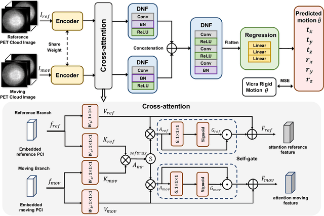
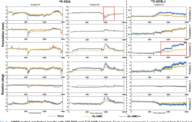
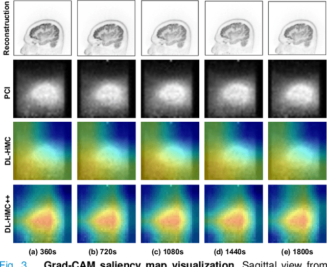
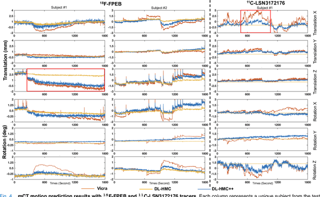
Head movement poses a significant challenge in brain positron emission tomography (PET) imaging, resulting in image artifacts and tracer uptake quantification inaccuracies. Effective head motion estimation and correction are crucial for precise quantitative image analysis and accurate diagnosis of neurological disorders. Hardware-based motion tracking (HMT) has limited applicability in real-world clinical practice. To overcome this limitation, we propose a deep-learning head motion correction approach with cross-attention (DL-HMC++) to predict rigid head motion from one-second 3D PET raw data. DL-HMC++ is trained in a supervised manner by leveraging existing dynamic PET scans with gold-standard motion measurements from external HMT. We evaluate DL-HMC++ on two PET scanners (HRRT and mCT) and four radiotracers (18F-FDG, 18F-FPEB, 11C-UCB-J, and 11C-LSN3172176) to demonstrate the effectiveness and generalization of the approach in large cohort PET studies. Quantitative and qualitative results demonstrate that DL-HMC++ consistently outperforms state-of-the-art data-driven motion estimation methods, producing motion-free images with clear delineation of brain structures and reduced motion artifacts that are indistinguishable from gold-standard HMT. Brain region of interest standard uptake value analysis exhibits average difference ratios between DL-HMC++ and gold-standard HMT to be 1.2 plus-minus 0.5% for HRRT and 0.5 plus-minus 0.2% for mCT. DL-HMC++ demonstrates the potential for data-driven PET head motion correction to remove the burden of HMT, making motion correction accessible to clinical populations beyond research settings. The code is available at https://github.com/maxxxxxxcai/DL-HMC-TMI.
Snap-and-tune: combining deep learning and test-time optimization for high-fidelity cardiovascular volumetric meshing
Jun 09, 2025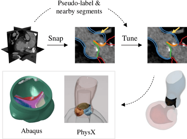

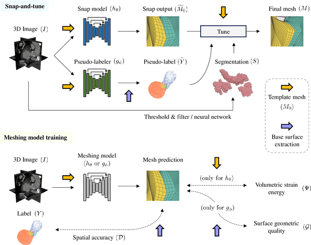
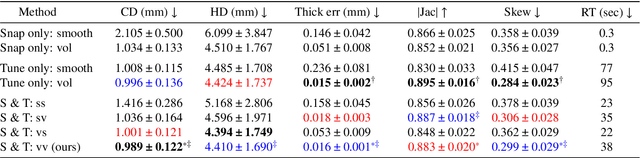
High-quality volumetric meshing from medical images is a key bottleneck for physics-based simulations in personalized medicine. For volumetric meshing of complex medical structures, recent studies have often utilized deep learning (DL)-based template deformation approaches to enable fast test-time generation with high spatial accuracy. However, these approaches still exhibit limitations, such as limited flexibility at high-curvature areas and unrealistic inter-part distances. In this study, we introduce a simple yet effective snap-and-tune strategy that sequentially applies DL and test-time optimization, which combines fast initial shape fitting with more detailed sample-specific mesh corrections. Our method provides significant improvements in both spatial accuracy and mesh quality, while being fully automated and requiring no additional training labels. Finally, we demonstrate the versatility and usefulness of our newly generated meshes via solid mechanics simulations in two different software platforms. Our code is available at https://github.com/danpak94/Deep-Cardiac-Volumetric-Mesh.
Using Foundation Models as Pseudo-Label Generators for Pre-Clinical 4D Cardiac CT Segmentation
May 14, 2025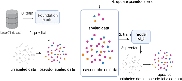



Cardiac image segmentation is an important step in many cardiac image analysis and modeling tasks such as motion tracking or simulations of cardiac mechanics. While deep learning has greatly advanced segmentation in clinical settings, there is limited work on pre-clinical imaging, notably in porcine models, which are often used due to their anatomical and physiological similarity to humans. However, differences between species create a domain shift that complicates direct model transfer from human to pig data. Recently, foundation models trained on large human datasets have shown promise for robust medical image segmentation; yet their applicability to porcine data remains largely unexplored. In this work, we investigate whether foundation models can generate sufficiently accurate pseudo-labels for pig cardiac CT and propose a simple self-training approach to iteratively refine these labels. Our method requires no manually annotated pig data, relying instead on iterative updates to improve segmentation quality. We demonstrate that this self-training process not only enhances segmentation accuracy but also smooths out temporal inconsistencies across consecutive frames. Although our results are encouraging, there remains room for improvement, for example by incorporating more sophisticated self-training strategies and by exploring additional foundation models and other cardiac imaging technologies.
Adapting Vision Foundation Models for Real-time Ultrasound Image Segmentation
Mar 31, 2025We propose a novel approach that adapts hierarchical vision foundation models for real-time ultrasound image segmentation. Existing ultrasound segmentation methods often struggle with adaptability to new tasks, relying on costly manual annotations, while real-time approaches generally fail to match state-of-the-art performance. To overcome these limitations, we introduce an adaptive framework that leverages the vision foundation model Hiera to extract multi-scale features, interleaved with DINOv2 representations to enhance visual expressiveness. These enriched features are then decoded to produce precise and robust segmentation. We conduct extensive evaluations on six public datasets and one in-house dataset, covering both cardiac and thyroid ultrasound segmentation. Experiments show that our approach outperforms state-of-the-art methods across multiple datasets and excels with limited supervision, surpassing nnUNet by over 20\% on average in the 1\% and 10\% data settings. Our method achieves $\sim$77 FPS inference speed with TensorRT on a single GPU, enabling real-time clinical applications.
Progressive Test Time Energy Adaptation for Medical Image Segmentation
Mar 20, 2025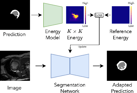
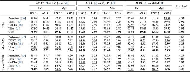
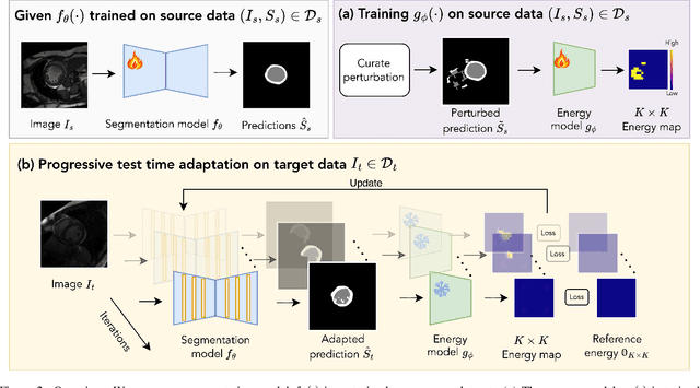
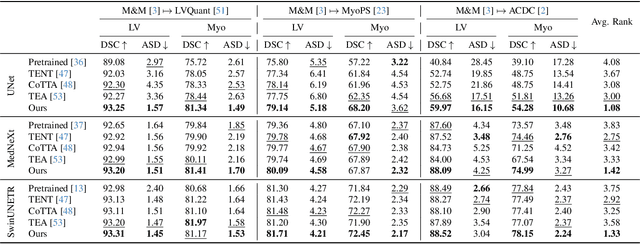
We propose a model-agnostic, progressive test-time energy adaptation approach for medical image segmentation. Maintaining model performance across diverse medical datasets is challenging, as distribution shifts arise from inconsistent imaging protocols and patient variations. Unlike domain adaptation methods that require multiple passes through target data - impractical in clinical settings - our approach adapts pretrained models progressively as they process test data. Our method leverages a shape energy model trained on source data, which assigns an energy score at the patch level to segmentation maps: low energy represents in-distribution (accurate) shapes, while high energy signals out-of-distribution (erroneous) predictions. By minimizing this energy score at test time, we refine the segmentation model to align with the target distribution. To validate the effectiveness and adaptability, we evaluated our framework on eight public MRI (bSSFP, T1- and T2-weighted) and X-ray datasets spanning cardiac, spinal cord, and lung segmentation. We consistently outperform baselines both quantitatively and qualitatively.