 Add to Chrome
 Add to Firefox
 Add to Edge
Super-Generalist: Towards Comprehensive and Accurate Medical Image Understanding via Generalist-Specialist Synergy
Jul 10, 2026
Medical images require comprehensive and accurate interpretation to support the diagnosis of diverse clincial conditions. Recent vision-language generalist models offer broad task coverage and promising zero-shot capabilities, yet often lack fine-grained anatomical and lesion awareness for reliable diagnosis and spatial interpretability. In contrast, supervised specialist models achieve strong performance on specific tasks but typically lack generalization across diseases and anatomies. In this work, we present SuG, a Super-Generalist framework that unifies generalist vision-language learning with specialist objectives, enabling both broad generalization and specialist-level diagnostic capability. We perform specialist-enhanced vision-language alignment in SuG by incorporating spatial priors from multiple segmentation experts, including anatomy, class-specific lesion and class-agnostic lesion segmentors that captures lesions beyond anatomies annotated during training. To improve lesion grounding capability, we leverage lesion masks as spatial priors to calibrate text-conditioned visual attention, encouraging disease-related semantics to focus on clinically relevant regions. We evaluate SuG on extensive chest and abdominal CT benchmarks, including CT-RATE, Merlin, MedVL-CT69K, and several in-house tumor datasets. SuG achieves state-of-the-art performance across a wide range of disease diagnosis tasks and surpasses specialist models on several critical tumor diagnosis benchmarks. Furthermore, SuG demonstrates strong lesion grounding capability, including robust generalization to lesion types lacking class-specific supervision.

Large-scale and Fine-grained Vision-language Pre-training for Enhanced CT Image Understanding
Jan 24, 2025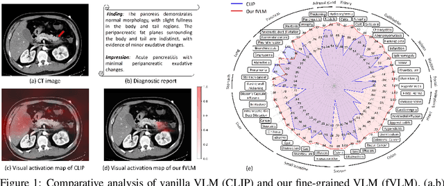
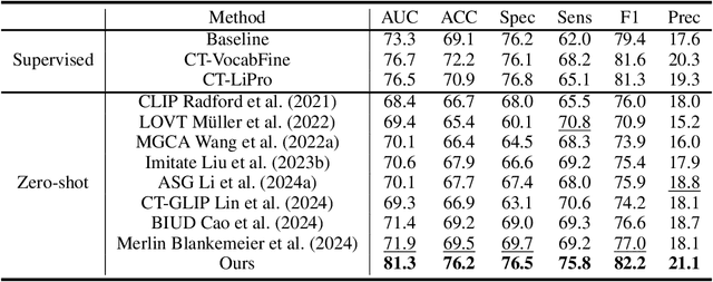
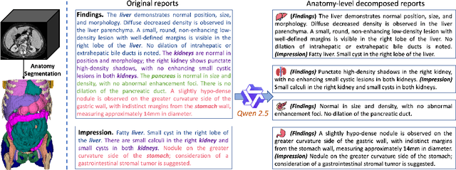
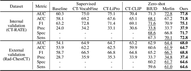
Artificial intelligence (AI) shows great potential in assisting radiologists to improve the efficiency and accuracy of medical image interpretation and diagnosis. However, a versatile AI model requires large-scale data and comprehensive annotations, which are often impractical in medical settings. Recent studies leverage radiology reports as a naturally high-quality supervision for medical images, using contrastive language-image pre-training (CLIP) to develop language-informed models for radiological image interpretation. Nonetheless, these approaches typically contrast entire images with reports, neglecting the local associations between imaging regions and report sentences, which may undermine model performance and interoperability. In this paper, we propose a fine-grained vision-language model (fVLM) for anatomy-level CT image interpretation. Specifically, we explicitly match anatomical regions of CT images with corresponding descriptions in radiology reports and perform contrastive pre-training for each anatomy individually. Fine-grained alignment, however, faces considerable false-negative challenges, mainly from the abundance of anatomy-level healthy samples and similarly diseased abnormalities. To tackle this issue, we propose identifying false negatives of both normal and abnormal samples and calibrating contrastive learning from patient-level to disease-aware pairing. We curated the largest CT dataset to date, comprising imaging and report data from 69,086 patients, and conducted a comprehensive evaluation of 54 major and important disease diagnosis tasks across 15 main anatomies. Experimental results demonstrate the substantial potential of fVLM in versatile medical image interpretation. In the zero-shot classification task, we achieved an average AUC of 81.3% on 54 diagnosis tasks, surpassing CLIP and supervised methods by 12.9% and 8.0%, respectively.
Deep learning to estimate the physical proportion of infected region of lung for COVID-19 pneumonia with CT image set
Jun 09, 2020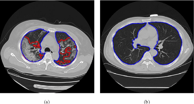
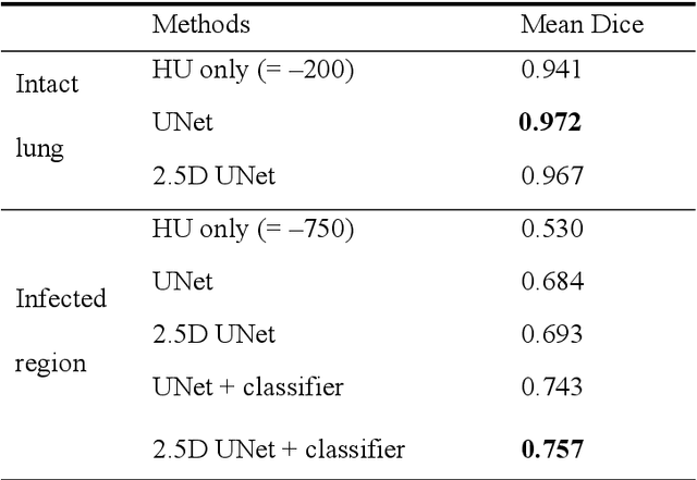
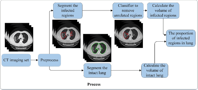
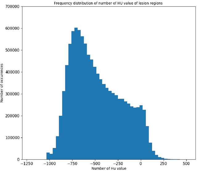
Utilizing computed tomography (CT) images to quickly estimate the severity of cases with COVID-19 is one of the most straightforward and efficacious methods. Two tasks were studied in this present paper. One was to segment the mask of intact lung in case of pneumonia. Another was to generate the masks of regions infected by COVID-19. The masks of these two parts of images then were converted to corresponding volumes to calculate the physical proportion of infected region of lung. A total of 129 CT image set were herein collected and studied. The intrinsic Hounsfiled value of CT images was firstly utilized to generate the initial dirty version of labeled masks both for intact lung and infected regions. Then, the samples were carefully adjusted and improved by two professional radiologists to generate the final training set and test benchmark. Two deep learning models were evaluated: UNet and 2.5D UNet. For the segment of infected regions, a deep learning based classifier was followed to remove unrelated blur-edged regions that were wrongly segmented out such as air tube and blood vessel tissue etc. For the segmented masks of intact lung and infected regions, the best method could achieve 0.972 and 0.757 measure in mean Dice similarity coefficient on our test benchmark. As the overall proportion of infected region of lung, the final result showed 0.961 (Pearson's correlation coefficient) and 11.7% (mean absolute percent error). The instant proportion of infected regions of lung could be used as a visual evidence to assist clinical physician to determine the severity of the case. Furthermore, a quantified report of infected regions can help predict the prognosis for COVID-19 cases which were scanned periodically within the treatment cycle.