 Add to Chrome
 Add to Firefox
 Add to Edge
CARVE: Cross-Slice Anisotropic Reallocation of Visual Evidence for Efficient 3D Medical Volume Understanding
Aug 05, 2026
Slice-based MLLMs leverage mature 2D encoders by representing 3D volumes as sequences of 2D slices. However, this slice-wise formulation produces thousands of visual tokens that burden the LLM backbone, many of which capture overlapping visual evidence across adjacent slices. To understand how effectively a growing visual token budget improves performance, we perform scaling analyses on two 3D medical VQA benchmarks and find diminishing returns: cost keeps rising while accuracy saturates, and improving in-plane resolution is more effective than adding slices at comparable budgets. The budget should therefore be allocated more selectively rather than simply enlarged, yet most token compression methods are designed for 2D images or videos, where redundancy arises from spatial layout or temporal motion rather than from near-duplicate content along the depth axis. We present CARVE, a training-free framework that compresses visual tokens prior to LLM inference and casts token reduction as budget-constrained 2.5D allocation. CARVE partitions the depth axis into coherent windows and allocates tokens non-uniformly according to normalized cross-slice evidence. Under a shared budget, CARVE builds spatial anchors on representative slices and retrieves locally varying evidence from the full volume, then merges remaining eligible tokens into nearby anchors within each window. Removing roughly 80% of the visual tokens on Hulu-Med-7B, CARVE leads all compression baselines on every AMOS-MM report-generation metric, with 6.2 points higher retention of full-token quality than the strongest baseline, and preserves 98.1% of full-token performance across three VQA benchmarks.

Two-Stage Cross-Domain Cervical Abnormality Screening with Cytopathological Image Synthesis and Knowledge Distillation
Jun 26, 2026Cross-domain diagnosis remains a major challenge in cervical cell pathology due to pronounced domain shifts across institutions and the subtle visual differences among disease stages, which jointly impair model generalization. To address these issues, this paper proposes a two-stage framework for cross-domain cervical cell detection. In the first stage, we propose the Spatially-Continuous Unpaired Neural Schrödinger Bridge (SC-UNSB), which constructs a synthetic intermediate domain to mitigate cross-domain distribution shifts by modeling image translation as an entropy-regularized optimal transport process. In the second stage, we propose a dual-level feature alignment strategy within a knowledge distillation, which progressively aligns shallow structural features and deep semantic representations to facilitate the transfer of domain-invariant knowledge from the source to the target model. Experimental results demonstrate that the proposed method effectively mitigates domain shift and category ambiguity, improving the cross-domain detection performance.
Brain-Adapter: A Dual-Stream Vision-Language MIL Framework for Comprehensive 3D CT Diagnosis of Acute Intracranial Pathologies
Jun 22, 2026Automated diagnosis of 3D brain CT scans is essential for critical care, yet it remains challenging due to the heavy reliance on manual annotations and the limited semantic understanding of conventional models. While 2D foundation vision-language models (VLMs) have shown remarkable generalization, effectively transferring their representational power to 3D volumes remains an open problem. In this paper, we propose Brain-Adapter, a novel dual-stream multiple instance learning (MIL) framework that leverages pre-trained 2D biomedical VLMs and raw diagnostic reports for robust scan-level multi-label classification. Specifically, we introduce a Text-Conditioned Attention (TCA) mechanism, utilizing raw diagnostic sentences as semantic queries to dynamically align visual cues with specific disease concepts. Concurrently, a parallel visual MIL stream captures global scan characteristics, supervised by structured labels extracted via a Large Language Model (LLM). To ensure representation coherence, a consistency constraint enforces synergy between the two streams. During inference, an Uncertainty-Aware Refinement (UAR) module dynamically calibrates and fuses these dual-stream predictions to resolve ambiguous cases. Extensive experiments demonstrate that our method significantly outperforms state-of-the-art 3D models and standard MIL approaches. By eliminating the reliance on dense annotations, Brain-Adapter provides a highly scalable and clinically viable solution for 3D acute intracranial pathology analysis.
PPGL-Swarm: Integrated Multimodal Risk Stratification and Hereditary Syndrome Detection in Pheochromocytoma and Paraganglioma
Mar 23, 2026Pheochromocytomas and paragangliomas (PPGLs) are rare neuroendocrine tumors, of which 15-25% develop metastatic disease with 5-year survival rates reported as low as 34%. PPGL may indicate hereditary syndromes requiring stricter, syndrome-specific treatment and surveillance, but clinicians often fail to recognize these associations in routine care. Clinical practice uses GAPP score for PPGL grading, but several limitations remain for PPGL diagnosis: (1) GAPP scoring demands a high workload for clinician because it requires the manual evaluation of six independent components; (2) key components such as cellularity and Ki-67 are often evaluated with subjective criteria; (3) several clinically relevant metastatic risk factors are not captured by GAPP, such as SDHB mutations, which have been associated with reported metastatic rates of 35-75%. Agent-driven diagnostic systems appear promising, but most lack traceable reasoning for decision-making and do not incorporate domain-specific knowledge such as PPGL genotype information. To address these limitations, we present PPGL-Swarm, an agentic PPGL diagnostic system that generates a comprehensive report, including automated GAPP scoring (with quantified cellularity and Ki-67), genotype risk alerts, and multimodal report with integrated evidence. The system provides an auditable reasoning trail by decomposing diagnosis into micro-tasks, each assigned to a specialized agent. The gene and table agents use knowledge enhancement to better interpret genotype and laboratory findings, and during training we use reinforcement learning to refine tool selection and task assignment.
LGAN: An Efficient High-Order Graph Neural Network via the Line Graph Aggregation
Dec 11, 2025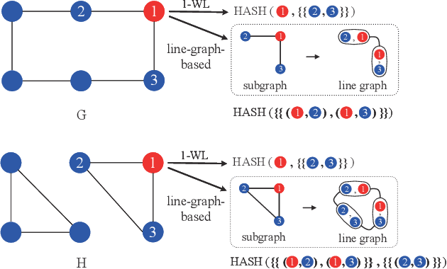
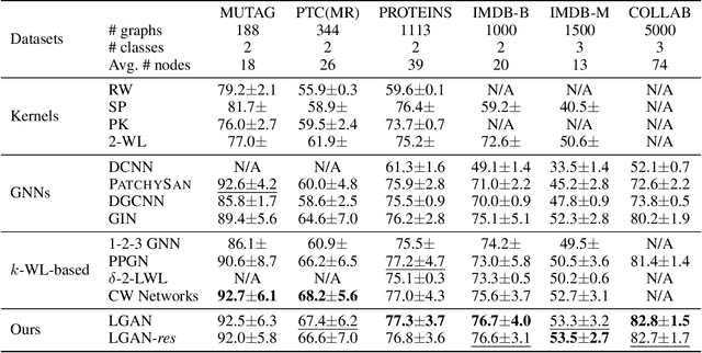
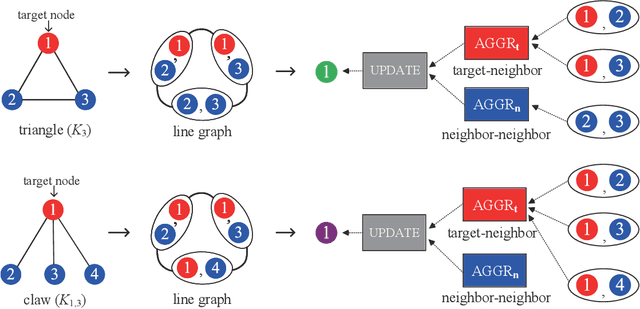
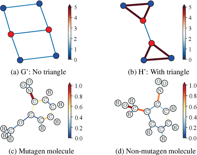
Graph Neural Networks (GNNs) have emerged as a dominant paradigm for graph classification. Specifically, most existing GNNs mainly rely on the message passing strategy between neighbor nodes, where the expressivity is limited by the 1-dimensional Weisfeiler-Lehman (1-WL) test. Although a number of k-WL-based GNNs have been proposed to overcome this limitation, their computational cost increases rapidly with k, significantly restricting the practical applicability. Moreover, since the k-WL models mainly operate on node tuples, these k-WL-based GNNs cannot retain fine-grained node- or edge-level semantics required by attribution methods (e.g., Integrated Gradients), leading to the less interpretable problem. To overcome the above shortcomings, in this paper, we propose a novel Line Graph Aggregation Network (LGAN), that constructs a line graph from the induced subgraph centered at each node to perform the higher-order aggregation. We theoretically prove that the LGAN not only possesses the greater expressive power than the 2-WL under injective aggregation assumptions, but also has lower time complexity. Empirical evaluations on benchmarks demonstrate that the LGAN outperforms state-of-the-art k-WL-based GNNs, while offering better interpretability.
Pathology Image Restoration via Mixture of Prompts
Mar 16, 2025In digital pathology, acquiring all-in-focus images is essential to high-quality imaging and high-efficient clinical workflow. Traditional scanners achieve this by scanning at multiple focal planes of varying depths and then merging them, which is relatively slow and often struggles with complex tissue defocus. Recent prevailing image restoration technique provides a means to restore high-quality pathology images from scans of single focal planes. However, existing image restoration methods are inadequate, due to intricate defocus patterns in pathology images and their domain-specific semantic complexities. In this work, we devise a two-stage restoration solution cascading a transformer and a diffusion model, to benefit from their powers in preserving image fidelity and perceptual quality, respectively. We particularly propose a novel mixture of prompts for the two-stage solution. Given initial prompt that models defocus in microscopic imaging, we design two prompts that describe the high-level image semantics from pathology foundation model and the fine-grained tissue structures via edge extraction. We demonstrate that, by feeding the prompt mixture to our method, we can restore high-quality pathology images from single-focal-plane scans, implying high potentials of the mixture of prompts to clinical usage. Code will be publicly available at https://github.com/caijd2000/MoP.
LSA: Latent Style Augmentation Towards Stain-Agnostic Cervical Cancer Screening
Mar 09, 2025The deployment of computer-aided diagnosis systems for cervical cancer screening using whole slide images (WSIs) faces critical challenges due to domain shifts caused by staining variations across different scanners and imaging environments. While existing stain augmentation methods improve patch-level robustness, they fail to scale to WSIs due to two key limitations: (1) inconsistent stain patterns when extending patch operations to gigapixel slides, and (2) prohibitive computational/storage costs from offline processing of augmented WSIs.To address this, we propose Latent Style Augmentation (LSA), a framework that performs efficient, online stain augmentation directly on WSI-level latent features. We first introduce WSAug, a WSI-level stain augmentation method ensuring consistent stain across patches within a WSI. Using offline-augmented WSIs by WSAug, we design and train Stain Transformer, which can simulate targeted style in the latent space, efficiently enhancing the robustness of the WSI-level classifier. We validate our method on a multi-scanner WSI dataset for cervical cancer diagnosis. Despite being trained on data from a single scanner, our approach achieves significant performance improvements on out-of-distribution data from other scanners. Code will be available at https://github.com/caijd2000/LSA.
Structure-Guided MR-to-CT Synthesis with Spatial and Semantic Alignments for Attenuation Correction of Whole-Body PET/MR Imaging
Nov 26, 2024Deep-learning-based MR-to-CT synthesis can estimate the electron density of tissues, thereby facilitating PET attenuation correction in whole-body PET/MR imaging. However, whole-body MR-to-CT synthesis faces several challenges including the issue of spatial misalignment and the complexity of intensity mapping, primarily due to the variety of tissues and organs throughout the whole body. Here we propose a novel whole-body MR-to-CT synthesis framework, which consists of three novel modules to tackle these challenges: (1) Structure-Guided Synthesis module leverages structure-guided attention gates to enhance synthetic image quality by diminishing unnecessary contours of soft tissues; (2) Spatial Alignment module yields precise registration between paired MR and CT images by taking into account the impacts of tissue volumes and respiratory movements, thus providing well-aligned ground-truth CT images during training; (3) Semantic Alignment module utilizes contrastive learning to constrain organ-related semantic information, thereby ensuring the semantic authenticity of synthetic CT images.We conduct extensive experiments to demonstrate that the proposed whole-body MR-to-CT framework can produce visually plausible and semantically realistic CT images, and validate its utility in PET attenuation correction.
REHRSeg: Unleashing the Power of Self-Supervised Super-Resolution for Resource-Efficient 3D MRI Segmentation
Oct 14, 2024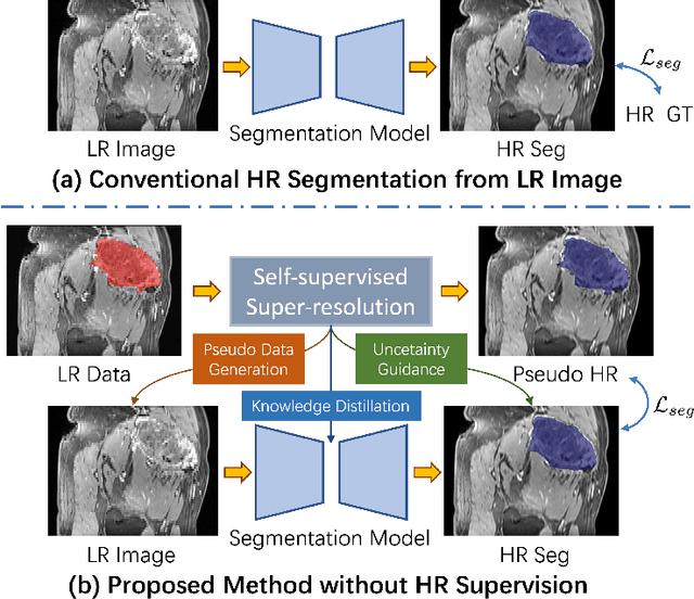
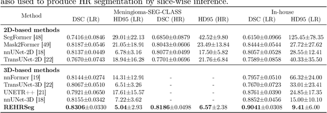
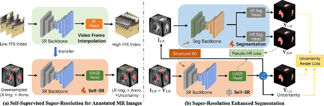
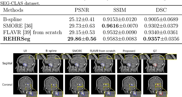
High-resolution (HR) 3D magnetic resonance imaging (MRI) can provide detailed anatomical structural information, enabling precise segmentation of regions of interest for various medical image analysis tasks. Due to the high demands of acquisition device, collection of HR images with their annotations is always impractical in clinical scenarios. Consequently, segmentation results based on low-resolution (LR) images with large slice thickness are often unsatisfactory for subsequent tasks. In this paper, we propose a novel Resource-Efficient High-Resolution Segmentation framework (REHRSeg) to address the above-mentioned challenges in real-world applications, which can achieve HR segmentation while only employing the LR images as input. REHRSeg is designed to leverage self-supervised super-resolution (self-SR) to provide pseudo supervision, therefore the relatively easier-to-acquire LR annotated images generated by 2D scanning protocols can be directly used for model training. The main contribution to ensure the effectiveness in self-SR for enhancing segmentation is three-fold: (1) We mitigate the data scarcity problem in the medical field by using pseudo-data for training the segmentation model. (2) We design an uncertainty-aware super-resolution (UASR) head in self-SR to raise the awareness of segmentation uncertainty as commonly appeared on the ROI boundaries. (3) We align the spatial features for self-SR and segmentation through structural knowledge distillation to enable a better capture of region correlations. Experimental results demonstrate that REHRSeg achieves high-quality HR segmentation without intensive supervision, while also significantly improving the baseline performance for LR segmentation.
Inter-slice Super-resolution of Magnetic Resonance Images by Pre-training and Self-supervised Fine-tuning
Jun 10, 2024
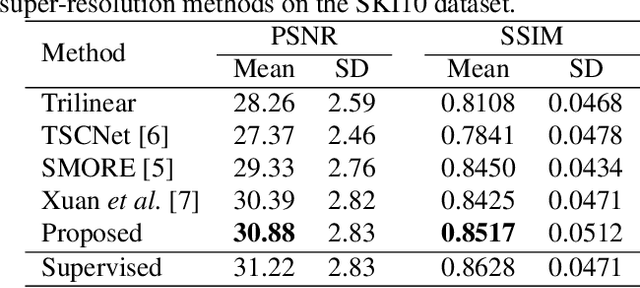
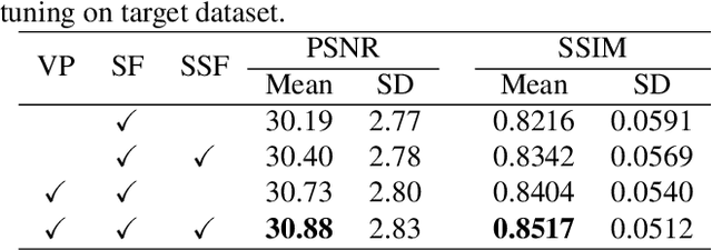
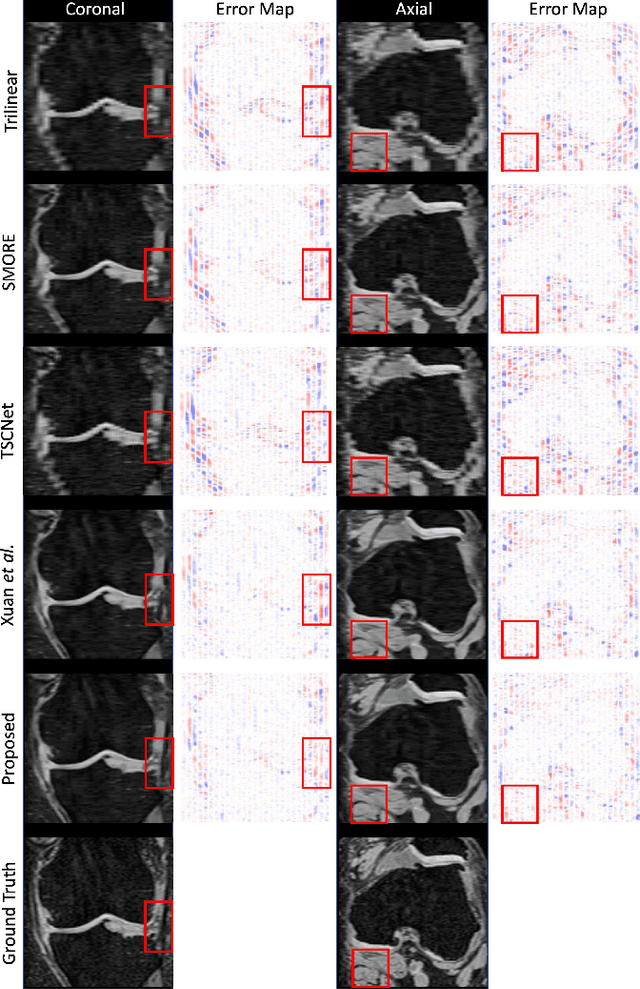
In clinical practice, 2D magnetic resonance (MR) sequences are widely adopted. While individual 2D slices can be stacked to form a 3D volume, the relatively large slice spacing can pose challenges for both image visualization and subsequent analysis tasks, which often require isotropic voxel spacing. To reduce slice spacing, deep-learning-based super-resolution techniques are widely investigated. However, most current solutions require a substantial number of paired high-resolution and low-resolution images for supervised training, which are typically unavailable in real-world scenarios. In this work, we propose a self-supervised super-resolution framework for inter-slice super-resolution of MR images. Our framework is first featured by pre-training on video dataset, as temporal correlation of videos is found beneficial for modeling the spatial relation among MR slices. Then, we use public high-quality MR dataset to fine-tune our pre-trained model, for enhancing awareness of our model to medical data. Finally, given a target dataset at hand, we utilize self-supervised fine-tuning to further ensure our model works well with user-specific super-resolution tasks. The proposed method demonstrates superior performance compared to other self-supervised methods and also holds the potential to benefit various downstream applications.