 Add to Chrome
 Add to Firefox
 Add to Edge
Dual Skipping Guidance for Document Retrieval with Learned Sparse Representations
Apr 23, 2022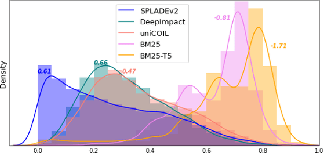
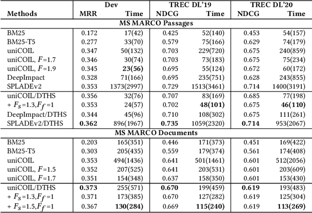
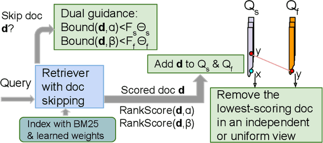
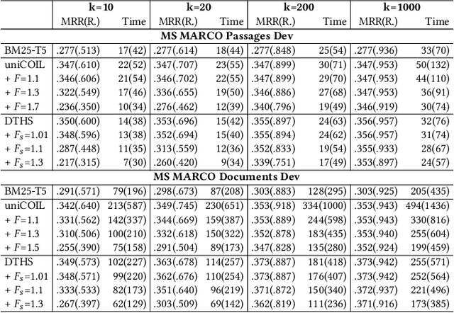
This paper proposes a dual skipping guidance scheme with hybrid scoring to accelerate document retrieval that uses learned sparse representations while still delivering a good relevance. This scheme uses both lexical BM25 and learned neural term weights to bound and compose the rank score of a candidate document separately for skipping and final ranking, and maintains two top-k thresholds during inverted index traversal. This paper evaluates time efficiency and ranking relevance of the proposed scheme in searching MS MARCO TREC datasets.
Pan-cancer computational histopathology reveals tumor mutational burden status through weakly-supervised deep learning
Apr 07, 2022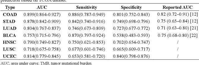
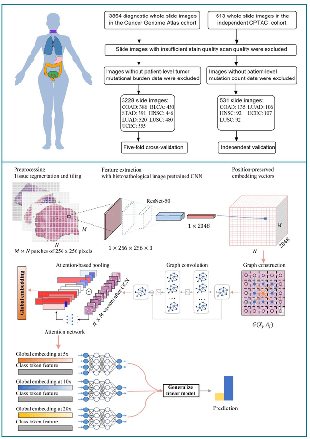
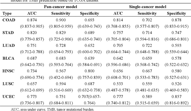
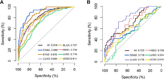
Tumor mutational burden (TMB) is a potential genomic biomarker that can help identify patients who will benefit from immunotherapy across a variety of cancers. We included whole slide images (WSIs) of 3228 diagnostic slides from the Cancer Genome Atlas and 531 WSIs from the Clinical Proteomic Tumor Analysis Consortium for the development and verification of a pan-cancer TMB prediction model (PC-TMB). We proposed a multiscale weakly-supervised deep learning framework for predicting TMB of seven types of tumors based only on routinely used hematoxylin-eosin (H&E)-stained WSIs. PC-TMB achieved a mean area under curve (AUC) of 0.818 (0.804-0.831) in the cross-validation cohort, which was superior to the best single-scale model. In comparison with the state-of-the-art TMB prediction model from previous publications, our multiscale model achieved better performance over previously reported models. In addition, the improvements of PC-TMB over the single-tumor models were also confirmed by the ablation tests on 10x magnification. The PC-TMB algorithm also exhibited good generalization on external validation cohort with AUC of 0.732 (0.683-0.761). PC-TMB possessed a comparable survival-risk stratification performance to the TMB measured by whole exome sequencing, but with low cost and being time-efficient for providing a prognostic biomarker of multiple solid tumors. Moreover, spatial heterogeneity of TMB within tumors was also identified through our PC-TMB, which might enable image-based screening for molecular biomarkers with spatial variation and potential exploring for genotype-spatial heterogeneity relationships.
Mitosis domain generalization in histopathology images -- The MIDOG challenge
Apr 06, 2022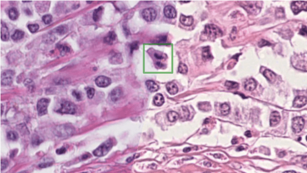
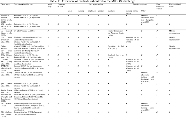
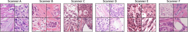
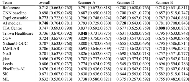
The density of mitotic figures within tumor tissue is known to be highly correlated with tumor proliferation and thus is an important marker in tumor grading. Recognition of mitotic figures by pathologists is known to be subject to a strong inter-rater bias, which limits the prognostic value. State-of-the-art deep learning methods can support the expert in this assessment but are known to strongly deteriorate when applied in a different clinical environment than was used for training. One decisive component in the underlying domain shift has been identified as the variability caused by using different whole slide scanners. The goal of the MICCAI MIDOG 2021 challenge has been to propose and evaluate methods that counter this domain shift and derive scanner-agnostic mitosis detection algorithms. The challenge used a training set of 200 cases, split across four scanning systems. As a test set, an additional 100 cases split across four scanning systems, including two previously unseen scanners, were given. The best approaches performed on an expert level, with the winning algorithm yielding an F_1 score of 0.748 (CI95: 0.704-0.781). In this paper, we evaluate and compare the approaches that were submitted to the challenge and identify methodological factors contributing to better performance.
A Deep Learning Framework for Nuclear Segmentation and Classification in Histopathological Images
Mar 04, 2022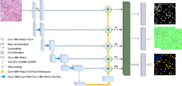
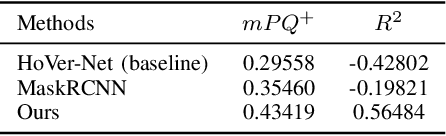
Nucleus segmentation and classification are the prerequisites in the workflow of digital pathology processing. However, it is very challenging due to its high-level heterogeneity and wide variations. This work proposes a deep neural network to simultaneously achieve nuclear classification and segmentation, which is designed using a unified framework with three different branches, including segmentation, HoVer mapping, and classification. The segmentation branch aims to generate the boundaries of each nucleus. The HoVer branch calculates the horizontal and vertical distances of nuclear pixels to their centres of mass. The nuclear classification branch is used to distinguish the class of pixels inside the nucleus obtained from segmentation.
Sk-Unet Model with Fourier Domain for Mitosis Detection
Sep 20, 2021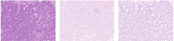
Mitotic count is the most important morphological feature of breast cancer grading. Many deep learning-based methods have been proposed but suffer from domain shift. In this work, we construct a Fourier-based segmentation model for mitosis detection to address the problem. Swapping the low-frequency spectrum of source and target images is shown effective to alleviate the discrepancy between different scanners. Our Fourier-based segmentation method can achieve F1 with 0.7456 on the preliminary test set.
Deep Learning methods for automatic evaluation of delayed enhancement-MRI. The results of the EMIDEC challenge
Aug 10, 2021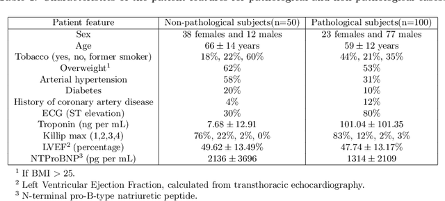
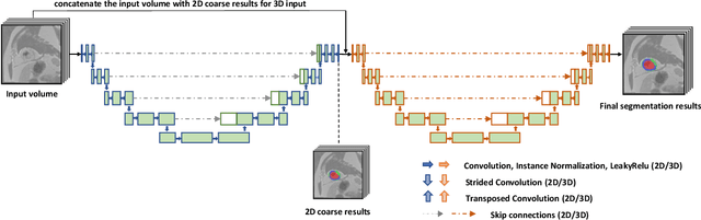
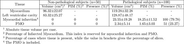
A key factor for assessing the state of the heart after myocardial infarction (MI) is to measure whether the myocardium segment is viable after reperfusion or revascularization therapy. Delayed enhancement-MRI or DE-MRI, which is performed several minutes after injection of the contrast agent, provides high contrast between viable and nonviable myocardium and is therefore a method of choice to evaluate the extent of MI. To automatically assess myocardial status, the results of the EMIDEC challenge that focused on this task are presented in this paper. The challenge's main objectives were twofold. First, to evaluate if deep learning methods can distinguish between normal and pathological cases. Second, to automatically calculate the extent of myocardial infarction. The publicly available database consists of 150 exams divided into 50 cases with normal MRI after injection of a contrast agent and 100 cases with myocardial infarction (and then with a hyperenhanced area on DE-MRI), whatever their inclusion in the cardiac emergency department. Along with MRI, clinical characteristics are also provided. The obtained results issued from several works show that the automatic classification of an exam is a reachable task (the best method providing an accuracy of 0.92), and the automatic segmentation of the myocardium is possible. However, the segmentation of the diseased area needs to be improved, mainly due to the small size of these areas and the lack of contrast with the surrounding structures.