 Add to Chrome
 Add to Firefox
 Add to Edge
Deep Learning-Based Airway Segmentation in Systemic Lupus Erythematosus Patients with Interstitial Lung Disease (SLE-ILD): A Comparative High-Resolution CT Analysis
Mar 18, 2026To characterize lobar and segmental airway volume differences between systemic lupus erythematosus (SLE) patients with interstitial lung disease (ILD) and those without ILD (non-ILD) using a deep learning-based approach on non-contrast chest high-resolution CT (HRCT). Methods: A retrospective analysis was conducted on 106 SLE patients (27 SLE-ILD, 79 SLE-non-ILD) who underwent HRCT. A customized deep learning framework based on the U-Net architecture was developed to automatically segment airway structures at the lobar and segmental levels via HRCT. Volumetric measurements of lung lobes and segments derived from the segmentations were statistically compared between the two groups using two-sample t-tests (significance threshold: p < 0.05). Results: At lobar level, significant airway volume enlargement in SLE-ILD patients was observed in the right upper lobe (p=0.009) and left upper lobe (p=0.039) compared to SLE-non-ILD. At the segmental level, significant differences were found in segments including R1 (p=0.016), R3 (p<0.001), and L3 (p=0.038), with the most marked changes in the upper lung zones, while lower zones showed non-significant trends. Conclusion: Our study demonstrates that an automated deep learning-based approach can effectively quantify airway volumes on HRCT scans and reveal significant, region-specific airway dilation in patients with SLE-ILD compared to those without ILD. The pattern of involvement, predominantly affecting the upper lobes and specific segments, highlights a distinct topographic phenotype of SLE-ILD and implicates airway structural alterations as a potential biomarker for disease presence. This AI-powered quantitative imaging biomarker holds promise for enhancing the early detection and monitoring of ILD in the SLE population, ultimately contributing to more personalized patient management.
Bronchovascular Tree-Guided Weakly Supervised Learning Method for Pulmonary Segment Segmentation
May 20, 2025


Pulmonary segment segmentation is crucial for cancer localization and surgical planning. However, the pixel-wise annotation of pulmonary segments is laborious, as the boundaries between segments are indistinguishable in medical images. To this end, we propose a weakly supervised learning (WSL) method, termed Anatomy-Hierarchy Supervised Learning (AHSL), which consults the precise clinical anatomical definition of pulmonary segments to perform pulmonary segment segmentation. Since pulmonary segments reside within the lobes and are determined by the bronchovascular tree, i.e., artery, airway and vein, the design of the loss function is founded on two principles. First, segment-level labels are utilized to directly supervise the output of the pulmonary segments, ensuring that they accurately encompass the appropriate bronchovascular tree. Second, lobe-level supervision indirectly oversees the pulmonary segment, ensuring their inclusion within the corresponding lobe. Besides, we introduce a two-stage segmentation strategy that incorporates bronchovascular priori information. Furthermore, a consistency loss is proposed to enhance the smoothness of segment boundaries, along with an evaluation metric designed to measure the smoothness of pulmonary segment boundaries. Visual inspection and evaluation metrics from experiments conducted on a private dataset demonstrate the effectiveness of our method.
Deep Learning-based Segmentation of Cerebral Aneurysms in 3D TOF-MRA using Coarse-to-Fine Framework
Oct 26, 2021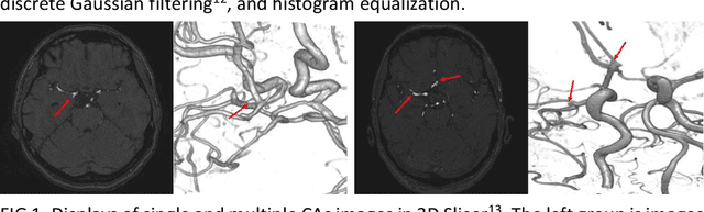
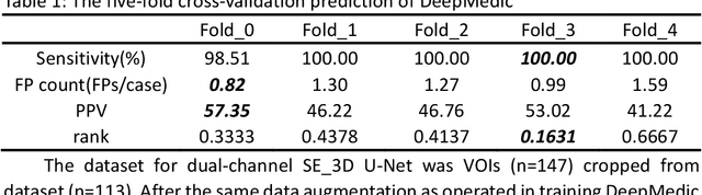
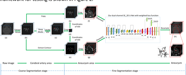
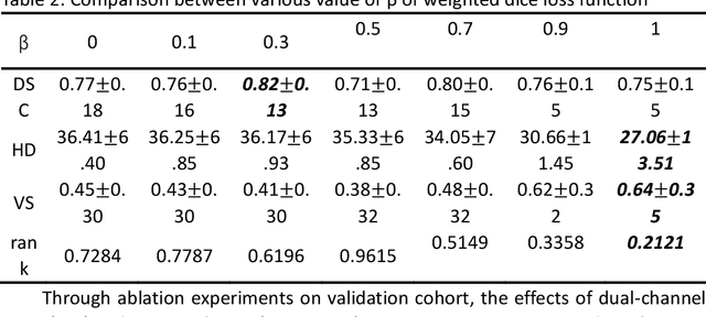
BACKGROUND AND PURPOSE: Cerebral aneurysm is one of the most common cerebrovascular diseases, and SAH caused by its rupture has a very high mortality and disability rate. Existing automatic segmentation methods based on DLMs with TOF-MRA modality could not segment edge voxels very well, so that our goal is to realize more accurate segmentation of cerebral aneurysms in 3D TOF-MRA with the help of DLMs. MATERIALS AND METHODS: In this research, we proposed an automatic segmentation framework of cerebral aneurysm in 3D TOF-MRA. The framework was composed of two segmentation networks ranging from coarse to fine. The coarse segmentation network, namely DeepMedic, completed the coarse segmentation of cerebral aneurysms, and the processed results were fed into the fine segmentation network, namely dual-channel SE_3D U-Net trained with weighted loss function, for fine segmentation. Images from ADAM2020 (n=113) were used for training and validation and images from another center (n=45) were used for testing. The segmentation metrics we used include DSC, HD, and VS. RESULTS: The trained cerebral aneurysm segmentation model achieved DSC of 0.75, HD of 1.52, and VS of 0.91 on validation cohort. On the totally independent test cohort, our method achieved the highest DSC of 0.12, the lowest HD of 11.61, and the highest VS of 0.16 in comparison with state-of-the-art segmentation networks. CONCLUSIONS: The coarse-to-fine framework, which composed of DeepMedic and dual-channel SE_3D U-Net can segment cerebral aneurysms in 3D TOF-MRA with a superior accuracy.