 Add to Chrome
 Add to Firefox
 Add to Edge
A New Angle on Bones: Robust Pose Estimation in X-Ray and Ultrasound
Jun 03, 2026Measuring the angle between bone structures is a routine task in medical image analysis and provides a key quantitative parameter for diagnosis and treatment planning. Automated methods can reduce time and cost while improving reproducibility. In this work, we address automatic bone pose estimation using a learning-based point candidate proposal followed by a line model to extract axis parameters. Since conventional line models such as least squares are sensitive to outliers, we incorporate false-positive reduction strategies and robust fitting techniques, such as RANSAC and Hough transforms, to improve robustness. We evaluate our method on three clinically relevant paediatric angle estimation tasks: fracture fragment assessment in radiographs and ultrasound and developmental dysplasia of the hip evaluation in ultrasound using the Graf method. Our approach achieves mean errors of $4.1^\circ$, $5.4^\circ$, and $5.51^\circ$, respectively, not only remaining within the expected clinical observer variability, but also significantly outperforming landmark-based methods. Our code and annotations for fracture angle assessment in radiographs are publicly available on GitHub.
CoRe: Joint Optimization with Contrastive Learning for Medical Image Registration
Mar 24, 2026Medical image registration is a fundamental task in medical image analysis, enabling the alignment of images from different modalities or time points. However, intensity inconsistencies and nonlinear tissue deformations pose significant challenges to the robustness of registration methods. Recent approaches leveraging self-supervised representation learning show promise by pre-training feature extractors to generate robust anatomical embeddings, that farther used for the registration. In this work, we propose a novel framework that integrates equivariant contrastive learning directly into the registration model. Our approach leverages the power of contrastive learning to learn robust feature representations that are invariant to tissue deformations. By jointly optimizing the contrastive and registration objectives, we ensure that the learned representations are not only informative but also suitable for the registration task. We evaluate our method on abdominal and thoracic image registration tasks, including both intra-patient and inter-patient scenarios. Experimental results demonstrate that the integration of contrastive learning directly into the registration framework significantly improves performance, surpassing strong baseline methods.
Effective Feature Learning for 3D Medical Registration via Domain-Specialized DINO Pretraining
Mar 14, 2026Medical image registration is a critical component of clinical imaging workflows, enabling accurate longitudinal assessment, multi-modal data fusion, and image-guided interventions. Intensity-based approaches often struggle with interscanner variability and complex anatomical deformations, whereas feature-based methods offer improved robustness by leveraging semantically informed representations. In this work, we investigate DINO-style self-supervised pretraining directly on 3D medical imaging data, aiming to learn dense volumetric features well suited for deformable registration. We assess the resulting representations on challenging interpatient abdominal registration task across both MRI and CT modalities. Our domain-specialized pretraining outperforms the DINOv2 model trained on a large-scale collection of natural images, while requiring substantially lower computational resources at inference time. Moreover, it surpasses established registration models under out-of-domain evaluation, demonstrating the value of task-agnostic yet medical imaging-focused pretraining for robust and efficient 3D image registration.
Depth to Anatomy: Learning Internal Organ Locations from Surface Depth Images
Jan 26, 2026Automated patient positioning plays an important role in optimizing scanning procedure and improving patient throughput. Leveraging depth information captured by RGB-D cameras presents a promising approach for estimating internal organ positions, thereby enabling more accurate and efficient positioning. In this work, we propose a learning-based framework that directly predicts the 3D locations and shapes of multiple internal organs from single 2D depth images of the body surface. Utilizing a large-scale dataset of full-body MRI scans, we synthesize depth images paired with corresponding anatomical segmentations to train a unified convolutional neural network architecture. Our method accurately localizes a diverse set of anatomical structures, including bones and soft tissues, without requiring explicit surface reconstruction. Experimental results demonstrate the potential of integrating depth sensors into radiology workflows to streamline scanning procedures and enhance patient experience through automated patient positioning.
Fracture Morphology Classification: Local Multiclass Modeling for Multilabel Complexity
Dec 16, 2025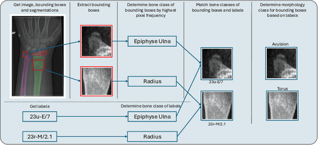

Between $15\,\%$ and $45\,\%$ of children experience a fracture during their growth years, making accurate diagnosis essential. Fracture morphology, alongside location and fragment angle, is a key diagnostic feature. In this work, we propose a method to extract fracture morphology by assigning automatically global AO codes to corresponding fracture bounding boxes. This approach enables the use of public datasets and reformulates the global multilabel task into a local multiclass one, improving the average F1 score by $7.89\,\%$. However, performance declines when using imperfect fracture detectors, highlighting challenges for real-world deployment. Our code is available on GitHub.
PrIINeR: Towards Prior-Informed Implicit Neural Representations for Accelerated MRI
Aug 11, 2025Accelerating Magnetic Resonance Imaging (MRI) reduces scan time but often degrades image quality. While Implicit Neural Representations (INRs) show promise for MRI reconstruction, they struggle at high acceleration factors due to weak prior constraints, leading to structural loss and aliasing artefacts. To address this, we propose PrIINeR, an INR-based MRI reconstruction method that integrates prior knowledge from pre-trained deep learning models into the INR framework. By combining population-level knowledge with instance-based optimization and enforcing dual data consistency, PrIINeR aligns both with the acquired k-space data and the prior-informed reconstruction. Evaluated on the NYU fastMRI dataset, our method not only outperforms state-of-the-art INR-based approaches but also improves upon several learning-based state-of-the-art methods, significantly improving structural preservation and fidelity while effectively removing aliasing artefacts.PrIINeR bridges deep learning and INR-based techniques, offering a more reliable solution for high-quality, accelerated MRI reconstruction. The code is publicly available on https://github.com/multimodallearning/PrIINeR.
Beyond the LUMIR challenge: The pathway to foundational registration models
May 30, 2025


Medical image challenges have played a transformative role in advancing the field, catalyzing algorithmic innovation and establishing new performance standards across diverse clinical applications. Image registration, a foundational task in neuroimaging pipelines, has similarly benefited from the Learn2Reg initiative. Building on this foundation, we introduce the Large-scale Unsupervised Brain MRI Image Registration (LUMIR) challenge, a next-generation benchmark designed to assess and advance unsupervised brain MRI registration. Distinct from prior challenges that leveraged anatomical label maps for supervision, LUMIR removes this dependency by providing over 4,000 preprocessed T1-weighted brain MRIs for training without any label maps, encouraging biologically plausible deformation modeling through self-supervision. In addition to evaluating performance on 590 held-out test subjects, LUMIR introduces a rigorous suite of zero-shot generalization tasks, spanning out-of-domain imaging modalities (e.g., FLAIR, T2-weighted, T2*-weighted), disease populations (e.g., Alzheimer's disease), acquisition protocols (e.g., 9.4T MRI), and species (e.g., macaque brains). A total of 1,158 subjects and over 4,000 image pairs were included for evaluation. Performance was assessed using both segmentation-based metrics (Dice coefficient, 95th percentile Hausdorff distance) and landmark-based registration accuracy (target registration error). Across both in-domain and zero-shot tasks, deep learning-based methods consistently achieved state-of-the-art accuracy while producing anatomically plausible deformation fields. The top-performing deep learning-based models demonstrated diffeomorphic properties and inverse consistency, outperforming several leading optimization-based methods, and showing strong robustness to most domain shifts, the exception being a drop in performance on out-of-domain contrasts.
OncoReg: Medical Image Registration for Oncological Challenges
Apr 01, 2025In modern cancer research, the vast volume of medical data generated is often underutilised due to challenges related to patient privacy. The OncoReg Challenge addresses this issue by enabling researchers to develop and validate image registration methods through a two-phase framework that ensures patient privacy while fostering the development of more generalisable AI models. Phase one involves working with a publicly available dataset, while phase two focuses on training models on a private dataset within secure hospital networks. OncoReg builds upon the foundation established by the Learn2Reg Challenge by incorporating the registration of interventional cone-beam computed tomography (CBCT) with standard planning fan-beam CT (FBCT) images in radiotherapy. Accurate image registration is crucial in oncology, particularly for dynamic treatment adjustments in image-guided radiotherapy, where precise alignment is necessary to minimise radiation exposure to healthy tissues while effectively targeting tumours. This work details the methodology and data behind the OncoReg Challenge and provides a comprehensive analysis of the competition entries and results. Findings reveal that feature extraction plays a pivotal role in this registration task. A new method emerging from this challenge demonstrated its versatility, while established approaches continue to perform comparably to newer techniques. Both deep learning and classical approaches still play significant roles in image registration, with the combination of methods - particularly in feature extraction - proving most effective.
Internal Organ Localization Using Depth Images
Mar 30, 2025Automated patient positioning is a crucial step in streamlining MRI workflows and enhancing patient throughput. RGB-D camera-based systems offer a promising approach to automate this process by leveraging depth information to estimate internal organ positions. This paper investigates the feasibility of a learning-based framework to infer approximate internal organ positions from the body surface. Our approach utilizes a large-scale dataset of MRI scans to train a deep learning model capable of accurately predicting organ positions and shapes from depth images alone. We demonstrate the effectiveness of our method in localization of multiple internal organs, including bones and soft tissues. Our findings suggest that RGB-D camera-based systems integrated into MRI workflows have the potential to streamline scanning procedures and improve patient experience by enabling accurate and automated patient positioning.
PULPo: Probabilistic Unsupervised Laplacian Pyramid Registration
Jul 15, 2024


Deformable image registration is fundamental to many medical imaging applications. Registration is an inherently ambiguous task often admitting many viable solutions. While neural network-based registration techniques enable fast and accurate registration, the majority of existing approaches are not able to estimate uncertainty. Here, we present PULPo, a method for probabilistic deformable registration capable of uncertainty quantification. PULPo probabilistically models the distribution of deformation fields on different hierarchical levels combining them using Laplacian pyramids. This allows our method to model global as well as local aspects of the deformation field. We evaluate our method on two widely used neuroimaging datasets and find that it achieves high registration performance as well as substantially better calibrated uncertainty quantification compared to the current state-of-the-art.