 Add to Chrome
 Add to Firefox
 Add to Edge
Cross-Modal MRI Ovary Segmentation in Endometriosis Using Unpaired TVUS Prototype Priors
Aug 19, 2026
Transvaginal ultrasound (TVUS) and magnetic resonance imaging (MRI) provide complementary information for endometriosis image analysis, yet existing studies mainly focus on single-modality analysis or disease classification, leaving cross-modal ovarian segmentation largely unexplored. In this work, to tackle the increased difficulty of ovary segmentation in MRI due to ovaries' small target size and ambiguous boundaries with surrounding pelvic structures, we propose a dual branch framework for ovary segmentation across TVUS and MRI. More specifically, by adapting MedSAM3 with TVUS-derived prototype bank, we aim to align anatomically consistent feature representations across both modalities. Extensive experiments are conducted on endometriosis-related TVUS and MRI datasets. We observe quantitative and qualitative improvements of over 5 percentage points for the proposed dual-branch approach compared with multiple state-of-the-art methods. Furthermore, our ablation study shows the contribution of individual components such as the prototype bank and the importance of warm-up pretraining in the source TVUS domain.

A Compact Top-Loading Robot for Endovascular Interventions: Design, Control and Evaluation
Jul 13, 2026Robot-assisted endovascular intervention can potentially reduce radiation exposure, improve surgeon ergonomics, enable telesurgery, support active assistance and autonomy, and enhance procedural precision. However, existing systems often suffer from limited procedural coverage because constrained patient-side setups, restricted flexibility, and complex instrument exchange hinder clinical workflow integration. This work presents a compact robotic system for endovascular interventions that enables continuous translational and rotational manipulation of standard endovascular instruments. The system consists of two alternating carts with pneumatically actuated membrane grippers integrated into rotating gripper gears. Its top-loading design allows rapid exchange of instruments such as guidewires and catheters without changing the robotic setup. A leader-follower control strategy enables continuous motion despite the finite stroke of each cart. The system was evaluated in motion-tracking experiments with guidewires and catheters and in an in vitro vascular phantom. The motion-tracking experiments showed generally smooth translational and rotational motion profiles. Across all tested guidewire and catheter experiments, the mean relative tracking errors were 3.6% for translational motion and 4.1% for rotational motion. In the vascular phantom, robot-assisted navigation reached the target in most trials, demonstrating the feasibility of the proposed manipulation concept under in vitro conditions. The presented robotic system demonstrates technical feasibility for continuous manipulation of standard endovascular instruments in bench-top and in vitro experiments. The compact top-loading design may ease instrument exchange and clinical workflow integration. Future work will focus on improving gripping performance, actuation speed, force feedback, and evaluation in more clinically realistic settings.
Comparing Commercial Depth Sensor Accuracy for Medical Applications
Jun 11, 2026Depth estimation has numerous medical and surgical applications. We benchmark four depth sensors on a porcine bone specimen, a porcine belly specimen, and a silicone kidney phantom using stylus-sampled references. These objects contain several real-world challenges, including homogeneous surfaces, specular surfaces, and subsurface scattering. The comparison includes stereo, structured-light, and time-of-flight sensors at a distance of approximately 50 cm. Specifically, the Intel RealSense D405 (Intel RealSense, United States), PMD Flexx2 (pmdtechnologies, Germany), Stereolabs ZED 2i (Stereolabs, France), and Zivid 2M+ 60 (Zivid, Norway) are compared. The Zivid 2M+ 60 performed best across all objects and metrics considered in this work. The ZED ranked second for real tissue, but last on the phantom.
Point Cloud Segmentation for Autonomous Clip Positioning in Laparoscopic Cholecystectomy on a Phantom
Jun 10, 2026High-risk applications in robotics, such as robot-assisted surgery, present unique challenges. These systems must be both highly precise and interpretable in order to be deployed in environments with very low tolerance for error or unsafe exploration. We present the first robotic system to demonstrate autonomous clip positioning on a physical phantom in laparoscopic surgery, one of the most common interventions in general surgery. After segmentation of a colorless point cloud from a single camera, target positions for the clips are extracted using spline interpolation, and can then be adjusted by the human operator. The segmentation model is trained on only 60 hand-labeled real point clouds, reflecting data scarcity in the surgical domain. We overcome this with a combination of pre-training on 128,000 synthetic point clouds and two novel data augmentation techniques. The motion of the end-effector to each target is visualized for the operator, satisfying the unique motion constraints of minimally-invasive surgery while ensuring that the robot's actions are verifiable and interpretable. In real robot experiments, our system localizes targets with the required precision of 0.75mm at a 95% success rate and executes autonomous clip positioning with a 100% success rate. We provide insights that are applicable to many other surgical and non-surgical tasks that require identifying and navigating to a precise target. Source code and project page: https://github.com/balazsgyenes/kirurc
* 8 pages, 5 figures, accepted to IEEE Robotics and Automation Letters (RAL)
A Position Statement on Endovascular Models and Effectiveness Metrics for Mechanical Thrombectomy Navigation, on behalf of the Stakeholder Taskforce for AI-assisted Robotic Thrombectomy (START)
Mar 30, 2026While we are making progress in overcoming infectious diseases and cancer; one of the major medical challenges of the mid-21st century will be the rising prevalence of stroke. Large vessels occlusions are especially debilitating, yet effective treatment (needed within hours to achieve best outcomes) remains limited due to geography. One solution for improving timely access to mechanical thrombectomy in geographically diverse populations is the deployment of robotic surgical systems. Artificial intelligence (AI) assistance may enable the upskilling of operators in this emerging therapeutic delivery approach. Our aim was to establish consensus frameworks for developing and validating AI-assisted robots for thrombectomy. Objectives included standardizing effectiveness metrics and defining reference testbeds across in silico, in vitro, ex vivo, and in vivo environments. To achieve this, we convened experts in neurointervention, robotics, data science, health economics, policy, statistics, and patient advocacy. Consensus was built through an incubator day, a Delphi process, and a final Position Statement. We identified that the four essential testbed environments each had distinct validation roles. Realism requirements vary: simpler testbeds should include realistic vessel anatomy compatible with guidewire and catheter use, while standard testbeds should incorporate deformable vessels. More advanced testbeds should include blood flow, pulsatility, and disease features. There are two macro-classes of effectiveness metrics: one for in silico, in vitro, and ex vivo stages focusing on technical navigation, and another for in vivo stages, focused on clinical outcomes. Patient safety is central to this technology's development. One requisite patient safety task needed now is to correlate in vitro measurements to in vivo complications.
* Published in Journal of the American Heart Association
Ensemble Learning and 3D Pix2Pix for Comprehensive Brain Tumor Analysis in Multimodal MRI
Dec 16, 2024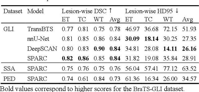
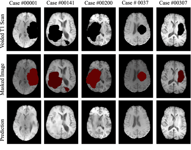
Motivated by the need for advanced solutions in the segmentation and inpainting of glioma-affected brain regions in multi-modal magnetic resonance imaging (MRI), this study presents an integrated approach leveraging the strengths of ensemble learning with hybrid transformer models and convolutional neural networks (CNNs), alongside the innovative application of 3D Pix2Pix Generative Adversarial Network (GAN). Our methodology combines robust tumor segmentation capabilities, utilizing axial attention and transformer encoders for enhanced spatial relationship modeling, with the ability to synthesize biologically plausible brain tissue through 3D Pix2Pix GAN. This integrated approach addresses the BraTS 2023 cluster challenges by offering precise segmentation and realistic inpainting, tailored for diverse tumor types and sub-regions. The results demonstrate outstanding performance, evidenced by quantitative evaluations such as the Dice Similarity Coefficient (DSC), Hausdorff Distance (HD95) for segmentation, and Structural Similarity Index Measure (SSIM), Peak Signal-to-Noise Ratio (PSNR), and Mean-Square Error (MSE) for inpainting. Qualitative assessments further validate the high-quality, clinically relevant outputs. In conclusion, this study underscores the potential of combining advanced machine learning techniques for comprehensive brain tumor analysis, promising significant advancements in clinical decision-making and patient care within the realm of medical imaging.
Unified HT-CNNs Architecture: Transfer Learning for Segmenting Diverse Brain Tumors in MRI from Gliomas to Pediatric Tumors
Dec 11, 2024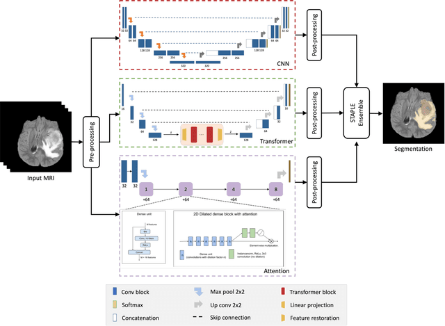
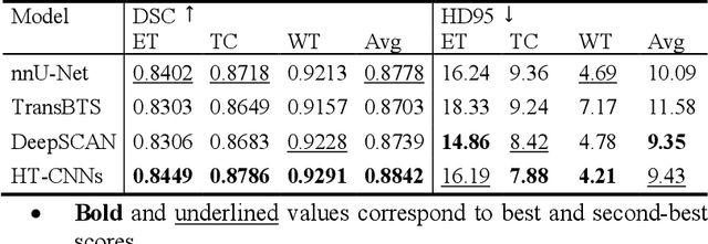
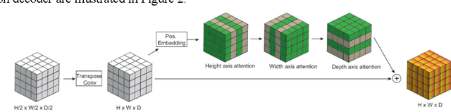
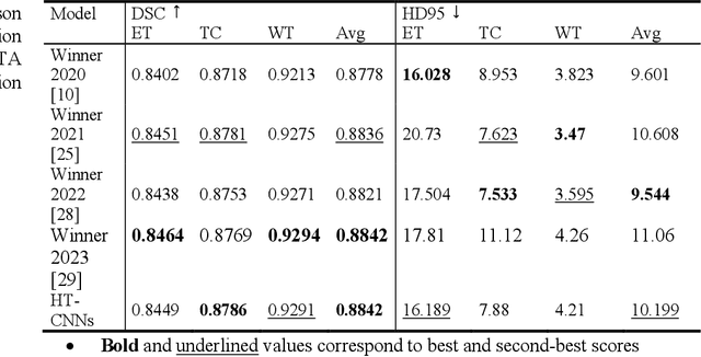
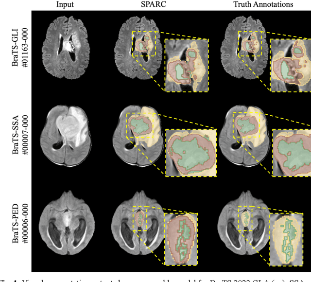
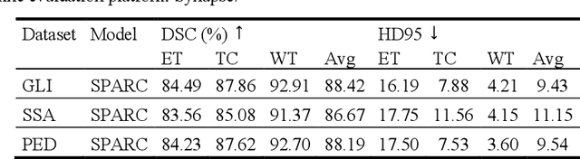
Accurate segmentation of brain tumors from 3D multimodal MRI is vital for diagnosis and treatment planning across diverse brain tumors. This paper addresses the challenges posed by the BraTS 2023, presenting a unified transfer learning approach that applies to a broader spectrum of brain tumors. We introduce HT-CNNs, an ensemble of Hybrid Transformers and Convolutional Neural Networks optimized through transfer learning for varied brain tumor segmentation. This method captures spatial and contextual details from MRI data, fine-tuned on diverse datasets representing common tumor types. Through transfer learning, HT-CNNs utilize the learned representations from one task to improve generalization in another, harnessing the power of pre-trained models on large datasets and fine-tuning them on specific tumor types. We preprocess diverse datasets from multiple international distributions, ensuring representativeness for the most common brain tumors. Our rigorous evaluation employs standardized quantitative metrics across all tumor types, ensuring robustness and generalizability. The proposed ensemble model achieves superior segmentation results across the BraTS validation datasets over the previous winning methods. Comprehensive quantitative evaluations using the DSC and HD95 demonstrate the effectiveness of our approach. Qualitative segmentation predictions further validate the high-quality outputs produced by our model. Our findings underscore the potential of transfer learning and ensemble approaches in medical image segmentation, indicating a substantial enhancement in clinical decision-making and patient care. Despite facing challenges related to post-processing and domain gaps, our study sets a new precedent for future research for brain tumor segmentation. The docker image for the code and models has been made publicly available, https://hub.docker.com/r/razeineldin/ht-cnns.
LUDO: Low-Latency Understanding of Highly Deformable Objects using Point Cloud Occupancy Functions
Nov 13, 2024Accurately determining the shape and location of internal structures within deformable objects is crucial for medical tasks that require precise targeting, such as robotic biopsies. We introduce LUDO, a method for accurate low-latency understanding of deformable objects. LUDO reconstructs objects in their deformed state, including their internal structures, from a single-view point cloud observation in under 30 ms using occupancy networks. We demonstrate LUDO's abilities for autonomous targeting of internal regions of interest (ROIs) in highly deformable objects. Additionally, LUDO provides uncertainty estimates and explainability for its predictions, both of which are important in safety-critical applications such as surgical interventions. We evaluate LUDO in real-world robotic experiments, achieving a success rate of 98.9% for puncturing various ROIs inside highly deformable objects. LUDO demonstrates the potential to interact with deformable objects without the need for deformable registration methods.
Tracking Tumors under Deformation from Partial Point Clouds using Occupancy Networks
Nov 04, 2024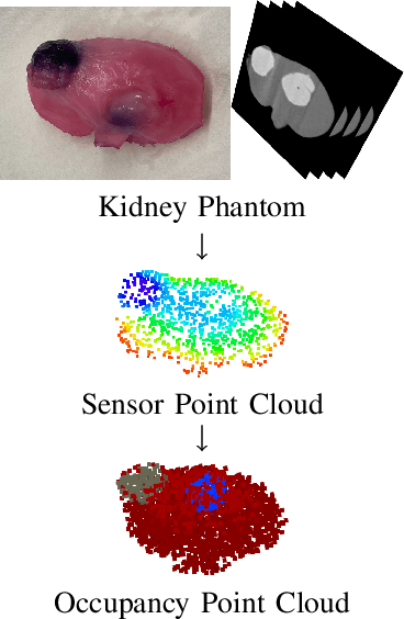
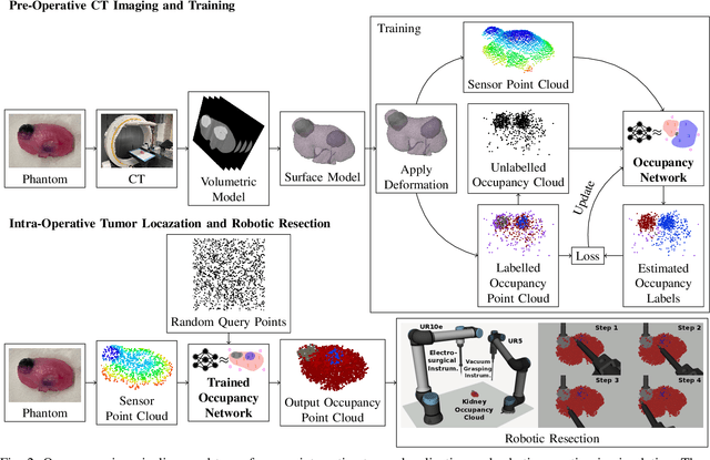
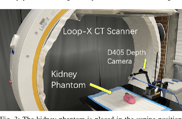

To track tumors during surgery, information from preoperative CT scans is used to determine their position. However, as the surgeon operates, the tumor may be deformed which presents a major hurdle for accurately resecting the tumor, and can lead to surgical inaccuracy, increased operation time, and excessive margins. This issue is particularly pronounced in robot-assisted partial nephrectomy (RAPN), where the kidney undergoes significant deformations during operation. Toward addressing this, we introduce a occupancy network-based method for the localization of tumors within kidney phantoms undergoing deformations at interactive speeds. We validate our method by introducing a 3D hydrogel kidney phantom embedded with exophytic and endophytic renal tumors. It closely mimics real tissue mechanics to simulate kidney deformation during in vivo surgery, providing excellent contrast and clear delineation of tumor margins to enable automatic threshold-based segmentation. Our findings indicate that the proposed method can localize tumors in moderately deforming kidneys with a margin of 6mm to 10mm, while providing essential volumetric 3D information at over 60Hz. This capability directly enables downstream tasks such as robotic resection.
Learning-Based Autonomous Navigation, Benchmark Environments and Simulation Framework for Endovascular Interventions
Oct 02, 2024
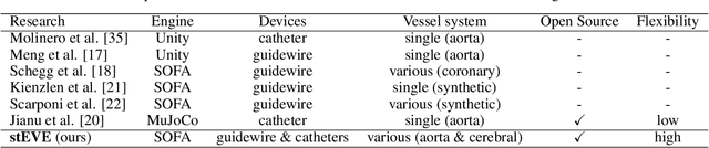
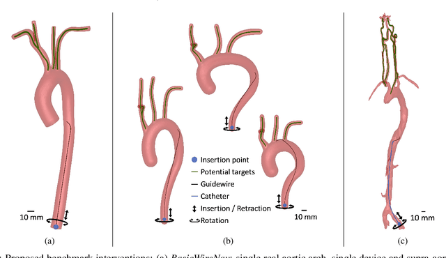
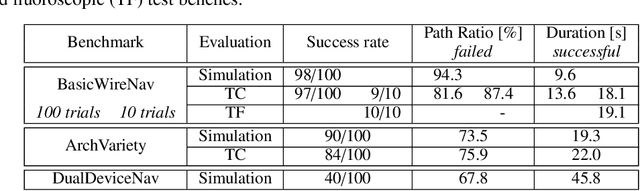
Endovascular interventions are a life-saving treatment for many diseases, yet suffer from drawbacks such as radiation exposure and potential scarcity of proficient physicians. Robotic assistance during these interventions could be a promising support towards these problems. Research focusing on autonomous endovascular interventions utilizing artificial intelligence-based methodologies is gaining popularity. However, variability in assessment environments hinders the ability to compare and contrast the efficacy of different approaches, primarily due to each study employing a unique evaluation framework. In this study, we present deep reinforcement learning-based autonomous endovascular device navigation on three distinct digital benchmark interventions: BasicWireNav, ArchVariety, and DualDeviceNav. The benchmark interventions were implemented with our modular simulation framework stEVE (simulated EndoVascular Environment). Autonomous controllers were trained solely in simulation and evaluated in simulation and on physical test benches with camera and fluoroscopy feedback. Autonomous control for BasicWireNav and ArchVariety reached high success rates and was successfully transferred from the simulated training environment to the physical test benches, while autonomous control for DualDeviceNav reached a moderate success rate. The experiments demonstrate the feasibility of stEVE and its potential for transferring controllers trained in simulation to real-world scenarios. Nevertheless, they also reveal areas that offer opportunities for future research. This study demonstrates the transferability of autonomous controllers from simulation to the real world in endovascular navigation and lowers the entry barriers and increases the comparability of research on endovascular assistance systems by providing open-source training scripts, benchmarks and the stEVE framework.