 Add to Chrome
 Add to Firefox
 Add to Edge
Brain Tumor Segmentation From Mri Using Deep Learning
Papers and Code
InfiltrNet: Dual-Branch CNN-Transformer Architecture for Brain Tumor Infiltration Risk Prediction
May 04, 2026Gliomas are aggressive brain tumors that infiltrate surrounding tissue beyond the visible tumor margins observed on Magnetic Resonance Imaging (MRI). Predicting the spatial extent of this infiltration is essential for surgical planning and radiation therapy, yet existing deep learning approaches focus on segmenting the visible tumor rather than estimating infiltration risk in the surrounding tissue. This paper presents InfiltrNet, a novel dual-branch architecture that combines a convolutional neural network (CNN) encoder with a Swin Transformer encoder through cross-attention fusion modules to predict three-zone infiltration risk maps from multimodal MRI. A label generation strategy based on distance transforms is proposed to derive reproducible infiltration risk zones from standard Brain Tumor Segmentation (BraTS) annotations. InfiltrNet is trained with a combined Dice-CrossEntropy and boundary-aware loss augmented by auxiliary supervision heads at intermediate decoder levels. Extensive experiments on BraTS 2020 and BraTS 2025 demonstrate that InfiltrNet outperforms five established baselines. Explainability analysis using GradCAM++ and Occlusion sensitivity confirms that the model attends to clinically relevant peritumoral regions.
Karhunen-Loève Expansion-Based Residual Anomaly Map for Resource-Efficient Glioma MRI Segmentation
Jan 21, 2026Accurate segmentation of brain tumors is essential for clinical diagnosis and treatment planning. Deep learning is currently the state-of-the-art for brain tumor segmentation, yet it requires either large datasets or extensive computational resources that are inaccessible in most areas. This makes the problem increasingly difficult: state-of-the-art models use thousands of training cases and vast computational power, where performance drops sharply when either is limited. The top performer in the Brats GLI 2023 competition relied on supercomputers trained on over 92,000 augmented MRI scans using an AMD EPYC 7402 CPU, six NVIDIA RTX 6000 GPUs (48GB VRAM each), and 1024GB of RAM over multiple weeks. To address this, the Karhunen--Loève Expansion (KLE) was implemented as a feature extraction step on downsampled, z-score normalized MRI volumes. Each 240$\times$240$\times$155 multi-modal scan is reduced to four $48^3$ channels and compressed into 32 KL coefficients. The resulting approximate reconstruction enables a residual-based anomaly map, which is upsampled and added as a fifth channel to a compact 3D U-Net. All experiments were run on a consumer workstation (AMD Ryzen 5 7600X CPU, RTX 4060Ti (8GB VRAM), and 64GB RAM while using far fewer training cases. This model achieves post-processed Dice scores of 0.929 (WT), 0.856 (TC), and 0.821 (ET), with HD95 distances of 2.93, 6.78, and 10.35 voxels. These results are significantly better than the winning BraTS 2023 methodology for HD95 distances and WT dice scores. This demonstrates that a KLE-based residual anomaly map can dramatically reduce computational cost and data requirements while retaining state-of-the-art performance.
Improving Pre-trained Segmentation Models using Post-Processing
Dec 16, 2025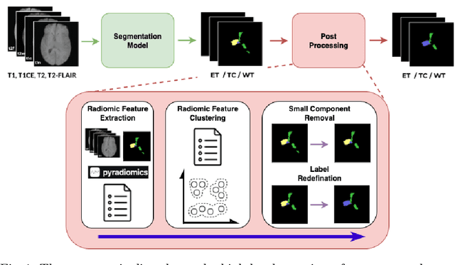
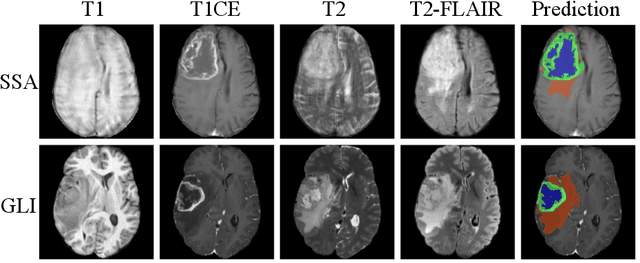
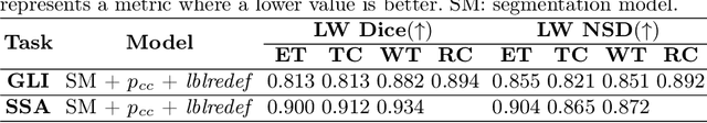
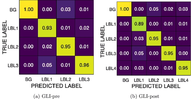
Gliomas are the most common malignant brain tumors in adults and are among the most lethal. Despite aggressive treatment, the median survival rate is less than 15 months. Accurate multiparametric MRI (mpMRI) tumor segmentation is critical for surgical planning, radiotherapy, and disease monitoring. While deep learning models have improved the accuracy of automated segmentation, large-scale pre-trained models generalize poorly and often underperform, producing systematic errors such as false positives, label swaps, and slice discontinuities in slices. These limitations are further compounded by unequal access to GPU resources and the growing environmental cost of large-scale model training. In this work, we propose adaptive post-processing techniques to refine the quality of glioma segmentations produced by large-scale pretrained models developed for various types of tumors. We demonstrated the techniques in multiple BraTS 2025 segmentation challenge tasks, with the ranking metric improving by 14.9 % for the sub-Saharan Africa challenge and 0.9% for the adult glioma challenge. This approach promotes a shift in brain tumor segmentation research from increasingly complex model architectures to efficient, clinically aligned post-processing strategies that are precise, computationally fair, and sustainable.
Enhancing Neuro-Oncology Through Self-Assessing Deep Learning Models for Brain Tumor Unified Model for MRI Segmentation
Nov 16, 2025Accurate segmentation of brain tumors is vital for diagnosis, surgical planning, and treatment monitoring. Deep learning has advanced on benchmarks, but two issues limit clinical use: no uncertainty estimates for errors and no segmentation of healthy brain structures around tumors for surgery. Current methods fail to unify tumor localization with anatomical context and lack confidence scores. This study presents an uncertainty-aware framework augmenting nnUNet with a channel for voxel-wise uncertainty. Trained on BraTS2023, it yields a correlation of 0.750 and RMSD of 0.047 for uncertainty without hurting tumor accuracy. It predicts uncertainty in one pass, with no extra networks or inferences, aiding clinical decisions. For whole-brain context, a unified model combines normal and cancer datasets, achieving a DSC of 0.81 for brain structures and 0.86 for tumor, with robust key-region performance. Combining both innovations gives the first model outputting tumor in natural surroundings plus an overlaid uncertainty map. Visual checks of outputs show uncertainty offers key insights to evaluate predictions and fix errors, helping informed surgical decisions from AI.
Demystifying Deep Learning-based Brain Tumor Segmentation with 3D UNets and Explainable AI (XAI): A Comparative Analysis
Oct 09, 2025The current study investigated the use of Explainable Artificial Intelligence (XAI) to improve the accuracy of brain tumor segmentation in MRI images, with the goal of assisting physicians in clinical decision-making. The study focused on applying UNet models for brain tumor segmentation and using the XAI techniques of Gradient-weighted Class Activation Mapping (Grad-CAM) and attention-based visualization to enhance the understanding of these models. Three deep learning models - UNet, Residual UNet (ResUNet), and Attention UNet (AttUNet) - were evaluated to identify the best-performing model. XAI was employed with the aims of clarifying model decisions and increasing physicians' trust in these models. We compared the performance of two UNet variants (ResUNet and AttUNet) with the conventional UNet in segmenting brain tumors from the BraTS2020 public dataset and analyzed model predictions with Grad-CAM and attention-based visualization. Using the latest computer hardware, we trained and validated each model using the Adam optimizer and assessed their performance with respect to: (i) training, validation, and inference times, (ii) segmentation similarity coefficients and loss functions, and (iii) classification performance. Notably, during the final testing phase, ResUNet outperformed the other models with respect to Dice and Jaccard similarity scores, as well as accuracy, recall, and F1 scores. Grad-CAM provided visuospatial insights into the tumor subregions each UNet model focused on while attention-based visualization provided valuable insights into the working mechanisms of AttUNet's attention modules. These results demonstrated ResUNet as the best-performing model and we conclude by recommending its use for automated brain tumor segmentation in future clinical assessments. Our source code and checkpoint are available at https://github.com/ethanong98/MultiModel-XAI-Brats2020
Validation of Various Normalization Methods for Brain Tumor Segmentation: Can Federated Learning Overcome This Heterogeneity?
Oct 08, 2025Deep learning (DL) has been increasingly applied in medical imaging, however, it requires large amounts of data, which raises many challenges related to data privacy, storage, and transfer. Federated learning (FL) is a training paradigm that overcomes these issues, though its effectiveness may be reduced when dealing with non-independent and identically distributed (non-IID) data. This study simulates non-IID conditions by applying different MRI intensity normalization techniques to separate data subsets, reflecting a common cause of heterogeneity. These subsets are then used for training and testing models for brain tumor segmentation. The findings provide insights into the influence of the MRI intensity normalization methods on segmentation models, both training and inference. Notably, the FL methods demonstrated resilience to inconsistently normalized data across clients, achieving the 3D Dice score of 92%, which is comparable to a centralized model (trained using all data). These results indicate that FL is a solution to effectively train high-performing models without violating data privacy, a crucial concern in medical applications. The code is available at: https://github.com/SanoScience/fl-varying-normalization.
No Modality Left Behind: Adapting to Missing Modalities via Knowledge Distillation for Brain Tumor Segmentation
Sep 18, 2025


Accurate brain tumor segmentation is essential for preoperative evaluation and personalized treatment. Multi-modal MRI is widely used due to its ability to capture complementary tumor features across different sequences. However, in clinical practice, missing modalities are common, limiting the robustness and generalizability of existing deep learning methods that rely on complete inputs, especially under non-dominant modality combinations. To address this, we propose AdaMM, a multi-modal brain tumor segmentation framework tailored for missing-modality scenarios, centered on knowledge distillation and composed of three synergistic modules. The Graph-guided Adaptive Refinement Module explicitly models semantic associations between generalizable and modality-specific features, enhancing adaptability to modality absence. The Bi-Bottleneck Distillation Module transfers structural and textural knowledge from teacher to student models via global style matching and adversarial feature alignment. The Lesion-Presence-Guided Reliability Module predicts prior probabilities of lesion types through an auxiliary classification task, effectively suppressing false positives under incomplete inputs. Extensive experiments on the BraTS 2018 and 2024 datasets demonstrate that AdaMM consistently outperforms existing methods, exhibiting superior segmentation accuracy and robustness, particularly in single-modality and weak-modality configurations. In addition, we conduct a systematic evaluation of six categories of missing-modality strategies, confirming the superiority of knowledge distillation and offering practical guidance for method selection and future research. Our source code is available at https://github.com/Quanato607/AdaMM.
TissUnet: Improved Extracranial Tissue and Cranium Segmentation for Children through Adulthood
Jun 06, 2025


Extracranial tissues visible on brain magnetic resonance imaging (MRI) may hold significant value for characterizing health conditions and clinical decision-making, yet they are rarely quantified. Current tools have not been widely validated, particularly in settings of developing brains or underlying pathology. We present TissUnet, a deep learning model that segments skull bone, subcutaneous fat, and muscle from routine three-dimensional T1-weighted MRI, with or without contrast enhancement. The model was trained on 155 paired MRI-computed tomography (CT) scans and validated across nine datasets covering a wide age range and including individuals with brain tumors. In comparison to AI-CT-derived labels from 37 MRI-CT pairs, TissUnet achieved a median Dice coefficient of 0.79 [IQR: 0.77-0.81] in a healthy adult cohort. In a second validation using expert manual annotations, median Dice was 0.83 [IQR: 0.83-0.84] in healthy individuals and 0.81 [IQR: 0.78-0.83] in tumor cases, outperforming previous state-of-the-art method. Acceptability testing resulted in an 89% acceptance rate after adjudication by a tie-breaker(N=108 MRIs), and TissUnet demonstrated excellent performance in the blinded comparative review (N=45 MRIs), including both healthy and tumor cases in pediatric populations. TissUnet enables fast, accurate, and reproducible segmentation of extracranial tissues, supporting large-scale studies on craniofacial morphology, treatment effects, and cardiometabolic risk using standard brain T1w MRI.
Synthetic Poisoning Attacks: The Impact of Poisoned MRI Image on U-Net Brain Tumor Segmentation
Feb 06, 2025Deep learning-based medical image segmentation models, such as U-Net, rely on high-quality annotated datasets to achieve accurate predictions. However, the increasing use of generative models for synthetic data augmentation introduces potential risks, particularly in the absence of rigorous quality control. In this paper, we investigate the impact of synthetic MRI data on the robustness and segmentation accuracy of U-Net models for brain tumor segmentation. Specifically, we generate synthetic T1-contrast-enhanced (T1-Ce) MRI scans using a GAN-based model with a shared encoding-decoding framework and shortest-path regularization. To quantify the effect of synthetic data contamination, we train U-Net models on progressively "poisoned" datasets, where synthetic data proportions range from 16.67% to 83.33%. Experimental results on a real MRI validation set reveal a significant performance degradation as synthetic data increases, with Dice coefficients dropping from 0.8937 (33.33% synthetic) to 0.7474 (83.33% synthetic). Accuracy and sensitivity exhibit similar downward trends, demonstrating the detrimental effect of synthetic data on segmentation robustness. These findings underscore the importance of quality control in synthetic data integration and highlight the risks of unregulated synthetic augmentation in medical image analysis. Our study provides critical insights for the development of more reliable and trustworthy AI-driven medical imaging systems.
Here Comes the Explanation: A Shapley Perspective on Multi-contrast Medical Image Segmentation
Apr 06, 2025


Deep learning has been successfully applied to medical image segmentation, enabling accurate identification of regions of interest such as organs and lesions. This approach works effectively across diverse datasets, including those with single-image contrast, multi-contrast, and multimodal imaging data. To improve human understanding of these black-box models, there is a growing need for Explainable AI (XAI) techniques for model transparency and accountability. Previous research has primarily focused on post hoc pixel-level explanations, using methods gradient-based and perturbation-based apporaches. These methods rely on gradients or perturbations to explain model predictions. However, these pixel-level explanations often struggle with the complexity inherent in multi-contrast magnetic resonance imaging (MRI) segmentation tasks, and the sparsely distributed explanations have limited clinical relevance. In this study, we propose using contrast-level Shapley values to explain state-of-the-art models trained on standard metrics used in brain tumor segmentation. Our results demonstrate that Shapley analysis provides valuable insights into different models' behavior used for tumor segmentation. We demonstrated a bias for U-Net towards over-weighing T1-contrast and FLAIR, while Swin-UNETR provided a cross-contrast understanding with balanced Shapley distribution.