 Add to Chrome
 Add to Firefox
 Add to Edge
From "What-If" to "What-Is": Counterfactual Thinking-Inspired Semantic Alignment for Visual Brain Decoding
Aug 15, 2026
Visual brain decoding reconstructs visual content perceived by a person from neural measurements such as fMRI, providing a computational approach to studying how visual information is represented in the brain. Recent multimodal representations and diffusion priors have improved reconstruction realism. However, visually plausible reconstructions may contain incorrect objects, attributes, or relations because a strong generative prior can complete content not sufficiently specified by the decoded representation. Conventional reconstruction metrics mainly assess the final image and may therefore obscure such semantic errors. We propose ConceptAlign, a counterfactual semantic alignment framework for visual brain decoding. ConceptAlign pools decoded visual tokens and projects them into a frozen text-embedding space, aligning the representation with the ground-truth caption while separating it from scene-preserving near-miss alternatives. Generated offline by an LLM, these alternatives modify one critical object, attribute, or relation while retaining the scene. A margin-based objective learns fine-grained semantic boundaries between the observed stimulus and plausible but incorrect interpretations without requiring LLM calls during inference. We introduce a systematic three-level semantic evaluation framework covering foundational discriminability, counterfactual description discrimination, and representational geometry. Experiments on the Natural Scenes Dataset show that ConceptAlign improves reconstruction measures, counterfactual semantic discrimination, and representational alignment over the MindEye2 backbone. Matched negative-source ablations, independent LLM and human-written alternatives, and human evaluation support the effectiveness and robustness of the supervision, with favorable patterns in fine-grained conflicts, limited-data decoding, and cross-subject structure.

Fundus Image-based Glaucoma Screening via Retinal Knowledge-Oriented Dynamic Multi-Level Feature Integration
Apr 14, 2026Automated diagnosis based on color fundus photography is essential for large-scale glaucoma screening. However, existing deep learning models are typically data-driven and lack explicit integration of retinal anatomical knowledge, which limits their robustness across heterogeneous clinical datasets. Moreover, pathological cues in fundus images may appear beyond predefined anatomical regions, making fixed-region feature extraction insufficient for reliable diagnosis. To address these challenges, we propose a retinal knowledge-oriented glaucoma screening framework that integrates dynamic multi-scale feature learning with domain-specific retinal priors. The framework adopts a tri-branch structure to capture complementary retinal representations, including global retinal context, structural features of the optic disc/cup, and dynamically localized pathological regions. A Dynamic Window Mechanism is devised to adaptively identify diagnostically informative regions, while a Knowledge-Enhanced Convolutional Attention Module incorporates retinal priors extracted from a pre-trained foundation model to guide attention learning. Extensive experiments on the large-scale AIROGS dataset demonstrate that the proposed method outperforms diverse baselines, achieving an AUC of 98.5% and an accuracy of 94.6%. Additional evaluations on multiple datasets from the SMDG-19 benchmark further confirm its strong cross-domain generalization capability, indicating that knowledge-guided attention combined with adaptive lesion localization can significantly improve the robustness of automated glaucoma screening systems.
Label uncertainty-guided multi-stream model for disease screening
Jan 28, 2022
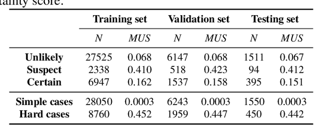
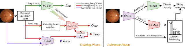
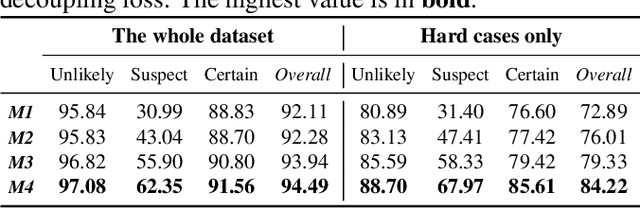
The annotation of disease severity for medical image datasets often relies on collaborative decisions from multiple human graders. The intra-observer variability derived from individual differences always persists in this process, yet the influence is often underestimated. In this paper, we cast the intra-observer variability as an uncertainty problem and incorporate the label uncertainty information as guidance into the disease screening model to improve the final decision. The main idea is dividing the images into simple and hard cases by uncertainty information, and then developing a multi-stream network to deal with different cases separately. Particularly, for hard cases, we strengthen the network's capacity in capturing the correct disease features and resisting the interference of uncertainty. Experiments on a fundus image-based glaucoma screening case study show that the proposed model outperforms several baselines, especially in screening hard cases.