 Add to Chrome
 Add to Firefox
 Add to Edge
Fundus Image-based Glaucoma Screening via Retinal Knowledge-Oriented Dynamic Multi-Level Feature Integration
Apr 14, 2026
Automated diagnosis based on color fundus photography is essential for large-scale glaucoma screening. However, existing deep learning models are typically data-driven and lack explicit integration of retinal anatomical knowledge, which limits their robustness across heterogeneous clinical datasets. Moreover, pathological cues in fundus images may appear beyond predefined anatomical regions, making fixed-region feature extraction insufficient for reliable diagnosis. To address these challenges, we propose a retinal knowledge-oriented glaucoma screening framework that integrates dynamic multi-scale feature learning with domain-specific retinal priors. The framework adopts a tri-branch structure to capture complementary retinal representations, including global retinal context, structural features of the optic disc/cup, and dynamically localized pathological regions. A Dynamic Window Mechanism is devised to adaptively identify diagnostically informative regions, while a Knowledge-Enhanced Convolutional Attention Module incorporates retinal priors extracted from a pre-trained foundation model to guide attention learning. Extensive experiments on the large-scale AIROGS dataset demonstrate that the proposed method outperforms diverse baselines, achieving an AUC of 98.5% and an accuracy of 94.6%. Additional evaluations on multiple datasets from the SMDG-19 benchmark further confirm its strong cross-domain generalization capability, indicating that knowledge-guided attention combined with adaptive lesion localization can significantly improve the robustness of automated glaucoma screening systems.

Enhancing Fundus Image-based Glaucoma Screening via Dynamic Global-Local Feature Integration
Apr 01, 2025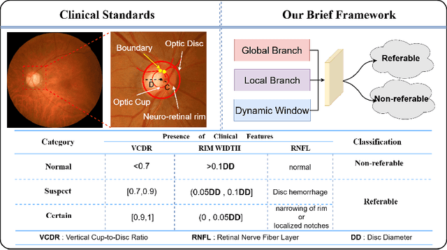
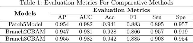
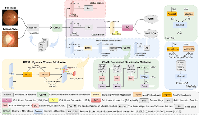
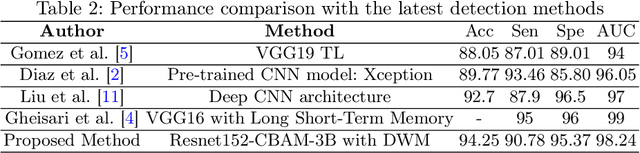
With the advancements in medical artificial intelligence (AI), fundus image classifiers are increasingly being applied to assist in ophthalmic diagnosis. While existing classification models have achieved high accuracy on specific fundus datasets, they struggle to address real-world challenges such as variations in image quality across different imaging devices, discrepancies between training and testing images across different racial groups, and the uncertain boundaries due to the characteristics of glaucomatous cases. In this study, we aim to address the above challenges posed by image variations by highlighting the importance of incorporating comprehensive fundus image information, including the optic cup (OC) and optic disc (OD) regions, and other key image patches. Specifically, we propose a self-adaptive attention window that autonomously determines optimal boundaries for enhanced feature extraction. Additionally, we introduce a multi-head attention mechanism to effectively fuse global and local features via feature linear readout, improving the model's discriminative capability. Experimental results demonstrate that our method achieves superior accuracy and robustness in glaucoma classification.