 Add to Chrome
 Add to Firefox
 Add to Edge
Progressive Experience Fusion for Multi-Task World Model Control in Endovascular Navigation
Aug 19, 2026
Autonomous endovascular navigation could support the delivery of mechanical thrombectomy to underserved areas, but controllers must navigate long, multi-stage paths across varying vascular anatomies. This study investigates Progressive Experience Fusion (PEF) to train a multi-task TD-MPC2 controller. We additionally evaluate a heuristic that changes the Model Predictive Path Integral planning horizon using residual action-sequence dispersion, and fine-tuning in a patient-specific simulation. Across five subtasks in ten known training anatomies with held-out targets, PEF achieved a mean success rate of 74%, compared with 37% for Soft Actor-Critic (p < 0.001) and 65% for base TD-MPC2 (p = 0.053). A PEF controller with adaptive-horizon planning trained on 30 vasculatures achieved a mean success rate of 90% in ten held-out vasculatures. The PEF agent successfully transferred to an unseen in vitro stroke patient vasculature under fluoroscopy, achieving a mean path ratio improvement from 63% to 80% with fine-tuning (p < 0.001), following 40x103 fine-tuning steps (corresponding to approximately 107 min of clinical inter-hospital transfer time). This work represents a proof of concept for multi-vasculature training and patient-specific adaptation, while further validation is required before clinical deployment.

Vascular Geometry Characterization for AI-Based Endovascular Navigation
Jul 10, 2026Mechanical thrombectomy (MT) is a time-critical intervention for acute ischemic stroke; however, access remains limited due to a shortage of neuroradiologists and specialized centers. Reinforcement learning (RL) offers potential to automate endovascular navigation and improve accessibility, yet current models lack standardized frameworks to assess navigation difficulty for model training and evaluation. This study aims to identify vascular metrics associated with navigation difficulty and to develop an automated pipeline for quantitative vascular feature extraction, enabling future complexity grading. Vascular trees were segmented from computed tomography angiograms from 61 patients, and vascular metrics including aortic arch type, presence of bovine arch, vessel length, tortuosity, take-off angle, number of reverse curves, were measured using a custom pipeline. A Soft Actor-Critic RL algorithm was used for 120 s autonomous navigation. Outcomes were analyzed using both mixed effects linear and logistic regression. On the left side, the presence of a bovine arch and aortic arch type II/III increased navigation time by 30.19 s and 37.92 s, respectively, while greater tortuosity (\b{eta} = 118.20) further prolonged the procedure and reduced success probability. On the right side, type II/III arches extended procedure time by 45.94 s, while each additional reverse curve was associated with 3.96 s longer navigation time and lower probability of success. These findings demonstrate for the first time that MT agent navigation difficulty is strongly influenced by vascular geometry. The proposed automated pipeline enables objective and quantitative characterization of vascular features, providing a foundation for future development of standardized complexity grading and RL model evaluation, without aiming to demonstrate clinically generalizable autonomous navigation.
Manual, Joystick, or Haptic Control? An In Vitro Comparison of Navigation Strategies for Robotic Interventional Neuroradiology Procedures
Jul 08, 2026Objective: To evaluate robotic controller interfaces for interventional neuroradiology procedures in-vitro incorporating a force-sensing platform to assess safety. Methods: A custom endovascular robot, device-mimicking controller, and sensorized neurovascular phantom were developed. Ten interventional neuroradiologists (4 novices, 6 experts) performed simulated navigations using four control modalities: device-mimicking controllers with and without haptic feedback, joystick-based input, and manual navigation. Navigation time, peak vessel-wall forces, incorrect catheterisations, and prolapse events were assessed, alongside user analyses. Results: Manual navigation was fastest (mean 47.7 s) compared to haptic-on (248.7 s), haptic-off (314.7 s), and joystick (392.6 s) modalities (p<0.001). Regardless of controller type, vessel-wall forces were below the 0.70 N puncture threshold; therefore all modalities were considered safe. Joystick produced significantly more prolapse events than manual control (1.56 vs 0.13; p=0.018). Operator experience was relevant to performance: experts made fewer incorrect catheterisations than novices (0.25 vs 0.62; p=0.035) and applied less vessel-wall force (p<0.0005); these effects were sustained across controllers but accentuated when haptics were on. Users perceived haptic on and haptic off as similarly intuitive, and more intuitive than joystick (p=0.033). Conclusion: Device-mimicking robotic controllers outperform joystick interfaces on most metrics; haptic feedback shows promising but non-significant performance benefits.
AA-ViT: Anatomically Aware Vision Transformer with Structural and Frequency Guidance for Contrast Enhanced Brain MRI Synthesis
Jul 08, 2026Accurate tumour localization and diagnosis is a critical component of clinical care for brain cancers. Magnetic Resonance Imaging (MRI) is the most commonly used imaging modality due to its superior soft-tissue contrast. However, standard MRI often exhibits limited contrast and imaging artifacts, which necessitates the use of contrast agents to enhance lesion visibility. The administration of chemical contrast agents is not always feasible and may be contraindicated in patients with renal impairment or other health conditions. As a result, developing accurate and non-invasive contrast enhanced MRI (CEMRI) synthesis methods has clinical importance. In recent years, numerous approaches for CEMRI synthesis have been proposed, predominantly relying on generative artificial intelligence models. While these methods demonstrate promising performance, their dependence on implicit feature learning often limits their ability to preserve anatomical boundaries and tumour-specific fine structures. To address these challenges, we propose an anatomically aware frequency-and-structure-guided vision transformer (AA-ViT), for CEMRI synthesis using pre-contrast MRI modalities (T1, T2, and FLAIR). Experiments on the BraTS 2021 dataset demonstrate that the proposed method preserves anatomical and lesion boundaries, achieving higher PSNR and SSIM than state-of-the-art approaches. Clinical evaluation by three neuroradiologists and a neurosurgeon on 19 randomly selected cases across diverse gliomas yielded a mean score of 3.94/5, providing preliminary clinical validation rarely seen in prior studies. Synthetic post-contrast scans from our model could lower scanning costs, shorten imaging time, and avoid the potential risks of using gadolinium-based contrast agents.
Toward Safe Autonomous Robotic Endovascular Interventions using World Models
Apr 22, 2026Autonomous mechanical thrombectomy (MT) presents substantial challenges due to highly variable vascular geometries and the requirements for accurate, real-time control. While reinforcement learning (RL) has emerged as a promising paradigm for the automation of endovascular navigation, existing approaches often show limited robustness when faced with diverse patient anatomies or extended navigation horizons. In this work, we investigate a world-model-based framework for autonomous endovascular navigation built on TD-MPC2, a model-based RL method that integrates planning and learned dynamics. We evaluate a TD-MPC2 agent trained on multiple navigation tasks across hold out patient-specific vasculatures and benchmark its performance against the state-of-the-art Soft Actor-Critic (SAC) algorithm agent. Both approaches are further validated in vitro using patient-specific vascular phantoms under fluoroscopic guidance. In simulation, TD-MPC2 demonstrates a significantly higher mean success rate than SAC (58% vs. 36%, p < 0.001), and mean tip contact forces of 0.15 N, well below the proposed 1.5 N vessel rupture threshold. Mean success rates for TD-MPC2 (68%) were comparable to SAC (60%) in vitro, but TD-MPC2 achieved superior path ratios (p = 0.017) at the cost of longer procedure times (p < 0.001). Together, these results provide the first demonstration of autonomous MT navigation validated across both hold out in silico data and fluoroscopy-guided in vitro experiments, highlighting the promise of world models for safe and generalizable AI-assisted endovascular interventions.
Reinforcement Learning for Safe Autonomous Two Device Navigation of Cerebral Vessels in Mechanical Thrombectomy
Mar 31, 2025Purpose: Autonomous systems in mechanical thrombectomy (MT) hold promise for reducing procedure times, minimizing radiation exposure, and enhancing patient safety. However, current reinforcement learning (RL) methods only reach the carotid arteries, are not generalizable to other patient vasculatures, and do not consider safety. We propose a safe dual-device RL algorithm that can navigate beyond the carotid arteries to cerebral vessels. Methods: We used the Simulation Open Framework Architecture to represent the intricacies of cerebral vessels, and a modified Soft Actor-Critic RL algorithm to learn, for the first time, the navigation of micro-catheters and micro-guidewires. We incorporate patient safety metrics into our reward function by integrating guidewire tip forces. Inverse RL is used with demonstrator data on 12 patient-specific vascular cases. Results: Our simulation demonstrates successful autonomous navigation within unseen cerebral vessels, achieving a 96% success rate, 7.0s procedure time, and 0.24 N mean forces, well below the proposed 1.5 N vessel rupture threshold. Conclusion: To the best of our knowledge, our proposed autonomous system for MT two-device navigation reaches cerebral vessels, considers safety, and is generalizable to unseen patient-specific cases for the first time. We envisage future work will extend the validation to vasculatures of different complexity and on in vitro models. While our contributions pave the way towards deploying agents in clinical settings, safety and trustworthiness will be crucial elements to consider when proposing new methodology.
Machine learning algorithms to predict the risk of rupture of intracranial aneurysms: a systematic review
Dec 06, 2024Purpose: Subarachnoid haemorrhage is a potentially fatal consequence of intracranial aneurysm rupture, however, it is difficult to predict if aneurysms will rupture. Prophylactic treatment of an intracranial aneurysm also involves risk, hence identifying rupture-prone aneurysms is of substantial clinical importance. This systematic review aims to evaluate the performance of machine learning algorithms for predicting intracranial aneurysm rupture risk. Methods: MEDLINE, Embase, Cochrane Library and Web of Science were searched until December 2023. Studies incorporating any machine learning algorithm to predict the risk of rupture of an intracranial aneurysm were included. Risk of bias was assessed using the Prediction Model Risk of Bias Assessment Tool (PROBAST). PROSPERO registration: CRD42023452509. Results: Out of 10,307 records screened, 20 studies met the eligibility criteria for this review incorporating a total of 20,286 aneurysm cases. The machine learning models gave a 0.66-0.90 range for performance accuracy. The models were compared to current clinical standards in six studies and gave mixed results. Most studies posed high or unclear risks of bias and concerns for applicability, limiting the inferences that can be drawn from them. There was insufficient homogenous data for a meta-analysis. Conclusions: Machine learning can be applied to predict the risk of rupture for intracranial aneurysms. However, the evidence does not comprehensively demonstrate superiority to existing practice, limiting its role as a clinical adjunct. Further prospective multicentre studies of recent machine learning tools are needed to prove clinical validation before they are implemented in the clinic.
Artificial intelligence for abnormality detection in high volume neuroimaging: a systematic review and meta-analysis
May 09, 2024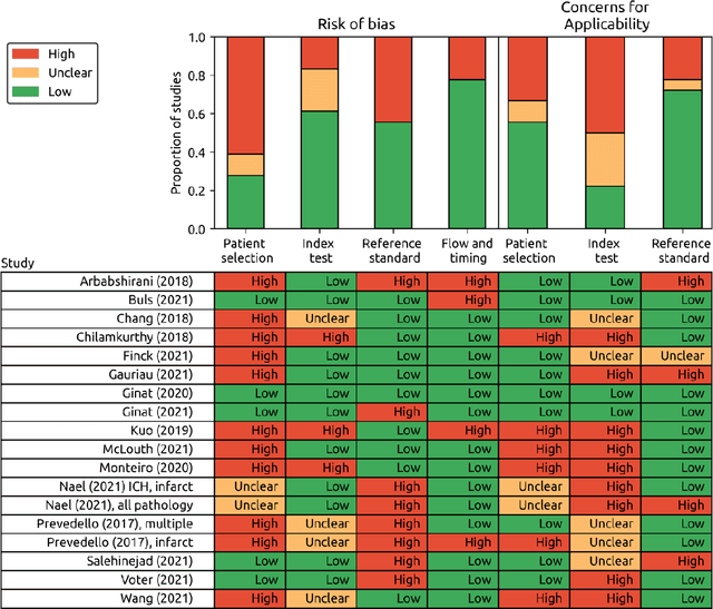
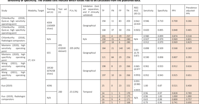
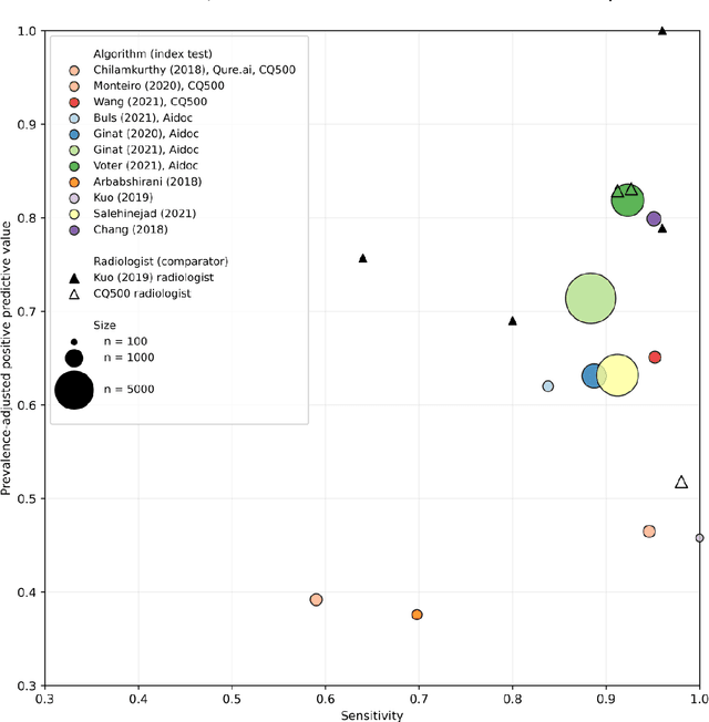
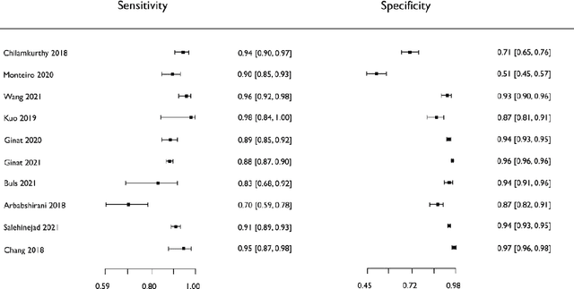
Purpose: Most studies evaluating artificial intelligence (AI) models that detect abnormalities in neuroimaging are either tested on unrepresentative patient cohorts or are insufficiently well-validated, leading to poor generalisability to real-world tasks. The aim was to determine the diagnostic test accuracy and summarise the evidence supporting the use of AI models performing first-line, high-volume neuroimaging tasks. Methods: Medline, Embase, Cochrane library and Web of Science were searched until September 2021 for studies that temporally or externally validated AI capable of detecting abnormalities in first-line CT or MR neuroimaging. A bivariate random-effects model was used for meta-analysis where appropriate. PROSPERO: CRD42021269563. Results: Only 16 studies were eligible for inclusion. Included studies were not compromised by unrepresentative datasets or inadequate validation methodology. Direct comparison with radiologists was available in 4/16 studies. 15/16 had a high risk of bias. Meta-analysis was only suitable for intracranial haemorrhage detection in CT imaging (10/16 studies), where AI systems had a pooled sensitivity and specificity 0.90 (95% CI 0.85 - 0.94) and 0.90 (95% CI 0.83 - 0.95) respectively. Other AI studies using CT and MRI detected target conditions other than haemorrhage (2/16), or multiple target conditions (4/16). Only 3/16 studies implemented AI in clinical pathways, either for pre-read triage or as post-read discrepancy identifiers. Conclusion: The paucity of eligible studies reflects that most abnormality detection AI studies were not adequately validated in representative clinical cohorts. The few studies describing how abnormality detection AI could impact patients and clinicians did not explore the full ramifications of clinical implementation.
Letter to the Editor: What are the legal and ethical considerations of submitting radiology reports to ChatGPT?
May 09, 2024This letter critically examines the recent article by Infante et al. assessing the utility of large language models (LLMs) like GPT-4, Perplexity, and Bard in identifying urgent findings in emergency radiology reports. While acknowledging the potential of LLMs in generating labels for computer vision, concerns are raised about the ethical implications of using patient data without explicit approval, highlighting the necessity of stringent data protection measures under GDPR.
Overcoming challenges of translating deep-learning models for glioblastoma: the ZGBM consortium
May 07, 2024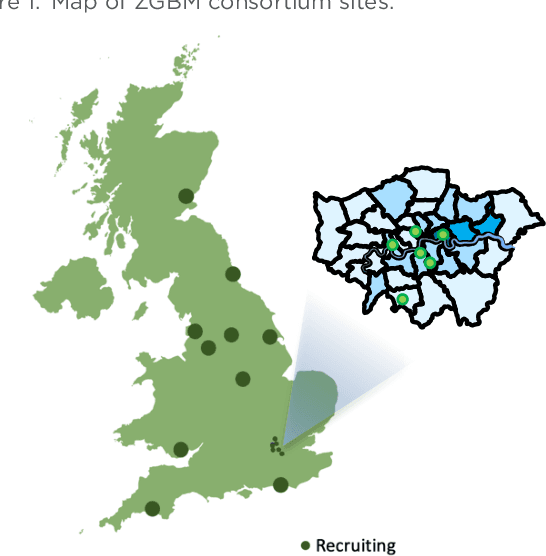
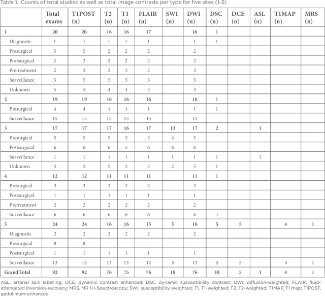
Objective: To report imaging protocol and scheduling variance in routine care of glioblastoma patients in order to demonstrate challenges of integrating deep-learning models in glioblastoma care pathways. Additionally, to understand the most common imaging studies and image contrasts to inform the development of potentially robust deep-learning models. Methods: MR imaging data were analysed from a random sample of five patients from the prospective cohort across five participating sites of the ZGBM consortium. Reported clinical and treatment data alongside DICOM header information were analysed to understand treatment pathway imaging schedules. Results: All sites perform all structural imaging at every stage in the pathway except for the presurgical study, where in some sites only contrast-enhanced T1-weighted imaging is performed. Diffusion MRI is the most common non-structural imaging type, performed at every site. Conclusion: The imaging protocol and scheduling varies across the UK, making it challenging to develop machine-learning models that could perform robustly at other centres. Structural imaging is performed most consistently across all centres. Advances in knowledge: Successful translation of deep-learning models will likely be based on structural post-treatment imaging unless there is significant effort made to standardise non-structural or peri-operative imaging protocols and schedules.