 Add to Chrome
 Add to Firefox
 Add to Edge
Manual, Joystick, or Haptic Control? An In Vitro Comparison of Navigation Strategies for Robotic Interventional Neuroradiology Procedures
Jul 08, 2026
Objective: To evaluate robotic controller interfaces for interventional neuroradiology procedures in-vitro incorporating a force-sensing platform to assess safety. Methods: A custom endovascular robot, device-mimicking controller, and sensorized neurovascular phantom were developed. Ten interventional neuroradiologists (4 novices, 6 experts) performed simulated navigations using four control modalities: device-mimicking controllers with and without haptic feedback, joystick-based input, and manual navigation. Navigation time, peak vessel-wall forces, incorrect catheterisations, and prolapse events were assessed, alongside user analyses. Results: Manual navigation was fastest (mean 47.7 s) compared to haptic-on (248.7 s), haptic-off (314.7 s), and joystick (392.6 s) modalities (p<0.001). Regardless of controller type, vessel-wall forces were below the 0.70 N puncture threshold; therefore all modalities were considered safe. Joystick produced significantly more prolapse events than manual control (1.56 vs 0.13; p=0.018). Operator experience was relevant to performance: experts made fewer incorrect catheterisations than novices (0.25 vs 0.62; p=0.035) and applied less vessel-wall force (p<0.0005); these effects were sustained across controllers but accentuated when haptics were on. Users perceived haptic on and haptic off as similarly intuitive, and more intuitive than joystick (p=0.033). Conclusion: Device-mimicking robotic controllers outperform joystick interfaces on most metrics; haptic feedback shows promising but non-significant performance benefits.

Labelling imaging datasets on the basis of neuroradiology reports: a validation study
Jul 08, 2020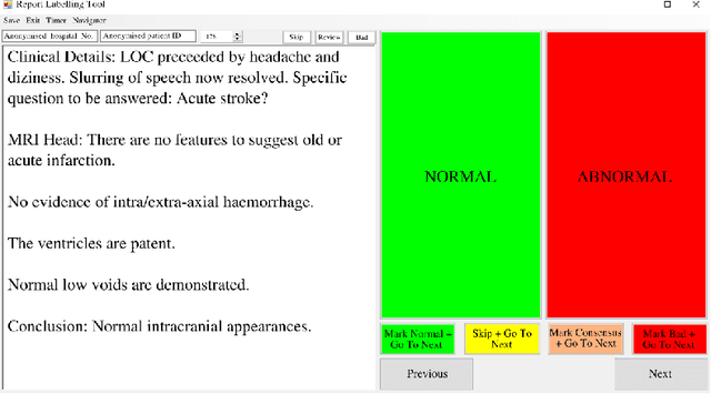

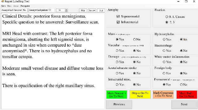
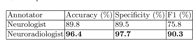
Natural language processing (NLP) shows promise as a means to automate the labelling of hospital-scale neuroradiology magnetic resonance imaging (MRI) datasets for computer vision applications. To date, however, there has been no thorough investigation into the validity of this approach, including determining the accuracy of report labels compared to image labels as well as examining the performance of non-specialist labellers. In this work, we draw on the experience of a team of neuroradiologists who labelled over 5000 MRI neuroradiology reports as part of a project to build a dedicated deep learning-based neuroradiology report classifier. We show that, in our experience, assigning binary labels (i.e. normal vs abnormal) to images from reports alone is highly accurate. In contrast to the binary labels, however, the accuracy of more granular labelling is dependent on the category, and we highlight reasons for this discrepancy. We also show that downstream model performance is reduced when labelling of training reports is performed by a non-specialist. To allow other researchers to accelerate their research, we make our refined abnormality definitions and labelling rules available, as well as our easy-to-use radiology report labelling app which helps streamline this process.