 Add to Chrome
 Add to Firefox
 Add to Edge
From Lines to Shapes: Geometric-Constrained Segmentation of X-Ray Collimators via Hough Transform
Sep 04, 2025Collimation in X-ray imaging restricts exposure to the region-of-interest (ROI) and minimizes the radiation dose applied to the patient. The detection of collimator shadows is an essential image-based preprocessing step in digital radiography posing a challenge when edges get obscured by scattered X-ray radiation. Regardless, the prior knowledge that collimation forms polygonal-shaped shadows is evident. For this reason, we introduce a deep learning-based segmentation that is inherently constrained to its geometry. We achieve this by incorporating a differentiable Hough transform-based network to detect the collimation borders and enhance its capability to extract the information about the ROI center. During inference, we combine the information of both tasks to enable the generation of refined, line-constrained segmentation masks. We demonstrate robust reconstruction of collimated regions achieving median Hausdorff distances of 4.3-5.0mm on diverse test sets of real Xray images. While this application involves at most four shadow borders, our method is not fundamentally limited by a specific number of edges.
A Realistic Collimated X-Ray Image Simulation Pipeline
Nov 15, 2024Collimator detection remains a challenging task in X-ray systems with unreliable or non-available information about the detectors position relative to the source. This paper presents a physically motivated image processing pipeline for simulating the characteristics of collimator shadows in X-ray images. By generating randomized labels for collimator shapes and locations, incorporating scattered radiation simulation, and including Poisson noise, the pipeline enables the expansion of limited datasets for training deep neural networks. We validate the proposed pipeline by a qualitative and quantitative comparison against real collimator shadows. Furthermore, it is demonstrated that utilizing simulated data within our deep learning framework not only serves as a suitable substitute for actual collimators but also enhances the generalization performance when applied to real-world data.
An Interpretable X-ray Style Transfer via Trainable Local Laplacian Filter
Nov 11, 2024


Radiologists have preferred visual impressions or 'styles' of X-ray images that are manually adjusted to their needs to support their diagnostic performance. In this work, we propose an automatic and interpretable X-ray style transfer by introducing a trainable version of the Local Laplacian Filter (LLF). From the shape of the LLF's optimized remap function, the characteristics of the style transfer can be inferred and reliability of the algorithm can be ensured. Moreover, we enable the LLF to capture complex X-ray style features by replacing the remap function with a Multi-Layer Perceptron (MLP) and adding a trainable normalization layer. We demonstrate the effectiveness of the proposed method by transforming unprocessed mammographic X-ray images into images that match the style of target mammograms and achieve a Structural Similarity Index (SSIM) of 0.94 compared to 0.82 of the baseline LLF style transfer method from Aubry et al.
StyleX: A Trainable Metric for X-ray Style Distances
May 23, 2024


The progression of X-ray technology introduces diverse image styles that need to be adapted to the preferences of radiologists. To support this task, we introduce a novel deep learning-based metric that quantifies style differences of non-matching image pairs. At the heart of our metric is an encoder capable of generating X-ray image style representations. This encoder is trained without any explicit knowledge of style distances by exploiting Simple Siamese learning. During inference, the style representations produced by the encoder are used to calculate a distance metric for non-matching image pairs. Our experiments investigate the proposed concept for a disclosed reproducible and a proprietary image processing pipeline along two dimensions: First, we use a t-distributed stochastic neighbor embedding (t-SNE) analysis to illustrate that the encoder outputs provide meaningful and discriminative style representations. Second, the proposed metric calculated from the encoder outputs is shown to quantify style distances for non-matching pairs in good alignment with the human perception. These results confirm that our proposed method is a promising technique to quantify style differences, which can be used for guided style selection as well as automatic optimization of image pipeline parameters.
Metal-conscious Embedding for CBCT Projection Inpainting
Nov 29, 2022The existence of metallic implants in projection images for cone-beam computed tomography (CBCT) introduces undesired artifacts which degrade the quality of reconstructed images. In order to reduce metal artifacts, projection inpainting is an essential step in many metal artifact reduction algorithms. In this work, a hybrid network combining the shift window (Swin) vision transformer (ViT) and a convolutional neural network is proposed as a baseline network for the inpainting task. To incorporate metal information for the Swin ViT-based encoder, metal-conscious self-embedding and neighborhood-embedding methods are investigated. Both methods have improved the performance of the baseline network. Furthermore, by choosing appropriate window size, the model with neighborhood-embedding could achieve the lowest mean absolute error of 0.079 in metal regions and the highest peak signal-to-noise ratio of 42.346 in CBCT projections. At the end, the efficiency of metal-conscious embedding on both simulated and real cadaver CBCT data has been demonstrated, where the inpainting capability of the baseline network has been enhanced.
Simulation-Driven Training of Vision Transformers Enabling Metal Segmentation in X-Ray Images
Mar 17, 2022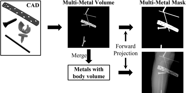
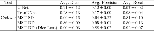
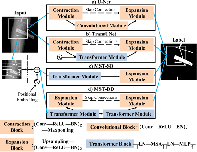
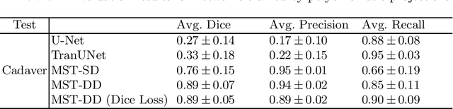
In several image acquisition and processing steps of X-ray radiography, knowledge of the existence of metal implants and their exact position is highly beneficial (e.g. dose regulation, image contrast adjustment). Another application which would benefit from an accurate metal segmentation is cone beam computed tomography (CBCT) which is based on 2D X-ray projections. Due to the high attenuation of metals, severe artifacts occur in the 3D X-ray acquisitions. The metal segmentation in CBCT projections usually serves as a prerequisite for metal artifact avoidance and reduction algorithms. Since the generation of high quality clinical training is a constant challenge, this study proposes to generate simulated X-ray images based on CT data sets combined with self-designed computer aided design (CAD) implants and make use of convolutional neural network (CNN) and vision transformer (ViT) for metal segmentation. Model test is performed on accurately labeled X-ray test datasets obtained from specimen scans. The CNN encoder-based network like U-Net has limited performance on cadaver test data with an average dice score below 0.30, while the metal segmentation transformer with dual decoder (MST-DD) shows high robustness and generalization on the segmentation task, with an average dice score of 0.90. Our study indicates that the CAD model-based data generation has high flexibility and could be a way to overcome the problem of shortage in clinical data sampling and labelling. Furthermore, the MST-DD approach generates a more reliable neural network in case of training on simulated data.
Automated detection and quantification of COVID-19 airspace disease on chest radiographs: A novel approach achieving radiologist-level performance using a CNN trained on digital reconstructed radiographs (DRRs) from CT-based ground-truth
Aug 13, 2020


Purpose: To leverage volumetric quantification of airspace disease (AD) derived from a superior modality (CT) serving as ground truth, projected onto digitally reconstructed radiographs (DRRs) to: 1) train a convolutional neural network to quantify airspace disease on paired CXRs; and 2) compare the DRR-trained CNN to expert human readers in the CXR evaluation of patients with confirmed COVID-19. Materials and Methods: We retrospectively selected a cohort of 86 COVID-19 patients (with positive RT-PCR), from March-May 2020 at a tertiary hospital in the northeastern USA, who underwent chest CT and CXR within 48 hrs. The ground truth volumetric percentage of COVID-19 related AD (POv) was established by manual AD segmentation on CT. The resulting 3D masks were projected into 2D anterior-posterior digitally reconstructed radiographs (DRR) to compute area-based AD percentage (POa). A convolutional neural network (CNN) was trained with DRR images generated from a larger-scale CT dataset of COVID-19 and non-COVID-19 patients, automatically segmenting lungs, AD and quantifying POa on CXR. CNN POa results were compared to POa quantified on CXR by two expert readers and to the POv ground-truth, by computing correlations and mean absolute errors. Results: Bootstrap mean absolute error (MAE) and correlations between POa and POv were 11.98% [11.05%-12.47%] and 0.77 [0.70-0.82] for average of expert readers, and 9.56%-9.78% [8.83%-10.22%] and 0.78-0.81 [0.73-0.85] for the CNN, respectively. Conclusion: Our CNN trained with DRR using CT-derived airspace quantification achieved expert radiologist level of accuracy in the quantification of airspace disease on CXR, in patients with positive RT-PCR for COVID-19.
Deep Learning-based Denoising of Mammographic Images using Physics-driven Data Augmentation
Dec 11, 2019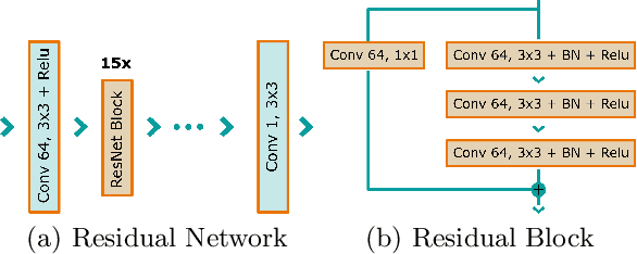

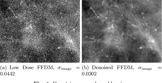
Mammography is using low-energy X-rays to screen the human breast and is utilized by radiologists to detect breast cancer. Typically radiologists require a mammogram with impeccable image quality for an accurate diagnosis. In this study, we propose a deep learning method based on Convolutional Neural Networks (CNNs) for mammogram denoising to improve the image quality. We first enhance the noise level and employ Anscombe Transformation (AT) to transform Poisson noise to white Gaussian noise. With this data augmentation, a deep residual network is trained to learn the noise map of the noisy images. We show, that the proposed method can remove not only simulated but also real noise. Furthermore, we also compare our results with state-of-the-art denoising methods, such as BM3D and DNCNN. In an early investigation, we achieved qualitatively better mammogram denoising results.