 Add to Chrome
 Add to Firefox
 Add to Edge
A Unified Model for Highly Accurate ECG-Free Dynamic Coronary Roadmapping Using Spatio-Temporal Transformers
Jul 09, 2026
Percutaneous Coronary Intervention (PCI) is a minimally invasive procedure used to restore coronary blood flow obstructed by atherosclerotic plaque. During PCI, repeated injections of iodine-based contrast agents are required to visualize the coronary arteries and guide interventional devices. However, frequent contrast injections increase radiation exposure and the risk of contrast-induced nephropathy, with acute kidney injury reported in up to 30% of patients with renal impairment. Dynamic Coronary Roadmapping (DRM) reduces these risks by overlaying a precomputed angiographic vessel map onto live fluoroscopy and continuously updating it throughout the procedure. Accurate DRM relies on precise cardiac phase matching between angiography and fluoroscopy, together with reliable catheter tip tracking for motion compensation. These tasks remain challenging in ECG-free settings and when only limited manual annotations are available. We present a unified DRM framework that simultaneously performs cardiac phase matching and catheter tip tracking for accurate real-time guidance. Our method employs a large-scale spatio-temporal encoder pretrained on 16 million X-ray frames to learn cardiac motion dynamics. To the best of our knowledge, this is the first application of large-scale spatio-temporal pretraining for motion compensation in DRM. We further introduce auxiliary tasks based on ECG R-peak detection and catheter tip tracking, improving optimization while eliminating the need for extensive catheter mask annotations. Finally, a majority-voting postprocessing strategy aggregates temporal predictions, improving robustness and providing a confidence score that correlates with phase-matching error. Comprehensive evaluation on clinical X-ray datasets demonstrates state-of-the-art performance, achieving low temporal misalignment and robust phase-matching accuracy suitable for real-time DRM.

Automated detection and quantification of COVID-19 airspace disease on chest radiographs: A novel approach achieving radiologist-level performance using a CNN trained on digital reconstructed radiographs (DRRs) from CT-based ground-truth
Aug 13, 2020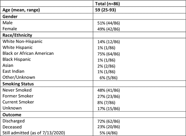
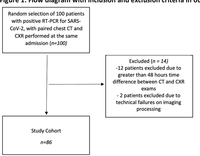


Purpose: To leverage volumetric quantification of airspace disease (AD) derived from a superior modality (CT) serving as ground truth, projected onto digitally reconstructed radiographs (DRRs) to: 1) train a convolutional neural network to quantify airspace disease on paired CXRs; and 2) compare the DRR-trained CNN to expert human readers in the CXR evaluation of patients with confirmed COVID-19. Materials and Methods: We retrospectively selected a cohort of 86 COVID-19 patients (with positive RT-PCR), from March-May 2020 at a tertiary hospital in the northeastern USA, who underwent chest CT and CXR within 48 hrs. The ground truth volumetric percentage of COVID-19 related AD (POv) was established by manual AD segmentation on CT. The resulting 3D masks were projected into 2D anterior-posterior digitally reconstructed radiographs (DRR) to compute area-based AD percentage (POa). A convolutional neural network (CNN) was trained with DRR images generated from a larger-scale CT dataset of COVID-19 and non-COVID-19 patients, automatically segmenting lungs, AD and quantifying POa on CXR. CNN POa results were compared to POa quantified on CXR by two expert readers and to the POv ground-truth, by computing correlations and mean absolute errors. Results: Bootstrap mean absolute error (MAE) and correlations between POa and POv were 11.98% [11.05%-12.47%] and 0.77 [0.70-0.82] for average of expert readers, and 9.56%-9.78% [8.83%-10.22%] and 0.78-0.81 [0.73-0.85] for the CNN, respectively. Conclusion: Our CNN trained with DRR using CT-derived airspace quantification achieved expert radiologist level of accuracy in the quantification of airspace disease on CXR, in patients with positive RT-PCR for COVID-19.
3D Tomographic Pattern Synthesis for Enhancing the Quantification of COVID-19
May 05, 2020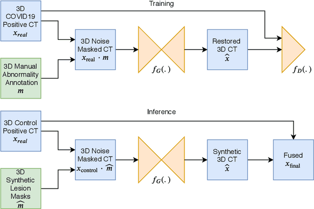
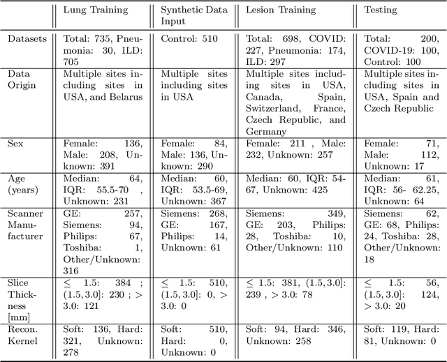
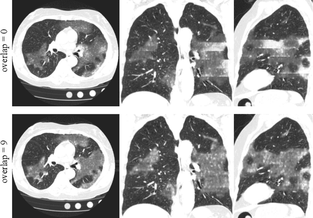
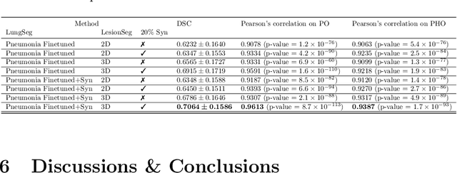
The Coronavirus Disease (COVID-19) has affected 1.8 million people and resulted in more than 110,000 deaths as of April 12, 2020. Several studies have shown that tomographic patterns seen on chest Computed Tomography (CT), such as ground-glass opacities, consolidations, and crazy paving pattern, are correlated with the disease severity and progression. CT imaging can thus emerge as an important modality for the management of COVID-19 patients. AI-based solutions can be used to support CT based quantitative reporting and make reading efficient and reproducible if quantitative biomarkers, such as the Percentage of Opacity (PO), can be automatically computed. However, COVID-19 has posed unique challenges to the development of AI, specifically concerning the availability of appropriate image data and annotations at scale. In this paper, we propose to use synthetic datasets to augment an existing COVID-19 database to tackle these challenges. We train a Generative Adversarial Network (GAN) to inpaint COVID-19 related tomographic patterns on chest CTs from patients without infectious diseases. Additionally, we leverage location priors derived from manually labeled COVID-19 chest CTs patients to generate appropriate abnormality distributions. Synthetic data are used to improve both lung segmentation and segmentation of COVID-19 patterns by adding 20% of synthetic data to the real COVID-19 training data. We collected 2143 chest CTs, containing 327 COVID-19 positive cases, acquired from 12 sites across 7 countries. By testing on 100 COVID-19 positive and 100 control cases, we show that synthetic data can help improve both lung segmentation (+6.02% lesion inclusion rate) and abnormality segmentation (+2.78% dice coefficient), leading to an overall more accurate PO computation (+2.82% Pearson coefficient).