 Add to Chrome
 Add to Firefox
 Add to Edge
Deep Generative Model-based Quality Control for Cardiac MRI Segmentation
Jun 23, 2020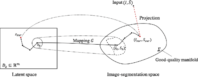
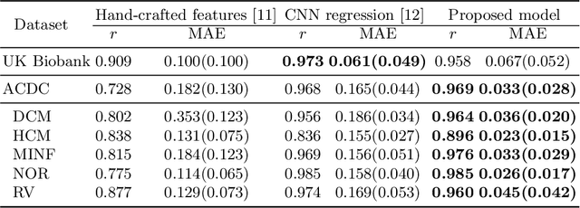
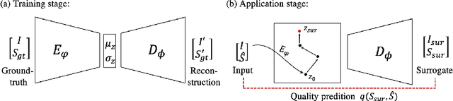
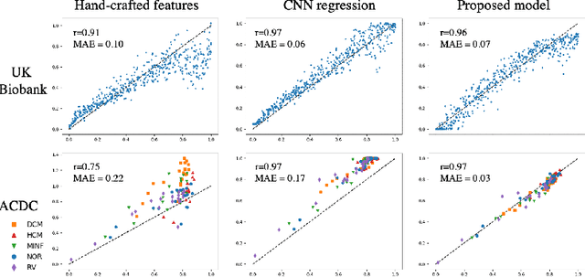
In recent years, convolutional neural networks have demonstrated promising performance in a variety of medical image segmentation tasks. However, when a trained segmentation model is deployed into the real clinical world, the model may not perform optimally. A major challenge is the potential poor-quality segmentations generated due to degraded image quality or domain shift issues. There is a timely need to develop an automated quality control method that can detect poor segmentations and feedback to clinicians. Here we propose a novel deep generative model-based framework for quality control of cardiac MRI segmentation. It first learns a manifold of good-quality image-segmentation pairs using a generative model. The quality of a given test segmentation is then assessed by evaluating the difference from its projection onto the good-quality manifold. In particular, the projection is refined through iterative search in the latent space. The proposed method achieves high prediction accuracy on two publicly available cardiac MRI datasets. Moreover, it shows better generalisation ability than traditional regression-based methods. Our approach provides a real-time and model-agnostic quality control for cardiac MRI segmentation, which has the potential to be integrated into clinical image analysis workflows.
Efficient Deep Representation Learning by Adaptive Latent Space Sampling
Apr 12, 2020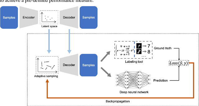
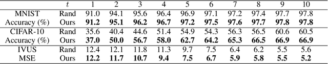
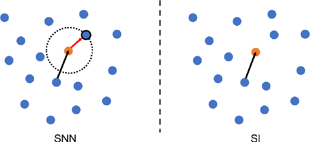
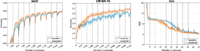
Supervised deep learning requires a large amount of training samples with annotations (e.g. label class for classification task, pixel- or voxel-wised label map for segmentation tasks), which are expensive and time-consuming to obtain. During the training of a deep neural network, the annotated samples are fed into the network in a mini-batch way, where they are often regarded of equal importance. However, some of the samples may become less informative during training, as the magnitude of the gradient start to vanish for these samples. In the meantime, other samples of higher utility or hardness may be more demanded for the training process to proceed and require more exploitation. To address the challenges of expensive annotations and loss of sample informativeness, here we propose a novel training framework which adaptively selects informative samples that are fed to the training process. The adaptive selection or sampling is performed based on a hardness-aware strategy in the latent space constructed by a generative model. To evaluate the proposed training framework, we perform experiments on three different datasets, including MNIST and CIFAR-10 for image classification task and a medical image dataset IVUS for biophysical simulation task. On all three datasets, the proposed framework outperforms a random sampling method, which demonstrates the effectiveness of proposed framework.
Automatic Brain Tumour Segmentation and Biophysics-Guided Survival Prediction
Nov 19, 2019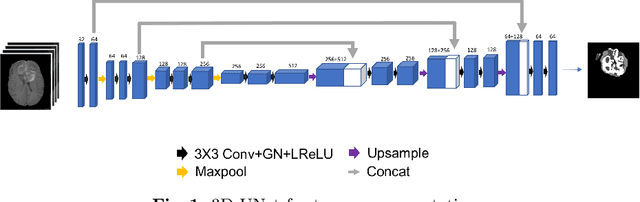
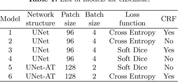
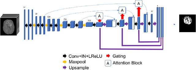
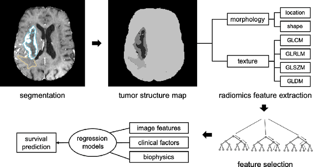
Gliomas are the most common malignant brain tumourswith intrinsic heterogeneity. Accurate segmentation of gliomas and theirsub-regions on multi-parametric magnetic resonance images (mpMRI)is of great clinical importance, which defines tumour size, shape andappearance and provides abundant information for preoperative diag-nosis, treatment planning and survival prediction. Recent developmentson deep learning have significantly improved the performance of auto-mated medical image segmentation. In this paper, we compare severalstate-of-the-art convolutional neural network models for brain tumourimage segmentation. Based on the ensembled segmentation, we presenta biophysics-guided prognostic model for patient overall survival predic-tion which outperforms a data-driven radiomics approach. Our methodwon the second place of the MICCAI 2019 BraTS Challenge for theoverall survival prediction.
Transfer Learning from Partial Annotations for Whole Brain Segmentation
Aug 28, 2019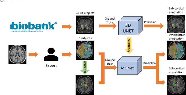

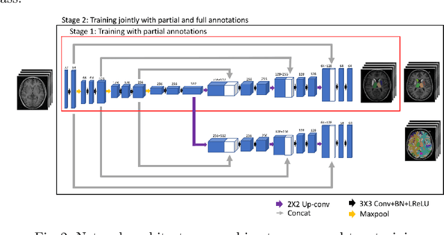
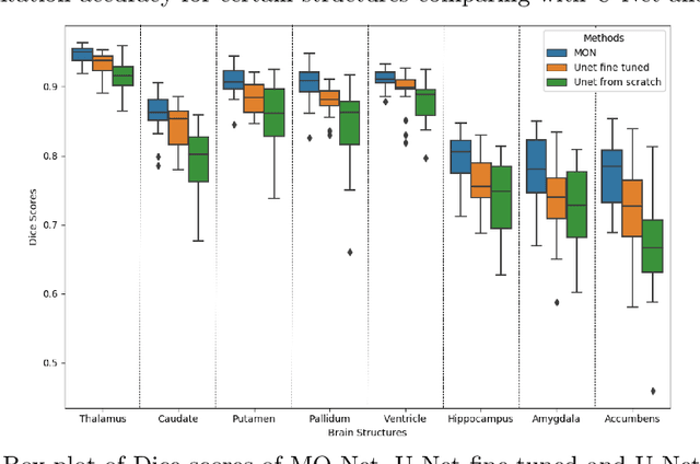
Brain MR image segmentation is a key task in neuroimaging studies. It is commonly conducted using standard computational tools, such as FSL, SPM, multi-atlas segmentation etc, which are often registration-based and suffer from expensive computation cost. Recently, there is an increased interest using deep neural networks for brain image segmentation, which have demonstrated advantages in both speed and performance. However, neural networks-based approaches normally require a large amount of manual annotations for optimising the massive amount of network parameters. For 3D networks used in volumetric image segmentation, this has become a particular challenge, as a 3D network consists of many more parameters compared to its 2D counterpart. Manual annotation of 3D brain images is extremely time-consuming and requires extensive involvement of trained experts. To address the challenge with limited manual annotations, here we propose a novel multi-task learning framework for brain image segmentation, which utilises a large amount of automatically generated partial annotations together with a small set of manually created full annotations for network training. Our method yields a high performance comparable to state-of-the-art methods for whole brain segmentation.
The Deep Poincaré Map: A Novel Approach for Left Ventricle Segmentation
Oct 30, 2018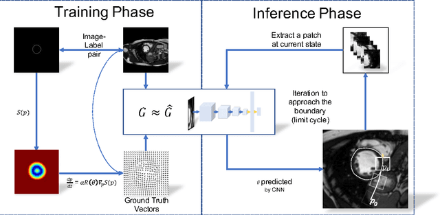
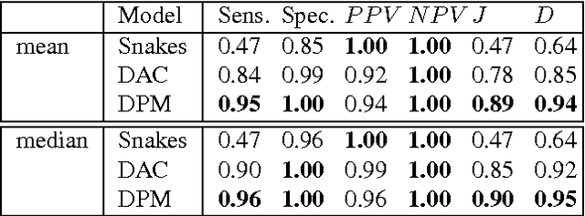
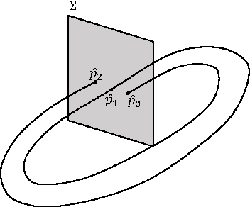
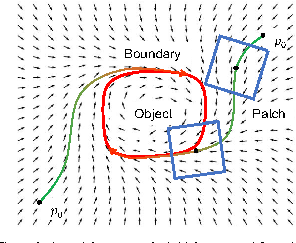
Precise segmentation of the left ventricle (LV) within cardiac MRI images is a prerequisite for the quantitative measurement of heart function. However, this task is challenging due to the limited availability of labeled data and motion artifacts from cardiac imaging. In this work, we present an iterative segmentation algorithm for LV delineation. By coupling deep learning with a novel dynamic-based labeling scheme, we present a new methodology where a policy model is learned to guide an agent to travel over the the image, tracing out a boundary of the ROI -- using the magnitude difference of the Poincar\'e map as a stopping criterion. Our method is evaluated on two datasets, namely the Sunnybrook Cardiac Dataset (SCD) and data from the STACOM 2011 LV segmentation challenge. Our method outperforms the previous research over many metrics. In order to demonstrate the transferability of our method we present encouraging results over the STACOM 2011 data, when using a model trained on the SCD dataset.
Automatic Brain Tumor Detection and Segmentation Using U-Net Based Fully Convolutional Networks
Jun 03, 2017
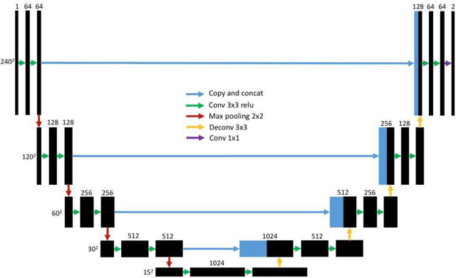
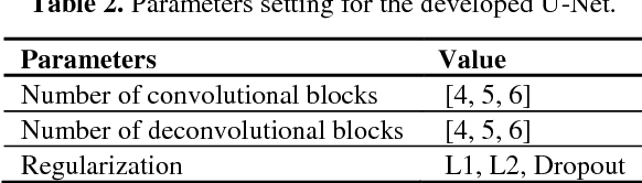
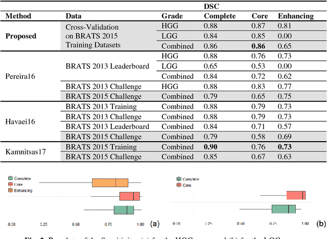
A major challenge in brain tumor treatment planning and quantitative evaluation is determination of the tumor extent. The noninvasive magnetic resonance imaging (MRI) technique has emerged as a front-line diagnostic tool for brain tumors without ionizing radiation. Manual segmentation of brain tumor extent from 3D MRI volumes is a very time-consuming task and the performance is highly relied on operator's experience. In this context, a reliable fully automatic segmentation method for the brain tumor segmentation is necessary for an efficient measurement of the tumor extent. In this study, we propose a fully automatic method for brain tumor segmentation, which is developed using U-Net based deep convolutional networks. Our method was evaluated on Multimodal Brain Tumor Image Segmentation (BRATS 2015) datasets, which contain 220 high-grade brain tumor and 54 low-grade tumor cases. Cross-validation has shown that our method can obtain promising segmentation efficiently.