 Add to Chrome
 Add to Firefox
 Add to Edge
X-GuideAR: An Augmented Reality Framework to Mitigate Radiation Exposure during Fluoroscopic Guidance
Jul 12, 2026
Achieving optimal screw placement for orthopedic surgeries requires frequent alignment checks and multiple anatomical views under X-ray -- a process known as "fluoro-hunting" that increases radiation exposure to patients and surgical teams. This work introduces X-GuideAR, an augmented reality (AR) framework for identifying optimal X-ray views, aimed at reducing radiation exposure while ensuring accurate screw placement. To exemplify the benefits of X-GuideAR, we focus on S2 alar-iliac (S2AI) screw placement. Our system provides radiation-free guidance for view acquisition and drilling by generating synthetic X-ray previews that accelerate fluoro-hunting. Once the required anatomical views are identified using these previews, a real X-ray is acquired, and the preview of the drilling trajectory is augmented onto it, facilitating precise screw placement with minimal additional radiation. A preliminary study involving eight S2AI trajectories performed by an expert spine surgeon demonstrated a 62.3% reduction in the number of X-rays. Post-procedure evaluations showed that trajectories done with X-GuideAR supported an average safe screw diameter of 12.95 mm compared to 5.9 mm under the conventional workflow, suggesting improved bony containment and potential biomechanical benefit. X-GuideAR shows great potential to reduce radiation exposure and streamline S2AI screw placement, offering a promising direction toward safer and more efficient surgeries.

StraightTrack: Towards Mixed Reality Navigation System for Percutaneous K-wire Insertion
Oct 02, 2024

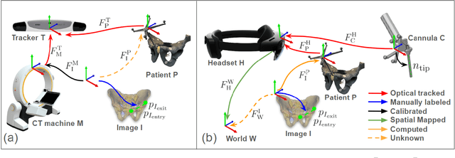

In percutaneous pelvic trauma surgery, accurate placement of Kirschner wires (K-wires) is crucial to ensure effective fracture fixation and avoid complications due to breaching the cortical bone along an unsuitable trajectory. Surgical navigation via mixed reality (MR) can help achieve precise wire placement in a low-profile form factor. Current approaches in this domain are as yet unsuitable for real-world deployment because they fall short of guaranteeing accurate visual feedback due to uncontrolled bending of the wire. To ensure accurate feedback, we introduce StraightTrack, an MR navigation system designed for percutaneous wire placement in complex anatomy. StraightTrack features a marker body equipped with a rigid access cannula that mitigates wire bending due to interactions with soft tissue and a covered bony surface. Integrated with an Optical See-Through Head-Mounted Display (OST HMD) capable of tracking the cannula body, StraightTrack offers real-time 3D visualization and guidance without external trackers, which are prone to losing line-of-sight. In phantom experiments with two experienced orthopedic surgeons, StraightTrack improves wire placement accuracy, achieving the ideal trajectory within $5.26 \pm 2.29$ mm and $2.88 \pm 1.49$ degree, compared to over 12.08 mm and 4.07 degree for comparable methods. As MR navigation systems continue to mature, StraightTrack realizes their potential for internal fracture fixation and other percutaneous orthopedic procedures.
Seamless Augmented Reality Integration in Arthroscopy: A Pipeline for Articular Reconstruction and Guidance
Oct 01, 2024Arthroscopy is a minimally invasive surgical procedure used to diagnose and treat joint problems. The clinical workflow of arthroscopy typically involves inserting an arthroscope into the joint through a small incision, during which surgeons navigate and operate largely by relying on their visual assessment through the arthroscope. However, the arthroscope's restricted field of view and lack of depth perception pose challenges in navigating complex articular structures and achieving surgical precision during procedures. Aiming at enhancing intraoperative awareness, we present a robust pipeline that incorporates simultaneous localization and mapping, depth estimation, and 3D Gaussian splatting to realistically reconstruct intra-articular structures solely based on monocular arthroscope video. Extending 3D reconstruction to Augmented Reality (AR) applications, our solution offers AR assistance for articular notch measurement and annotation anchoring in a human-in-the-loop manner. Compared to traditional Structure-from-Motion and Neural Radiance Field-based methods, our pipeline achieves dense 3D reconstruction and competitive rendering fidelity with explicit 3D representation in 7 minutes on average. When evaluated on four phantom datasets, our method achieves RMSE = 2.21mm reconstruction error, PSNR = 32.86 and SSIM = 0.89 on average. Because our pipeline enables AR reconstruction and guidance directly from monocular arthroscopy without any additional data and/or hardware, our solution may hold the potential for enhancing intraoperative awareness and facilitating surgical precision in arthroscopy. Our AR measurement tool achieves accuracy within 1.59 +/- 1.81mm and the AR annotation tool achieves a mIoU of 0.721.