 Add to Chrome
 Add to Firefox
 Add to Edge
Surgical Anatomy Recognition with Context Learning using Foundation Representations
Jun 20, 2026Accurate recognition of anatomical structures is essential for safe and effective minimally invasive surgery (MIS), yet it remains underexplored in surgical computer vision due to limited annotated data and methods tailored primarily to natural scenes. In this work, we present a combined dataset and model framework to advance anatomy-aware perception in MIS. First, we introduce ATLAS-120k, a large-scale clip-level semantic segmentation dataset comprising over 120,000 annotated frames from 100 surgical videos spanning 14 procedures and multiple modalities, including laparoscopic and robot-assisted surgery. The dataset captures substantial procedural variability and was created using a scalable annotation pipeline that integrates expert manual labeling, automated propagation, iterative refinement, and surgeon verification to ensure high-quality annotations. Second, we propose ATLAS (Anatomy Recognition with Context Learning using Foundation Representations), a video semantic segmentation model specifically designed for surgical anatomy recognition. Unlike conventional approaches that emphasize object tracking, ATLAS leverages foundation-model embeddings together with lightweight temporal reasoning to incorporate contextual cues such as procedure type, surgical phase, and short-term visual memory. This design enables temporally consistent and accurate predictions while maintaining real-time feasibility. Together, the dataset and model establish a practical foundation for robust surgical scene understanding and support the development of clinically applicable guidance systems for minimally invasive surgery. The models, dataset annotations and annotation platform are publicly available at: https://github.com/TimJaspers0801/ATLAS.
Object Tokens as a Bridge Between Segmentation and Visual Question Answering in Robotic Surgery
Jun 14, 2026Visual Question Answering (VQA) in robotic surgery, referred to as surgical VQA, requires high-level understanding of complex surgical scenes and the integration of visual perception with language reasoning, with the potential to support surgical training and intraoperative decision-making. Recent Vision-Language Models (VLMs) have shown promising performance through parameter-efficient fine-tuning; however, most existing approaches rely on coarse visual grounding, typically limited to bounding boxes, which fails to capture the fine-grained spatial structure of surgical objects. In this work, we propose a unified framework that jointly performs pixel-level segmentation and visual question answering within a single framework. Our approach integrates a VLM with a Segment Anything Model (SAM)-based decoder and represents scene elements as object tokens generated by the VLM. These object tokens guide answer prediction and are further projected to the SAM-based decoder to produce segmentation masks. By optimizing the object token embeddings through both segmentation and question answering objectives, the model learns spatially grounded representations that enhance visual reasoning while providing explicit pixel-level grounding. We evaluate the proposed method on the private RAMIE (Robot-Assisted Minimally Invasive Esophagectomy) dataset and the public EndoVis18 dataset, where it consistently outperforms baseline methods for surgical VQA. These results demonstrate that incorporating context-aware object tokens into vision-language models improves fine-grained surgical scene understanding.
Data-driven Synthesis of Magnetic Resonance Spectroscopy Data using a Variational Autoencoder
Feb 28, 2026The development of deep learning methods for magnetic resonance spectroscopy (MRS) is often hindered by limited availability of large, high-quality training datasets. While physics-based simulations are commonly used to mitigate this limitation, accurately modeling all in-vivo signal components remains challenging. In this work, we propose a data-driven framework for synthesizing in-vivo MRS data using a variational autoencoder (VAE) trained exclusively on measured single-voxel spectroscopy data. The model learns a low-dimensional latent representation of complex-valued spectra and enables generation of new samples through latent-space sampling and interpolation. The generative performance of the proposed approach is evaluated using a comprehensive set of complementary analyses, including reconstruction quality, feature-level similarity using low-dimensional embeddings, application-based signal quality metrics, and metabolite quantification agreement. The results demonstrate that the VAE accurately reconstructs dominant spectral patterns and generates synthetic spectra that occupy the same feature space as in-vivo data. In an example application targeting GABA-edited spectroscopy, augmenting limited subsets of transients with synthetic spectra improves signal quality metrics such as signal-to-noise ratio, linewidth, and shape scores. However, the results also reveal limitations of the generative approach, including under-representation of stochastic noise and reduced accuracy in absolute metabolite quantification, particularly for applications sensitive to concentration estimates. These findings highlight both potential and limitations of data-driven MRS synthesis. Beyond the proposed model, this study introduces a structured evaluation framework for generative MRS methods, emphasizing the importance of application-aware validation when synthetic data are used for downstream analysis.
SAM-Fed: SAM-Guided Federated Semi-Supervised Learning for Medical Image Segmentation
Nov 18, 2025Medical image segmentation is clinically important, yet data privacy and the cost of expert annotation limit the availability of labeled data. Federated semi-supervised learning (FSSL) offers a solution but faces two challenges: pseudo-label reliability depends on the strength of local models, and client devices often require compact or heterogeneous architectures due to limited computational resources. These constraints reduce the quality and stability of pseudo-labels, while large models, though more accurate, cannot be trained or used for routine inference on client devices. We propose SAM-Fed, a federated semi-supervised framework that leverages a high-capacity segmentation foundation model to guide lightweight clients during training. SAM-Fed combines dual knowledge distillation with an adaptive agreement mechanism to refine pixel-level supervision. Experiments on skin lesion and polyp segmentation across homogeneous and heterogeneous settings show that SAM-Fed consistently outperforms state-of-the-art FSSL methods.
Scaling up self-supervised learning for improved surgical foundation models
Jan 16, 2025


Foundation models have revolutionized computer vision by achieving vastly superior performance across diverse tasks through large-scale pretraining on extensive datasets. However, their application in surgical computer vision has been limited. This study addresses this gap by introducing SurgeNetXL, a novel surgical foundation model that sets a new benchmark in surgical computer vision. Trained on the largest reported surgical dataset to date, comprising over 4.7 million video frames, SurgeNetXL achieves consistent top-tier performance across six datasets spanning four surgical procedures and three tasks, including semantic segmentation, phase recognition, and critical view of safety (CVS) classification. Compared with the best-performing surgical foundation models, SurgeNetXL shows mean improvements of 2.4, 9.0, and 12.6 percent for semantic segmentation, phase recognition, and CVS classification, respectively. Additionally, SurgeNetXL outperforms the best-performing ImageNet-based variants by 14.4, 4.0, and 1.6 percent in the respective tasks. In addition to advancing model performance, this study provides key insights into scaling pretraining datasets, extending training durations, and optimizing model architectures specifically for surgical computer vision. These findings pave the way for improved generalizability and robustness in data-scarce scenarios, offering a comprehensive framework for future research in this domain. All models and a subset of the SurgeNetXL dataset, including over 2 million video frames, are publicly available at: https://github.com/TimJaspers0801/SurgeNet.
Benchmarking and Enhancing Surgical Phase Recognition Models for Robotic-Assisted Esophagectomy
Dec 05, 2024


Robotic-assisted minimally invasive esophagectomy (RAMIE) is a recognized treatment for esophageal cancer, offering better patient outcomes compared to open surgery and traditional minimally invasive surgery. RAMIE is highly complex, spanning multiple anatomical areas and involving repetitive phases and non-sequential phase transitions. Our goal is to leverage deep learning for surgical phase recognition in RAMIE to provide intraoperative support to surgeons. To achieve this, we have developed a new surgical phase recognition dataset comprising 27 videos. Using this dataset, we conducted a comparative analysis of state-of-the-art surgical phase recognition models. To more effectively capture the temporal dynamics of this complex procedure, we developed a novel deep learning model featuring an encoder-decoder structure with causal hierarchical attention, which demonstrates superior performance compared to existing models.
Benchmarking Pretrained Attention-based Models for Real-Time Recognition in Robot-Assisted Esophagectomy
Dec 04, 2024Esophageal cancer is among the most common types of cancer worldwide. It is traditionally treated using open esophagectomy, but in recent years, robot-assisted minimally invasive esophagectomy (RAMIE) has emerged as a promising alternative. However, robot-assisted surgery can be challenging for novice surgeons, as they often suffer from a loss of spatial orientation. Computer-aided anatomy recognition holds promise for improving surgical navigation, but research in this area remains limited. In this study, we developed a comprehensive dataset for semantic segmentation in RAMIE, featuring the largest collection of vital anatomical structures and surgical instruments to date. Handling this diverse set of classes presents challenges, including class imbalance and the recognition of complex structures such as nerves. This study aims to understand the challenges and limitations of current state-of-the-art algorithms on this novel dataset and problem. Therefore, we benchmarked eight real-time deep learning models using two pretraining datasets. We assessed both traditional and attention-based networks, hypothesizing that attention-based networks better capture global patterns and address challenges such as occlusion caused by blood or other tissues. The benchmark includes our RAMIE dataset and the publicly available CholecSeg8k dataset, enabling a thorough assessment of surgical segmentation tasks. Our findings indicate that pretraining on ADE20k, a dataset for semantic segmentation, is more effective than pretraining on ImageNet. Furthermore, attention-based models outperform traditional convolutional neural networks, with SegNeXt and Mask2Former achieving higher Dice scores, and Mask2Former additionally excelling in average symmetric surface distance.
Generative AI for Synthetic Data Across Multiple Medical Modalities: A Systematic Review of Recent Developments and Challenges
Jul 02, 2024


This paper presents a comprehensive systematic review of generative models (GANs, VAEs, DMs, and LLMs) used to synthesize various medical data types, including imaging (dermoscopic, mammographic, ultrasound, CT, MRI, and X-ray), text, time-series, and tabular data (EHR). Unlike previous narrowly focused reviews, our study encompasses a broad array of medical data modalities and explores various generative models. Our search strategy queries databases such as Scopus, PubMed, and ArXiv, focusing on recent works from January 2021 to November 2023, excluding reviews and perspectives. This period emphasizes recent advancements beyond GANs, which have been extensively covered previously. The survey reveals insights from three key aspects: (1) Synthesis applications and purpose of synthesis, (2) generation techniques, and (3) evaluation methods. It highlights clinically valid synthesis applications, demonstrating the potential of synthetic data to tackle diverse clinical requirements. While conditional models incorporating class labels, segmentation masks and image translations are prevalent, there is a gap in utilizing prior clinical knowledge and patient-specific context, suggesting a need for more personalized synthesis approaches and emphasizing the importance of tailoring generative approaches to the unique characteristics of medical data. Additionally, there is a significant gap in using synthetic data beyond augmentation, such as for validation and evaluation of downstream medical AI models. The survey uncovers that the lack of standardized evaluation methodologies tailored to medical images is a barrier to clinical application, underscoring the need for in-depth evaluation approaches, benchmarking, and comparative studies to promote openness and collaboration.
A Deep Learning Approach Utilizing Covariance Matrix Analysis for the ISBI Edited MRS Reconstruction Challenge
Jun 05, 2023This work proposes a method to accelerate the acquisition of high-quality edited magnetic resonance spectroscopy (MRS) scans using machine learning models taking the sample covariance matrix as input. The method is invariant to the number of transients and robust to noisy input data for both synthetic as well as in-vivo scenarios.
Pathology Synthesis of 3D Consistent Cardiac MR Im-ages Using 2D VAEs and GANs
Sep 09, 2022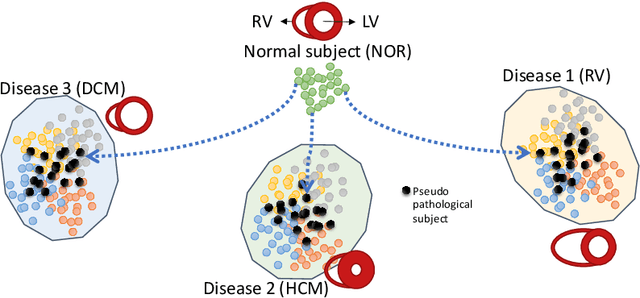
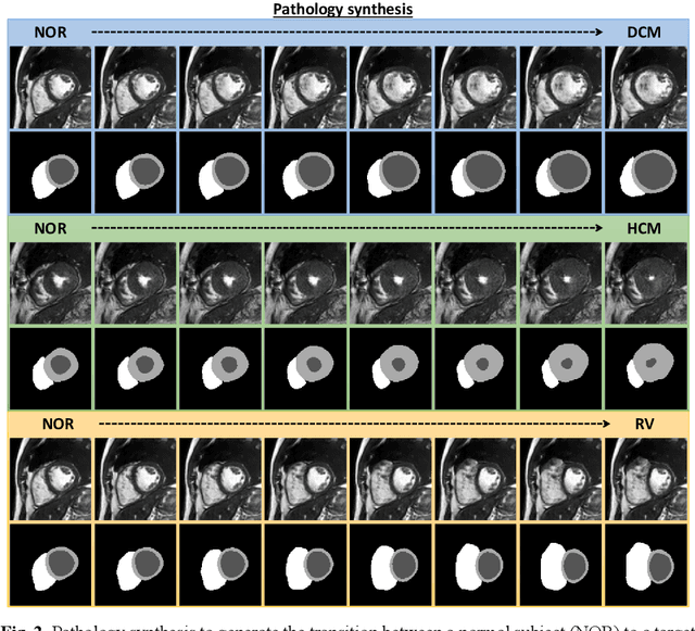
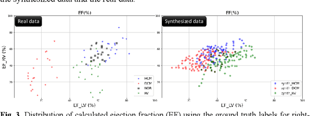
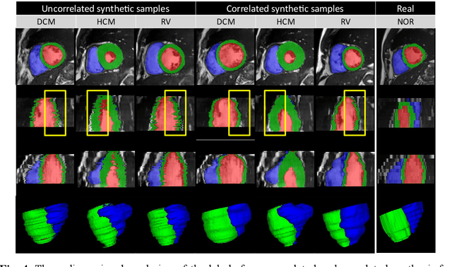
We propose a method for synthesizing cardiac MR images with plausible heart shapes and realistic appearances for the purpose of generating labeled data for deep-learning (DL) training. It breaks down the image synthesis into label deformation and label-to-image translation tasks. The former is achieved via latent space interpolation in a VAE model, while the latter is accomplished via a conditional GAN model. We devise an approach for label manipulation in the latent space of the trained VAE model, namely pathology synthesis, aiming to synthesize a series of pseudo-pathological synthetic subjects with characteristics of a desired heart disease. Furthermore, we propose to model the relationship between 2D slices in the latent space of the VAE via estimating the correlation coefficient matrix between the latent vectors and utilizing it to correlate elements of randomly drawn samples before decoding to image space. This simple yet effective approach results in generating 3D consistent subjects from 2D slice-by-slice generations. Such an approach could provide a solution to diversify and enrich the available database of cardiac MR images and to pave the way for the development of generalizable DL-based image analysis algorithms. The code will be available at https://github.com/sinaamirrajab/CardiacPathologySynthesis.