 Add to Chrome
 Add to Firefox
 Add to Edge
Dual Mixture-of-Experts Framework for Discrete-Time Survival Analysis
Oct 29, 2025Survival analysis is a task to model the time until an event of interest occurs, widely used in clinical and biomedical research. A key challenge is to model patient heterogeneity while also adapting risk predictions to both individual characteristics and temporal dynamics. We propose a dual mixture-of-experts (MoE) framework for discrete-time survival analysis. Our approach combines a feature-encoder MoE for subgroup-aware representation learning with a hazard MoE that leverages patient features and time embeddings to capture temporal dynamics. This dual-MoE design flexibly integrates with existing deep learning based survival pipelines. On METABRIC and GBSG breast cancer datasets, our method consistently improves performance, boosting the time-dependent C-index up to 0.04 on the test sets, and yields further gains when incorporated into the Consurv framework.
SDC-UDA: Volumetric Unsupervised Domain Adaptation Framework for Slice-Direction Continuous Cross-Modality Medical Image Segmentation
May 18, 2023Recent advances in deep learning-based medical image segmentation studies achieve nearly human-level performance in fully supervised manner. However, acquiring pixel-level expert annotations is extremely expensive and laborious in medical imaging fields. Unsupervised domain adaptation (UDA) can alleviate this problem, which makes it possible to use annotated data in one imaging modality to train a network that can successfully perform segmentation on target imaging modality with no labels. In this work, we propose SDC-UDA, a simple yet effective volumetric UDA framework for slice-direction continuous cross-modality medical image segmentation which combines intra- and inter-slice self-attentive image translation, uncertainty-constrained pseudo-label refinement, and volumetric self-training. Our method is distinguished from previous methods on UDA for medical image segmentation in that it can obtain continuous segmentation in the slice direction, thereby ensuring higher accuracy and potential in clinical practice. We validate SDC-UDA with multiple publicly available cross-modality medical image segmentation datasets and achieve state-of-the-art segmentation performance, not to mention the superior slice-direction continuity of prediction compared to previous studies.
COSMOS: Cross-Modality Unsupervised Domain Adaptation for 3D Medical Image Segmentation based on Target-aware Domain Translation and Iterative Self-Training
Mar 30, 2022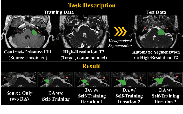
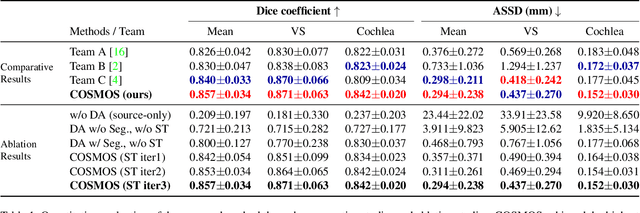
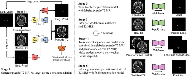
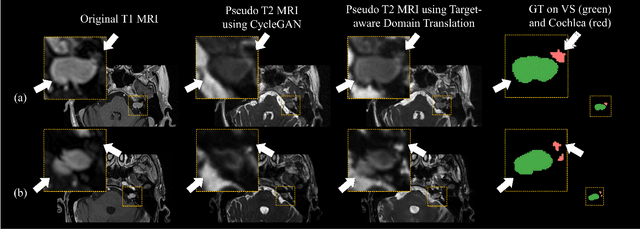
Recent advances in deep learning-based medical image segmentation studies achieve nearly human-level performance when in fully supervised condition. However, acquiring pixel-level expert annotations is extremely expensive and laborious in medical imaging fields. Unsupervised domain adaptation can alleviate this problem, which makes it possible to use annotated data in one imaging modality to train a network that can successfully perform segmentation on target imaging modality with no labels. In this work, we propose a self-training based unsupervised domain adaptation framework for 3D medical image segmentation named COSMOS and validate it with automatic segmentation of Vestibular Schwannoma (VS) and cochlea on high-resolution T2 Magnetic Resonance Images (MRI). Our target-aware contrast conversion network translates source domain annotated T1 MRI to pseudo T2 MRI to enable segmentation training on target domain, while preserving important anatomical features of interest in the converted images. Iterative self-training is followed to incorporate unlabeled data to training and incrementally improve the quality of pseudo-labels, thereby leading to improved performance of segmentation. COSMOS won the 1\textsuperscript{st} place in the Cross-Modality Domain Adaptation (crossMoDA) challenge held in conjunction with the 24th International Conference on Medical Image Computing and Computer Assisted Intervention (MICCAI 2021). It achieves mean Dice score and Average Symmetric Surface Distance of 0.871(0.063) and 0.437(0.270) for VS, and 0.842(0.020) and 0.152(0.030) for cochlea.
CrossMoDA 2021 challenge: Benchmark of Cross-Modality Domain Adaptation techniques for Vestibular Schwnannoma and Cochlea Segmentation
Jan 08, 2022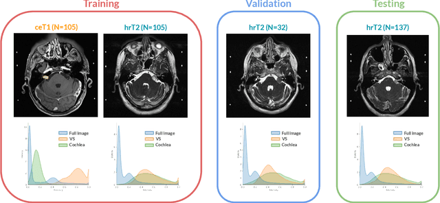
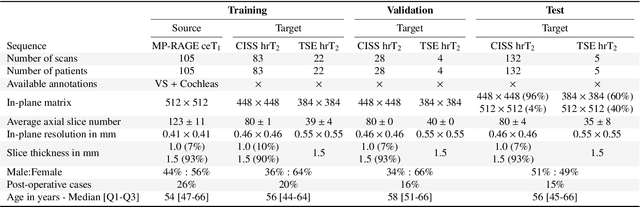
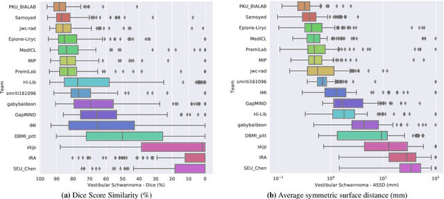
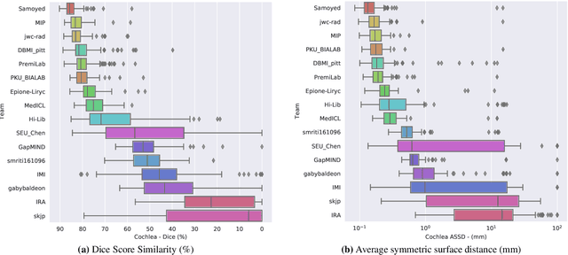
Domain Adaptation (DA) has recently raised strong interests in the medical imaging community. While a large variety of DA techniques has been proposed for image segmentation, most of these techniques have been validated either on private datasets or on small publicly available datasets. Moreover, these datasets mostly addressed single-class problems. To tackle these limitations, the Cross-Modality Domain Adaptation (crossMoDA) challenge was organised in conjunction with the 24th International Conference on Medical Image Computing and Computer Assisted Intervention (MICCAI 2021). CrossMoDA is the first large and multi-class benchmark for unsupervised cross-modality DA. The challenge's goal is to segment two key brain structures involved in the follow-up and treatment planning of vestibular schwannoma (VS): the VS and the cochleas. Currently, the diagnosis and surveillance in patients with VS are performed using contrast-enhanced T1 (ceT1) MRI. However, there is growing interest in using non-contrast sequences such as high-resolution T2 (hrT2) MRI. Therefore, we created an unsupervised cross-modality segmentation benchmark. The training set provides annotated ceT1 (N=105) and unpaired non-annotated hrT2 (N=105). The aim was to automatically perform unilateral VS and bilateral cochlea segmentation on hrT2 as provided in the testing set (N=137). A total of 16 teams submitted their algorithm for the evaluation phase. The level of performance reached by the top-performing teams is strikingly high (best median Dice - VS:88.4%; Cochleas:85.7%) and close to full supervision (median Dice - VS:92.5%; Cochleas:87.7%). All top-performing methods made use of an image-to-image translation approach to transform the source-domain images into pseudo-target-domain images. A segmentation network was then trained using these generated images and the manual annotations provided for the source image.
Self-Training Based Unsupervised Cross-Modality Domain Adaptation for Vestibular Schwannoma and Cochlea Segmentation
Sep 22, 2021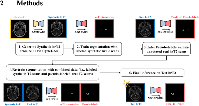
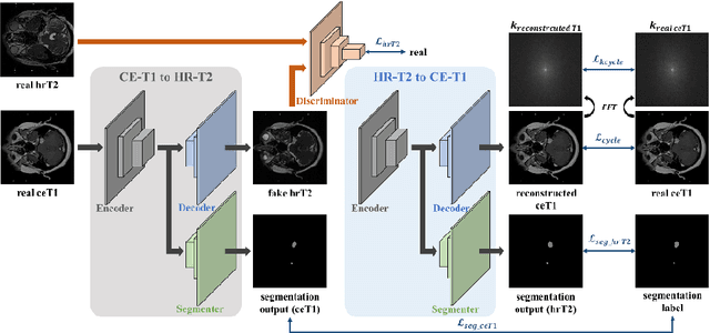
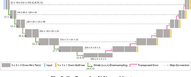
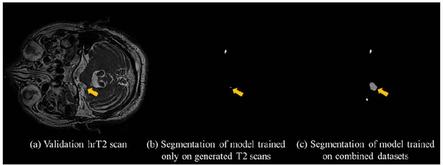
With the advances of deep learning, many medical image segmentation studies achieve human-level performance when in fully supervised condition. However, it is extremely expensive to acquire annotation on every data in medical fields, especially on magnetic resonance images (MRI) that comprise many different contrasts. Unsupervised methods can alleviate this problem; however, the performance drop is inevitable compared to fully supervised methods. In this work, we propose a self-training based unsupervised-learning framework that performs automatic segmentation of Vestibular Schwannoma (VS) and cochlea on high-resolution T2 scans. Our method consists of 4 main stages: 1) VS-preserving contrast conversion from contrast-enhanced T1 scan to high-resolution T2 scan, 2) training segmentation on generated T2 scans with annotations on T1 scans, and 3) Inferring pseudo-labels on non-annotated real T2 scans, and 4) boosting the generalizability of VS and cochlea segmentation by training with combined data (i.e., real T2 scans with pseudo-labels and generated T2 scans with true annotations). Our method showed mean Dice score and Average Symmetric Surface Distance (ASSD) of 0.8570 (0.0705) and 0.4970 (0.3391) for VS, 0.8446 (0.0211) and 0.1513 (0.0314) for Cochlea on CrossMoDA2021 challenge validation phase leaderboard, outperforming most other approaches.
State-of-the-Art Machine Learning MRI Reconstruction in 2020: Results of the Second fastMRI Challenge
Dec 28, 2020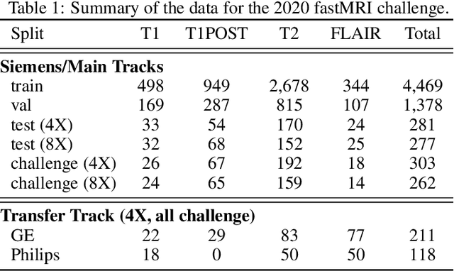
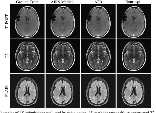
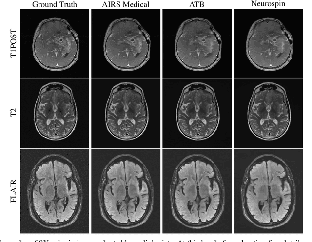
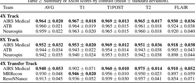
Accelerating MRI scans is one of the principal outstanding problems in the MRI research community. Towards this goal, we hosted the second fastMRI competition targeted towards reconstructing MR images with subsampled k-space data. We provided participants with data from 7,299 clinical brain scans (de-identified via a HIPAA-compliant procedure by NYU Langone Health), holding back the fully-sampled data from 894 of these scans for challenge evaluation purposes. In contrast to the 2019 challenge, we focused our radiologist evaluations on pathological assessment in brain images. We also debuted a new Transfer track that required participants to submit models evaluated on MRI scanners from outside the training set. We received 19 submissions from eight different groups. Results showed one team scoring best in both SSIM scores and qualitative radiologist evaluations. We also performed analysis on alternative metrics to mitigate the effects of background noise and collected feedback from the participants to inform future challenges. Lastly, we identify common failure modes across the submissions, highlighting areas of need for future research in the MRI reconstruction community.