 Add to Chrome
 Add to Firefox
 Add to Edge
TWINGS: Thin Plate Splines Warp-aligned Initialization for Sparse-View Gaussian Splatting
May 21, 2026Novel view synthesis from sparse-view inputs poses a significant challenge in 3D computer vision, particularly for achieving high-quality scene reconstructions with limited viewpoints. We introduce TWINGS, a framework that enhances 3D Gaussian Splatting (3DGS) by directly addressing point sparsity. We employ Thin Plate Splines (TPS), a smooth non-rigid deformation model that minimizes bending energy to estimate a globally coherent warp from control-point correspondences, to align backprojected points from estimated depth with triangulated 3D control points, yielding calibrated backprojected points. By sampling these calibrated points near the control points, TWINGS provides a fast and geometrically accurate initialization for 3DGS, ultimately improving structural detail preservation and color fidelity in reconstructed scenes. Extensive experiments on DTU, LLFF, and Mip-NeRF360 demonstrate that TWINGS consistently outperforms existing methods, delivering detailed and accurate reconstructions under sparse-view scenarios.
MUST: Modality-Specific Representation-Aware Transformer for Diffusion-Enhanced Survival Prediction with Missing Modality
Mar 27, 2026Accurate survival prediction from multimodal medical data is essential for precision oncology, yet clinical deployment faces a persistent challenge: modalities are frequently incomplete due to cost constraints, technical limitations, or retrospective data availability. While recent methods attempt to address missing modalities through feature alignment or joint distribution learning, they fundamentally lack explicit modeling of the unique contributions of each modality as opposed to the information derivable from other modalities. We propose MUST (Modality-Specific representation-aware Transformer), a novel framework that explicitly decomposes each modality's representation into modality-specific and cross-modal contextualized components through algebraic constraints in a learned low-rank shared subspace. This decomposition enables precise identification of what information is lost when a modality is absent. For the truly modality-specific information that cannot be inferred from available modalities, we employ conditional latent diffusion models to generate high-quality representations conditioned on recovered shared information and learned structural priors. Extensive experiments on five TCGA cancer datasets demonstrate that MUST achieves state-of-the-art performance with complete data while maintaining robust predictions in both missing pathology and missing genomics conditions, with clinically acceptable inference latency.
CD-Buffer: Complementary Dual-Buffer Framework for Test-Time Adaptation in Adverse Weather Object Detection
Mar 27, 2026Test-Time Adaptation (TTA) enables real-time adaptation to domain shifts without off-line retraining. Recent TTA methods have predominantly explored additive approaches that introduce lightweight modules for feature refinement. Recently, a subtractive approach that removes domain-sensitive channels has emerged as an alternative direction. We observe that these paradigms exhibit complementary effectiveness patterns: subtractive methods excel under severe shifts by removing corrupted features, while additive methods are effective under moderate shifts requiring refinement. However, each paradigm operates effectively only within limited shift severity ranges, failing to generalize across diverse corruption levels. This leads to the following question: can we adaptively balance both strategies based on measured feature-level domain shift? We propose CD-Buffer, a novel complementary dual-buffer framework where subtractive and additive mechanisms operate in opposite yet coordinated directions driven by a unified discrepancy metric. Our key innovation lies in the discrepancy-driven coupling: Our framework couples removal and refinement through a unified discrepancy metric, automatically balancing both strategies based on feature-level shift severity. This establishes automatic channel-wise balancing that adapts differentiated treatment to heterogeneous shift magnitudes without manual tuning. Extensive experiments on KITTI, Cityscapes, and ACDC datasets demonstrate state-of-the-art performance, consistently achieving superior results across diverse weather conditions and severity levels.
AcTTA: Rethinking Test-Time Adaptation via Dynamic Activation
Mar 27, 2026Test-time adaptation (TTA) aims to mitigate performance degradation under distribution shifts by updating model parameters during inference. Existing approaches have primarily framed adaptation around affine modulation, focusing on recalibrating normalization layers. This perspective, while effective, overlooks another influential component in representation dynamics: the activation function. We revisit this overlooked space and propose AcTTA, an activation-aware framework that reinterprets conventional activation functions from a learnable perspective and updates them adaptively at test time. AcTTA reformulates conventional activation functions (e.g., ReLU, GELU) into parameterized forms that shift their response threshold and modulate gradient sensitivity, enabling the network to adjust activation behavior under domain shifts. This functional reparameterization enables continuous adjustment of activation behavior without modifying network weights or requiring source data. Despite its simplicity, AcTTA achieves robust and stable adaptation across diverse corruptions. Across CIFAR10-C, CIFAR100-C, and ImageNet-C, AcTTA consistently surpasses normalization-based TTA methods. Our findings highlight activation adaptation as a compact and effective route toward domain-shift-robust test-time learning, broadening the prevailing affine-centric view of adaptation.
HP-GAN: Harnessing pretrained networks for GAN improvement with FakeTwins and discriminator consistency
Feb 03, 2026Generative Adversarial Networks (GANs) have made significant progress in enhancing the quality of image synthesis. Recent methods frequently leverage pretrained networks to calculate perceptual losses or utilize pretrained feature spaces. In this paper, we extend the capabilities of pretrained networks by incorporating innovative self-supervised learning techniques and enforcing consistency between discriminators during GAN training. Our proposed method, named HP-GAN, effectively exploits neural network priors through two primary strategies: FakeTwins and discriminator consistency. FakeTwins leverages pretrained networks as encoders to compute a self-supervised loss and applies this through the generated images to train the generator, thereby enabling the generation of more diverse and high quality images. Additionally, we introduce a consistency mechanism between discriminators that evaluate feature maps extracted from Convolutional Neural Network (CNN) and Vision Transformer (ViT) feature networks. Discriminator consistency promotes coherent learning among discriminators and enhances training robustness by aligning their assessments of image quality. Our extensive evaluation across seventeen datasets-including scenarios with large, small, and limited data, and covering a variety of image domains-demonstrates that HP-GAN consistently outperforms current state-of-the-art methods in terms of Fréchet Inception Distance (FID), achieving significant improvements in image diversity and quality. Code is available at: https://github.com/higun2/HP-GAN.
Large Language Models are Clinical Reasoners: Reasoning-Aware Diagnosis Framework with Prompt-Generated Rationales
Dec 12, 2023Machine reasoning has made great progress in recent years owing to large language models (LLMs). In the clinical domain, however, most NLP-driven projects mainly focus on clinical classification or reading comprehension, and under-explore clinical reasoning for disease diagnosis due to the expensive rationale annotation with clinicians. In this work, we present a ``reasoning-aware'' diagnosis framework that rationalizes the diagnostic process via prompt-based learning in a time- and labor-efficient manner, and learns to reason over the prompt-generated rationales. Specifically, we address the clinical reasoning for disease diagnosis, where the LLM generates diagnostic rationales providing its insight on presented patient data and the reasoning path towards the diagnosis, namely Clinical Chain-of-Thought (Clinical CoT). We empirically demonstrate LLMs/LMs' ability of clinical reasoning via extensive experiments and analyses on both rationale generation and disease diagnosis in various settings. We further propose a novel set of criteria for evaluating machine-generated rationales' potential for real-world clinical settings, facilitating and benefiting future research in this area.
SDC-UDA: Volumetric Unsupervised Domain Adaptation Framework for Slice-Direction Continuous Cross-Modality Medical Image Segmentation
May 18, 2023Recent advances in deep learning-based medical image segmentation studies achieve nearly human-level performance in fully supervised manner. However, acquiring pixel-level expert annotations is extremely expensive and laborious in medical imaging fields. Unsupervised domain adaptation (UDA) can alleviate this problem, which makes it possible to use annotated data in one imaging modality to train a network that can successfully perform segmentation on target imaging modality with no labels. In this work, we propose SDC-UDA, a simple yet effective volumetric UDA framework for slice-direction continuous cross-modality medical image segmentation which combines intra- and inter-slice self-attentive image translation, uncertainty-constrained pseudo-label refinement, and volumetric self-training. Our method is distinguished from previous methods on UDA for medical image segmentation in that it can obtain continuous segmentation in the slice direction, thereby ensuring higher accuracy and potential in clinical practice. We validate SDC-UDA with multiple publicly available cross-modality medical image segmentation datasets and achieve state-of-the-art segmentation performance, not to mention the superior slice-direction continuity of prediction compared to previous studies.
Evidence-empowered Transfer Learning for Alzheimer's Disease
Mar 03, 2023


Transfer learning has been widely utilized to mitigate the data scarcity problem in the field of Alzheimer's disease (AD). Conventional transfer learning relies on re-using models trained on AD-irrelevant tasks such as natural image classification. However, it often leads to negative transfer due to the discrepancy between the non-medical source and target medical domains. To address this, we present evidence-empowered transfer learning for AD diagnosis. Unlike conventional approaches, we leverage an AD-relevant auxiliary task, namely morphological change prediction, without requiring additional MRI data. In this auxiliary task, the diagnosis model learns the evidential and transferable knowledge from morphological features in MRI scans. Experimental results demonstrate that our framework is not only effective in improving detection performance regardless of model capacity, but also more data-efficient and faithful.
COSMOS: Cross-Modality Unsupervised Domain Adaptation for 3D Medical Image Segmentation based on Target-aware Domain Translation and Iterative Self-Training
Mar 30, 2022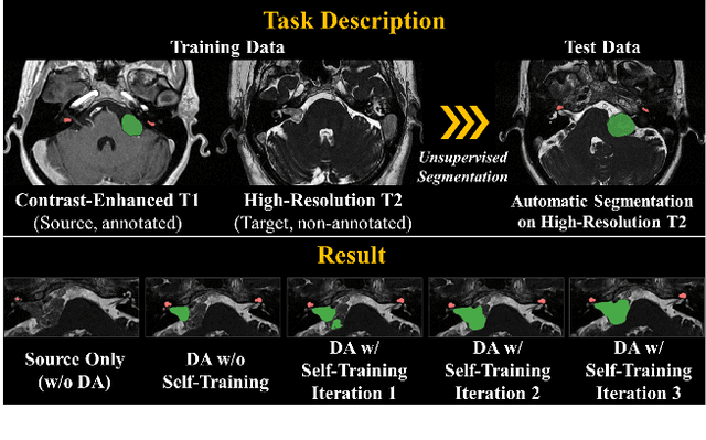
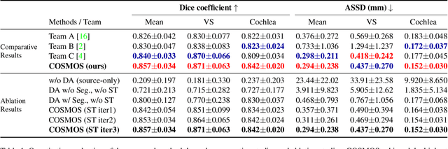
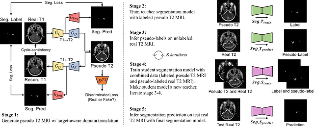
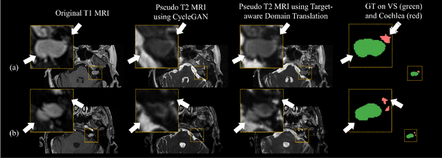
Recent advances in deep learning-based medical image segmentation studies achieve nearly human-level performance when in fully supervised condition. However, acquiring pixel-level expert annotations is extremely expensive and laborious in medical imaging fields. Unsupervised domain adaptation can alleviate this problem, which makes it possible to use annotated data in one imaging modality to train a network that can successfully perform segmentation on target imaging modality with no labels. In this work, we propose a self-training based unsupervised domain adaptation framework for 3D medical image segmentation named COSMOS and validate it with automatic segmentation of Vestibular Schwannoma (VS) and cochlea on high-resolution T2 Magnetic Resonance Images (MRI). Our target-aware contrast conversion network translates source domain annotated T1 MRI to pseudo T2 MRI to enable segmentation training on target domain, while preserving important anatomical features of interest in the converted images. Iterative self-training is followed to incorporate unlabeled data to training and incrementally improve the quality of pseudo-labels, thereby leading to improved performance of segmentation. COSMOS won the 1\textsuperscript{st} place in the Cross-Modality Domain Adaptation (crossMoDA) challenge held in conjunction with the 24th International Conference on Medical Image Computing and Computer Assisted Intervention (MICCAI 2021). It achieves mean Dice score and Average Symmetric Surface Distance of 0.871(0.063) and 0.437(0.270) for VS, and 0.842(0.020) and 0.152(0.030) for cochlea.
Self-Training Based Unsupervised Cross-Modality Domain Adaptation for Vestibular Schwannoma and Cochlea Segmentation
Sep 22, 2021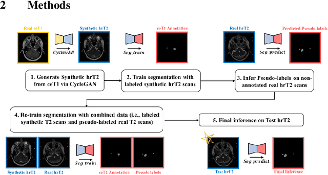
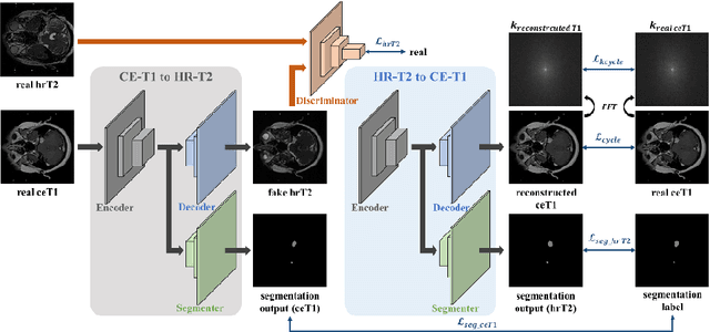
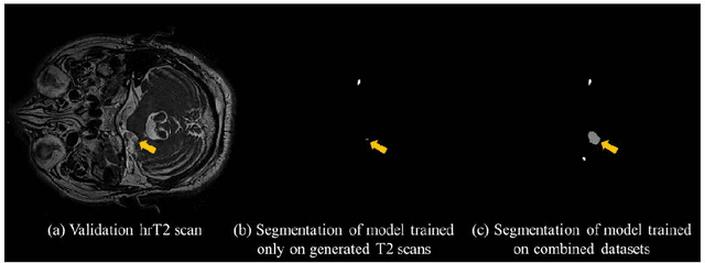
With the advances of deep learning, many medical image segmentation studies achieve human-level performance when in fully supervised condition. However, it is extremely expensive to acquire annotation on every data in medical fields, especially on magnetic resonance images (MRI) that comprise many different contrasts. Unsupervised methods can alleviate this problem; however, the performance drop is inevitable compared to fully supervised methods. In this work, we propose a self-training based unsupervised-learning framework that performs automatic segmentation of Vestibular Schwannoma (VS) and cochlea on high-resolution T2 scans. Our method consists of 4 main stages: 1) VS-preserving contrast conversion from contrast-enhanced T1 scan to high-resolution T2 scan, 2) training segmentation on generated T2 scans with annotations on T1 scans, and 3) Inferring pseudo-labels on non-annotated real T2 scans, and 4) boosting the generalizability of VS and cochlea segmentation by training with combined data (i.e., real T2 scans with pseudo-labels and generated T2 scans with true annotations). Our method showed mean Dice score and Average Symmetric Surface Distance (ASSD) of 0.8570 (0.0705) and 0.4970 (0.3391) for VS, 0.8446 (0.0211) and 0.1513 (0.0314) for Cochlea on CrossMoDA2021 challenge validation phase leaderboard, outperforming most other approaches.