 Add to Chrome
 Add to Firefox
 Add to Edge
Vision-Language Models as Zero-Annotation Oracles in Histopathology
Jun 15, 2026
Foreground segmentation is the critical first step of every computational pathology pipeline, yet existing methods rely on hand-tuned heuristics or supervised models that overfit to narrow stain and scanner distributions, failing silently on specialised stains such as Jones silver or Elastica van Gieson. We propose a coarse-to-fine approach that recasts foreground segmentation as a visual perception task and leverages general-purpose vision-language models (VLMs) as zero-annotation oracles. Our key insight is that tissue-versus-background discrimination is a natural-image recognition problem, not a histopathological one, so VLMs trained on internet-scale corpora generalise where domain-specific models cannot. We introduce Leica-75, a benchmark of 75 renal transplant whole-slide images spanning three stain families. On Leica-75, our method achieves the highest segmentation quality on out-of-distribution stains (Dice 0.858 +/- 0.027 on Jones, 0.853 +/- 0.041 on EVG) with 7x lower cross-stain variance than the best supervised baseline, while remaining competitive on in-distribution H&E. Few-shot prompting with automatically curated exemplars (Auto-context) rescues hard cases on Stress-32 (n=32), a curated stress-test subset (Dice 0.470 to 0.819 for the 2B model). VLM-based annotation review matches human expert consensus (kappa=0.989 for blur detection; mean precision/recall grading accuracy 0.708 vs. human 0.646 for segmentation mask review). The resulting pseudo-labels are used to distil lightweight student models that are as performant as the teacher model while running for a fraction of the cost. Our framework provides a principled, scalable solution to a persistent infrastructure bottleneck in digital pathology.

Graph Conditioned Diffusion for Controllable Histopathology Image Generation
Oct 08, 2025Recent advances in Diffusion Probabilistic Models (DPMs) have set new standards in high-quality image synthesis. Yet, controlled generation remains challenging, particularly in sensitive areas such as medical imaging. Medical images feature inherent structure such as consistent spatial arrangement, shape or texture, all of which are critical for diagnosis. However, existing DPMs operate in noisy latent spaces that lack semantic structure and strong priors, making it difficult to ensure meaningful control over generated content. To address this, we propose graph-based object-level representations for Graph-Conditioned-Diffusion. Our approach generates graph nodes corresponding to each major structure in the image, encapsulating their individual features and relationships. These graph representations are processed by a transformer module and integrated into a diffusion model via the text-conditioning mechanism, enabling fine-grained control over generation. We evaluate this approach using a real-world histopathology use case, demonstrating that our generated data can reliably substitute for annotated patient data in downstream segmentation tasks. The code is available here.
URCDM: Ultra-Resolution Image Synthesis in Histopathology
Jul 18, 2024Diagnosing medical conditions from histopathology data requires a thorough analysis across the various resolutions of Whole Slide Images (WSI). However, existing generative methods fail to consistently represent the hierarchical structure of WSIs due to a focus on high-fidelity patches. To tackle this, we propose Ultra-Resolution Cascaded Diffusion Models (URCDMs) which are capable of synthesising entire histopathology images at high resolutions whilst authentically capturing the details of both the underlying anatomy and pathology at all magnification levels. We evaluate our method on three separate datasets, consisting of brain, breast and kidney tissue, and surpass existing state-of-the-art multi-resolution models. Furthermore, an expert evaluation study was conducted, demonstrating that URCDMs consistently generate outputs across various resolutions that trained evaluators cannot distinguish from real images. All code and additional examples can be found on GitHub.
Ultra-Resolution Cascaded Diffusion Model for Gigapixel Image Synthesis in Histopathology
Dec 02, 2023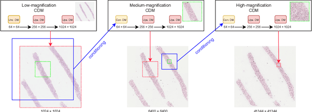
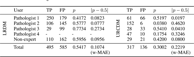
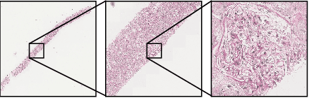
Diagnoses from histopathology images rely on information from both high and low resolutions of Whole Slide Images. Ultra-Resolution Cascaded Diffusion Models (URCDMs) allow for the synthesis of high-resolution images that are realistic at all magnification levels, focusing not only on fidelity but also on long-distance spatial coherency. Our model beats existing methods, improving the pFID-50k [2] score by 110.63 to 39.52 pFID-50k. Additionally, a human expert evaluation study was performed, reaching a weighted Mean Absolute Error (MAE) of 0.11 for the Lower Resolution Diffusion Models and a weighted MAE of 0.22 for the URCDM.
Realistic Data Enrichment for Robust Image Segmentation in Histopathology
Apr 19, 2023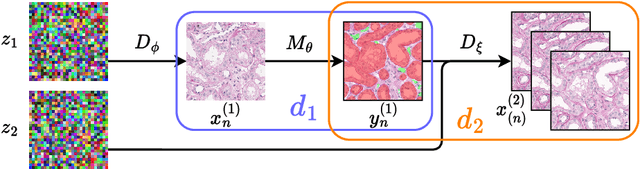
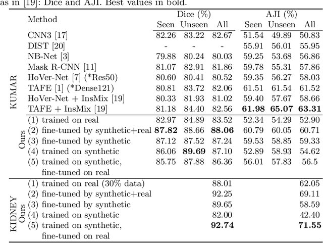
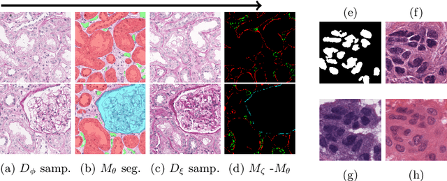
Poor performance of quantitative analysis in histopathological Whole Slide Images (WSI) has been a significant obstacle in clinical practice. Annotating large-scale WSIs manually is a demanding and time-consuming task, unlikely to yield the expected results when used for fully supervised learning systems. Rarely observed disease patterns and large differences in object scales are difficult to model through conventional patient intake. Prior methods either fall back to direct disease classification, which only requires learning a few factors per image, or report on average image segmentation performance, which is highly biased towards majority observations. Geometric image augmentation is commonly used to improve robustness for average case predictions and to enrich limited datasets. So far no method provided sampling of a realistic posterior distribution to improve stability, e.g. for the segmentation of imbalanced objects within images. Therefore, we propose a new approach, based on diffusion models, which can enrich an imbalanced dataset with plausible examples from underrepresented groups by conditioning on segmentation maps. Our method can simply expand limited clinical datasets making them suitable to train machine learning pipelines, and provides an interpretable and human-controllable way of generating histopathology images that are indistinguishable from real ones to human experts. We validate our findings on two datasets, one from the public domain and one from a Kidney Transplant study.