 Add to Chrome
 Add to Firefox
 Add to Edge
Vision-Language Models as Zero-Annotation Oracles in Histopathology
Jun 15, 2026
Foreground segmentation is the critical first step of every computational pathology pipeline, yet existing methods rely on hand-tuned heuristics or supervised models that overfit to narrow stain and scanner distributions, failing silently on specialised stains such as Jones silver or Elastica van Gieson. We propose a coarse-to-fine approach that recasts foreground segmentation as a visual perception task and leverages general-purpose vision-language models (VLMs) as zero-annotation oracles. Our key insight is that tissue-versus-background discrimination is a natural-image recognition problem, not a histopathological one, so VLMs trained on internet-scale corpora generalise where domain-specific models cannot. We introduce Leica-75, a benchmark of 75 renal transplant whole-slide images spanning three stain families. On Leica-75, our method achieves the highest segmentation quality on out-of-distribution stains (Dice 0.858 +/- 0.027 on Jones, 0.853 +/- 0.041 on EVG) with 7x lower cross-stain variance than the best supervised baseline, while remaining competitive on in-distribution H&E. Few-shot prompting with automatically curated exemplars (Auto-context) rescues hard cases on Stress-32 (n=32), a curated stress-test subset (Dice 0.470 to 0.819 for the 2B model). VLM-based annotation review matches human expert consensus (kappa=0.989 for blur detection; mean precision/recall grading accuracy 0.708 vs. human 0.646 for segmentation mask review). The resulting pseudo-labels are used to distil lightweight student models that are as performant as the teacher model while running for a fraction of the cost. Our framework provides a principled, scalable solution to a persistent infrastructure bottleneck in digital pathology.

Advances in Kidney Biopsy Structural Assessment through Dense Instance Segmentation
Sep 29, 2023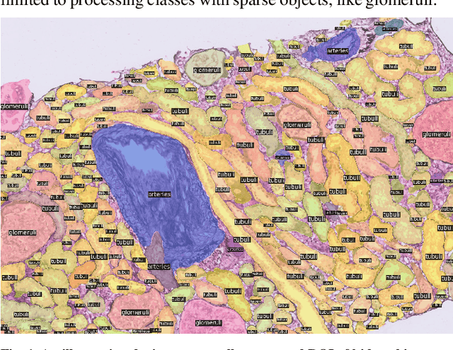

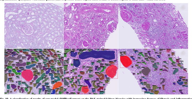
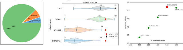
The kidney biopsy is the gold standard for the diagnosis of kidney diseases. Lesion scores made by expert renal pathologists are semi-quantitative and suffer from high inter-observer variability. Automatically obtaining statistics per segmented anatomical object, therefore, can bring significant benefits in reducing labor and this inter-observer variability. Instance segmentation for a biopsy, however, has been a challenging problem due to (a) the on average large number (around 300 to 1000) of densely touching anatomical structures, (b) with multiple classes (at least 3) and (c) in different sizes and shapes. The currently used instance segmentation models cannot simultaneously deal with these challenges in an efficient yet generic manner. In this paper, we propose the first anchor-free instance segmentation model that combines diffusion models, transformer modules, and RCNNs (regional convolution neural networks). Our model is trained on just one NVIDIA GeForce RTX 3090 GPU, but can efficiently recognize more than 500 objects with 3 common anatomical object classes in renal biopsies, i.e., glomeruli, tubuli, and arteries. Our data set consisted of 303 patches extracted from 148 Jones' silver-stained renal whole slide images (WSIs), where 249 patches were used for training and 54 patches for evaluation. In addition, without adjustment or retraining, the model can directly transfer its domain to generate decent instance segmentation results from PAS-stained WSIs. Importantly, it outperforms other baseline models and reaches an AP 51.7% in detection as the new state-of-the-art.