Alert button
Picture for Aikaterini Kotrotsou

Aikaterini Kotrotsou

Alert button

The Brain Tumor Segmentation (BraTS) Challenge 2023: Brain MR Image Synthesis for Tumor Segmentation (BraSyn)

May 20, 2023
Hongwei Bran Li, Syed Muhammad Anwar, Gian Marco Conte, Florian Kofler, Koen van Leemput, Marie Piraud, Ivan Ezhov, Felix Meissen, Maruf Adewole, Anastasia Janas, Anahita Fathi Kazerooni, Dominic LaBella, Ahmed W. Moawad, Keyvan Farahani, James Eddy, Timothy Bergquist, Verena Chung, Russell Takeshi Shinohara, Farouk Dako, Walter Wiggins, Zachary Reitman, Chunhao Wang, Xinyang Liu, Zhifan Jiang, Ariana Familiar, Elaine Johanson, Zeke Meier, Christos Davatzikos, John Freymann, Justin Kirby, Michel Bilello, Hassan M. Fathallah-Shaykh, Roland Wiest, Jan Kirschke, Rivka R. Colen, Aikaterini Kotrotsou, Pamela Lamontagne, Daniel Marcus, Mikhail Milchenko, Arash Nazeri, Marc André Weber, Abhishek Mahajan, Suyash Mohan, John Mongan, Christopher Hess, Soonmee Cha, Javier Villanueva, Meyer Errol Colak, Priscila Crivellaro, Andras Jakab, Jake Albrecht, Udunna Anazodo, Mariam Aboian, Thomas Yu, Verena Chung, Timothy Bergquist, James Eddy, Jake Albrecht, Ujjwal Baid, Spyridon Bakas, Marius George Linguraru, Bjoern Menze, Juan Eugenio Iglesias, Benedikt Wiestler

Figure 1 for The Brain Tumor Segmentation (BraTS) Challenge 2023: Brain MR Image Synthesis for Tumor Segmentation (BraSyn)

Automated brain tumor segmentation methods are well established, reaching performance levels with clear clinical utility. Most algorithms require four input magnetic resonance imaging (MRI) modalities, typically T1-weighted images with and without contrast enhancement, T2-weighted images, and FLAIR images. However, some of these sequences are often missing in clinical practice, e.g., because of time constraints and/or image artifacts (such as patient motion). Therefore, substituting missing modalities to recover segmentation performance in these scenarios is highly desirable and necessary for the more widespread adoption of such algorithms in clinical routine. In this work, we report the set-up of the Brain MR Image Synthesis Benchmark (BraSyn), organized in conjunction with the Medical Image Computing and Computer-Assisted Intervention (MICCAI) 2023. The objective of the challenge is to benchmark image synthesis methods that realistically synthesize missing MRI modalities given multiple available images to facilitate automated brain tumor segmentation pipelines. The image dataset is multi-modal and diverse, created in collaboration with various hospitals and research institutions.

* Technical report of BraSyn 
Viaarxiv icon

The Brain Tumor Segmentation (BraTS) Challenge 2023: Local Synthesis of Healthy Brain Tissue via Inpainting

May 15, 2023
Florian Kofler, Felix Meissen, Felix Steinbauer, Robert Graf, Eva Oswald, Ezequiel de da Rosa, Hongwei Bran Li, Ujjwal Baid, Florian Hoelzl, Oezguen Turgut, Izabela Horvath, Diana Waldmannstetter, Christina Bukas, Maruf Adewole, Syed Muhammad Anwar, Anastasia Janas, Anahita Fathi Kazerooni, Dominic LaBella, Ahmed W Moawad, Keyvan Farahani, James Eddy, Timothy Bergquist, Verena Chung, Russell Takeshi Shinohara, Farouk Dako, Walter Wiggins, Zachary Reitman, Chunhao Wang, Xinyang Liu, Zhifan Jiang, Ariana Familiar, Gian-Marco Conte, Elaine Johanson, Zeke Meier, Christos Davatzikos, John Freymann, Justin Kirby, Michel Bilello, Hassan M Fathallah-Shaykh, Roland Wiest, Jan Kirschke, Rivka R Colen, Aikaterini Kotrotsou, Pamela Lamontagne, Daniel Marcus, Mikhail Milchenko, Arash Nazeri, Marc-André Weber, Abhishek Mahajan, Suyash Mohan, John Mongan, Christopher Hess, Soonmee Cha, Javier Villanueva-Meyer, Errol Colak, Priscila Crivellaro, Andras Jakab, Jake Albrecht, Udunna Anazodo, Mariam Aboian, Juan Eugenio Iglesias, Koen Van Leemput, Spyridon Bakas, Daniel Rueckert, Benedikt Wiestler, Ivan Ezhov, Marie Piraud, Bjoern Menze

Figure 1 for The Brain Tumor Segmentation (BraTS) Challenge 2023: Local Synthesis of Healthy Brain Tissue via Inpainting

A myriad of algorithms for the automatic analysis of brain MR images is available to support clinicians in their decision-making. For brain tumor patients, the image acquisition time series typically starts with a scan that is already pathological. This poses problems, as many algorithms are designed to analyze healthy brains and provide no guarantees for images featuring lesions. Examples include but are not limited to algorithms for brain anatomy parcellation, tissue segmentation, and brain extraction. To solve this dilemma, we introduce the BraTS 2023 inpainting challenge. Here, the participants' task is to explore inpainting techniques to synthesize healthy brain scans from lesioned ones. The following manuscript contains the task formulation, dataset, and submission procedure. Later it will be updated to summarize the findings of the challenge. The challenge is organized as part of the BraTS 2023 challenge hosted at the MICCAI 2023 conference in Vancouver, Canada.

* 5 pages, 1 figure 
Viaarxiv icon

Federated Learning Enables Big Data for Rare Cancer Boundary Detection

Apr 25, 2022
Sarthak Pati, Ujjwal Baid, Brandon Edwards, Micah Sheller, Shih-Han Wang, G Anthony Reina, Patrick Foley, Alexey Gruzdev, Deepthi Karkada, Christos Davatzikos, Chiharu Sako, Satyam Ghodasara, Michel Bilello, Suyash Mohan, Philipp Vollmuth, Gianluca Brugnara, Chandrakanth J Preetha, Felix Sahm, Klaus Maier-Hein, Maximilian Zenk, Martin Bendszus, Wolfgang Wick, Evan Calabrese, Jeffrey Rudie, Javier Villanueva-Meyer, Soonmee Cha, Madhura Ingalhalikar, Manali Jadhav, Umang Pandey, Jitender Saini, John Garrett, Matthew Larson, Robert Jeraj, Stuart Currie, Russell Frood, Kavi Fatania, Raymond Y Huang, Ken Chang, Carmen Balana, Jaume Capellades, Josep Puig, Johannes Trenkler, Josef Pichler, Georg Necker, Andreas Haunschmidt, Stephan Meckel, Gaurav Shukla, Spencer Liem, Gregory S Alexander, Joseph Lombardo, Joshua D Palmer, Adam E Flanders, Adam P Dicker, Haris I Sair, Craig K Jones, Archana Venkataraman, Meirui Jiang, Tiffany Y So, Cheng Chen, Pheng Ann Heng, Qi Dou, Michal Kozubek, Filip Lux, Jan Michálek, Petr Matula, Miloš Keřkovský, Tereza Kopřivová, Marek Dostál, Václav Vybíhal, Michael A Vogelbaum, J Ross Mitchell, Joaquim Farinhas, Joseph A Maldjian, Chandan Ganesh Bangalore Yogananda, Marco C Pinho, Divya Reddy, James Holcomb, Benjamin C Wagner, Benjamin M Ellingson, Timothy F Cloughesy, Catalina Raymond, Talia Oughourlian, Akifumi Hagiwara, Chencai Wang, Minh-Son To, Sargam Bhardwaj, Chee Chong, Marc Agzarian, Alexandre Xavier Falcão, Samuel B Martins, Bernardo C A Teixeira, Flávia Sprenger, David Menotti, Diego R Lucio, Pamela LaMontagne, Daniel Marcus, Benedikt Wiestler, Florian Kofler, Ivan Ezhov, Marie Metz, Rajan Jain, Matthew Lee, Yvonne W Lui, Richard McKinley, Johannes Slotboom, Piotr Radojewski, Raphael Meier, Roland Wiest, Derrick Murcia, Eric Fu, Rourke Haas, John Thompson, David Ryan Ormond, Chaitra Badve, Andrew E Sloan, Vachan Vadmal, Kristin Waite, Rivka R Colen, Linmin Pei, Murat Ak, Ashok Srinivasan, J Rajiv Bapuraj, Arvind Rao, Nicholas Wang, Ota Yoshiaki, Toshio Moritani, Sevcan Turk, Joonsang Lee, Snehal Prabhudesai, Fanny Morón, Jacob Mandel, Konstantinos Kamnitsas, Ben Glocker, Luke V M Dixon, Matthew Williams, Peter Zampakis, Vasileios Panagiotopoulos, Panagiotis Tsiganos, Sotiris Alexiou, Ilias Haliassos, Evangelia I Zacharaki, Konstantinos Moustakas, Christina Kalogeropoulou, Dimitrios M Kardamakis, Yoon Seong Choi, Seung-Koo Lee, Jong Hee Chang, Sung Soo Ahn, Bing Luo, Laila Poisson, Ning Wen, Pallavi Tiwari, Ruchika Verma, Rohan Bareja, Ipsa Yadav, Jonathan Chen, Neeraj Kumar, Marion Smits, Sebastian R van der Voort, Ahmed Alafandi, Fatih Incekara, Maarten MJ Wijnenga, Georgios Kapsas, Renske Gahrmann, Joost W Schouten, Hendrikus J Dubbink, Arnaud JPE Vincent, Martin J van den Bent, Pim J French, Stefan Klein, Yading Yuan, Sonam Sharma, Tzu-Chi Tseng, Saba Adabi, Simone P Niclou, Olivier Keunen, Ann-Christin Hau, Martin Vallières, David Fortin, Martin Lepage, Bennett Landman, Karthik Ramadass, Kaiwen Xu, Silky Chotai, Lola B Chambless, Akshitkumar Mistry, Reid C Thompson, Yuriy Gusev, Krithika Bhuvaneshwar, Anousheh Sayah, Camelia Bencheqroun, Anas Belouali, Subha Madhavan, Thomas C Booth, Alysha Chelliah, Marc Modat, Haris Shuaib, Carmen Dragos, Aly Abayazeed, Kenneth Kolodziej, Michael Hill, Ahmed Abbassy, Shady Gamal, Mahmoud Mekhaimar, Mohamed Qayati, Mauricio Reyes, Ji Eun Park, Jihye Yun, Ho Sung Kim, Abhishek Mahajan, Mark Muzi, Sean Benson, Regina G H Beets-Tan, Jonas Teuwen, Alejandro Herrera-Trujillo, Maria Trujillo, William Escobar, Ana Abello, Jose Bernal, Jhon Gómez, Joseph Choi, Stephen Baek, Yusung Kim, Heba Ismael, Bryan Allen, John M Buatti, Aikaterini Kotrotsou, Hongwei Li, Tobias Weiss, Michael Weller, Andrea Bink, Bertrand Pouymayou, Hassan F Shaykh, Joel Saltz, Prateek Prasanna, Sampurna Shrestha, Kartik M Mani, David Payne, Tahsin Kurc, Enrique Pelaez, Heydy Franco-Maldonado, Francis Loayza, Sebastian Quevedo, Pamela Guevara, Esteban Torche, Cristobal Mendoza, Franco Vera, Elvis Ríos, Eduardo López, Sergio A Velastin, Godwin Ogbole, Dotun Oyekunle, Olubunmi Odafe-Oyibotha, Babatunde Osobu, Mustapha Shu'aibu, Adeleye Dorcas, Mayowa Soneye, Farouk Dako, Amber L Simpson, Mohammad Hamghalam, Jacob J Peoples, Ricky Hu, Anh Tran, Danielle Cutler, Fabio Y Moraes, Michael A Boss, James Gimpel, Deepak Kattil Veettil, Kendall Schmidt, Brian Bialecki, Sailaja Marella, Cynthia Price, Lisa Cimino, Charles Apgar, Prashant Shah, Bjoern Menze, Jill S Barnholtz-Sloan, Jason Martin, Spyridon Bakas

Although machine learning (ML) has shown promise in numerous domains, there are concerns about generalizability to out-of-sample data. This is currently addressed by centrally sharing ample, and importantly diverse, data from multiple sites. However, such centralization is challenging to scale (or even not feasible) due to various limitations. Federated ML (FL) provides an alternative to train accurate and generalizable ML models, by only sharing numerical model updates. Here we present findings from the largest FL study to-date, involving data from 71 healthcare institutions across 6 continents, to generate an automatic tumor boundary detector for the rare disease of glioblastoma, utilizing the largest dataset of such patients ever used in the literature (25,256 MRI scans from 6,314 patients). We demonstrate a 33% improvement over a publicly trained model to delineate the surgically targetable tumor, and 23% improvement over the tumor's entire extent. We anticipate our study to: 1) enable more studies in healthcare informed by large and diverse data, ensuring meaningful results for rare diseases and underrepresented populations, 2) facilitate further quantitative analyses for glioblastoma via performance optimization of our consensus model for eventual public release, and 3) demonstrate the effectiveness of FL at such scale and task complexity as a paradigm shift for multi-site collaborations, alleviating the need for data sharing.

* federated learning, deep learning, convolutional neural network, segmentation, brain tumor, glioma, glioblastoma, FeTS, BraTS 
Viaarxiv icon

QU-BraTS: MICCAI BraTS 2020 Challenge on Quantifying Uncertainty in Brain Tumor Segmentation -- Analysis of Ranking Metrics and Benchmarking Results

Dec 19, 2021
Raghav Mehta, Angelos Filos, Ujjwal Baid, Chiharu Sako, Richard McKinley, Michael Rebsamen, Katrin Dätwyler, Raphael Meier, Piotr Radojewski, Gowtham Krishnan Murugesan, Sahil Nalawade, Chandan Ganesh, Ben Wagner, Fang F. Yu, Baowei Fei, Ananth J. Madhuranthakam, Joseph A. Maldjian, Laura Daza, Catalina Gómez, Pablo Arbeláez, Chengliang Dai, Shuo Wang, Hadrien Raynaud, Yuanhan Mo, Elsa Angelini, Yike Guo, Wenjia Bai, Subhashis Banerjee, Linmin Pei, Murat AK, Sarahi Rosas-González, Illyess Zemmoura, Clovis Tauber, Minh H. Vu, Tufve Nyholm, Tommy Löfstedt, Laura Mora Ballestar, Veronica Vilaplana, Hugh McHugh, Gonzalo Maso Talou, Alan Wang, Jay Patel, Ken Chang, Katharina Hoebel, Mishka Gidwani, Nishanth Arun, Sharut Gupta, Mehak Aggarwal, Praveer Singh, Elizabeth R. Gerstner, Jayashree Kalpathy-Cramer, Nicolas Boutry, Alexis Huard, Lasitha Vidyaratne, Md Monibor Rahman, Khan M. Iftekharuddin, Joseph Chazalon, Elodie Puybareau, Guillaume Tochon, Jun Ma, Mariano Cabezas, Xavier Llado, Arnau Oliver, Liliana Valencia, Sergi Valverde, Mehdi Amian, Mohammadreza Soltaninejad, Andriy Myronenko, Ali Hatamizadeh, Xue Feng, Quan Dou, Nicholas Tustison, Craig Meyer, Nisarg A. Shah, Sanjay Talbar, Marc-Andr Weber, Abhishek Mahajan, Andras Jakab, Roland Wiest, Hassan M. Fathallah-Shaykh, Arash Nazeri, Mikhail Milchenko, Daniel Marcus, Aikaterini Kotrotsou, Rivka Colen, John Freymann, Justin Kirby, Christos Davatzikos, Bjoern Menze, Spyridon Bakas, Yarin Gal, Tal Arbel

Figure 1 for QU-BraTS: MICCAI BraTS 2020 Challenge on Quantifying Uncertainty in Brain Tumor Segmentation -- Analysis of Ranking Metrics and Benchmarking Results
Figure 2 for QU-BraTS: MICCAI BraTS 2020 Challenge on Quantifying Uncertainty in Brain Tumor Segmentation -- Analysis of Ranking Metrics and Benchmarking Results
Figure 3 for QU-BraTS: MICCAI BraTS 2020 Challenge on Quantifying Uncertainty in Brain Tumor Segmentation -- Analysis of Ranking Metrics and Benchmarking Results
Figure 4 for QU-BraTS: MICCAI BraTS 2020 Challenge on Quantifying Uncertainty in Brain Tumor Segmentation -- Analysis of Ranking Metrics and Benchmarking Results

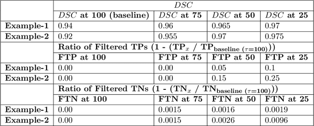
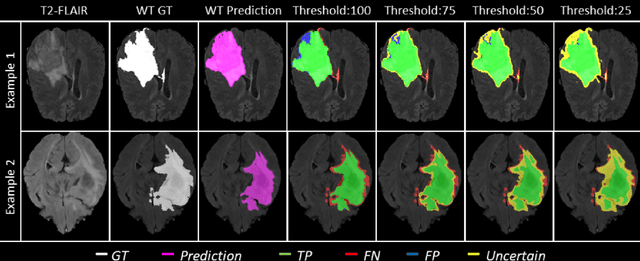
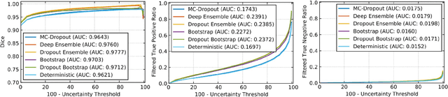
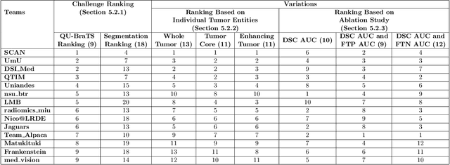
Deep learning (DL) models have provided the state-of-the-art performance in a wide variety of medical imaging benchmarking challenges, including the Brain Tumor Segmentation (BraTS) challenges. However, the task of focal pathology multi-compartment segmentation (e.g., tumor and lesion sub-regions) is particularly challenging, and potential errors hinder the translation of DL models into clinical workflows. Quantifying the reliability of DL model predictions in the form of uncertainties, could enable clinical review of the most uncertain regions, thereby building trust and paving the way towards clinical translation. Recently, a number of uncertainty estimation methods have been introduced for DL medical image segmentation tasks. Developing metrics to evaluate and compare the performance of uncertainty measures will assist the end-user in making more informed decisions. In this study, we explore and evaluate a metric developed during the BraTS 2019-2020 task on uncertainty quantification (QU-BraTS), and designed to assess and rank uncertainty estimates for brain tumor multi-compartment segmentation. This metric (1) rewards uncertainty estimates that produce high confidence in correct assertions, and those that assign low confidence levels at incorrect assertions, and (2) penalizes uncertainty measures that lead to a higher percentages of under-confident correct assertions. We further benchmark the segmentation uncertainties generated by 14 independent participating teams of QU-BraTS 2020, all of which also participated in the main BraTS segmentation task. Overall, our findings confirm the importance and complementary value that uncertainty estimates provide to segmentation algorithms, and hence highlight the need for uncertainty quantification in medical image analyses. Our evaluation code is made publicly available at https://github.com/RagMeh11/QU-BraTS.

* Under submission at MELBA journal 
Viaarxiv icon

The RSNA-ASNR-MICCAI BraTS 2021 Benchmark on Brain Tumor Segmentation and Radiogenomic Classification

Jul 05, 2021
Ujjwal Baid, Satyam Ghodasara, Michel Bilello, Suyash Mohan, Evan Calabrese, Errol Colak, Keyvan Farahani, Jayashree Kalpathy-Cramer, Felipe C. Kitamura, Sarthak Pati, Luciano M. Prevedello, Jeffrey D. Rudie, Chiharu Sako, Russell T. Shinohara, Timothy Bergquist, Rong Chai, James Eddy, Julia Elliott, Walter Reade, Thomas Schaffter, Thomas Yu, Jiaxin Zheng, BraTS Annotators, Christos Davatzikos, John Mongan, Christopher Hess, Soonmee Cha, Javier Villanueva-Meyer, John B. Freymann, Justin S. Kirby, Benedikt Wiestler, Priscila Crivellaro, Rivka R. Colen, Aikaterini Kotrotsou, Daniel Marcus, Mikhail Milchenko, Arash Nazeri, Hassan Fathallah-Shaykh, Roland Wiest, Andras Jakab, Marc-Andre Weber, Abhishek Mahajan, Bjoern Menze, Adam E. Flanders, Spyridon Bakas

Figure 1 for The RSNA-ASNR-MICCAI BraTS 2021 Benchmark on Brain Tumor Segmentation and Radiogenomic Classification
Figure 2 for The RSNA-ASNR-MICCAI BraTS 2021 Benchmark on Brain Tumor Segmentation and Radiogenomic Classification
Figure 3 for The RSNA-ASNR-MICCAI BraTS 2021 Benchmark on Brain Tumor Segmentation and Radiogenomic Classification

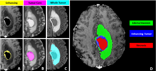
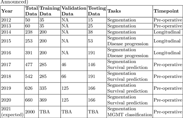
The BraTS 2021 challenge celebrates its 10th anniversary and is jointly organized by the Radiological Society of North America (RSNA), the American Society of Neuroradiology (ASNR), and the Medical Image Computing and Computer Assisted Interventions (MICCAI) society. Since its inception, BraTS has been focusing on being a common benchmarking venue for brain glioma segmentation algorithms, with well-curated multi-institutional multi-parametric magnetic resonance imaging (mpMRI) data. Gliomas are the most common primary malignancies of the central nervous system, with varying degrees of aggressiveness and prognosis. The RSNA-ASNR-MICCAI BraTS 2021 challenge targets the evaluation of computational algorithms assessing the same tumor compartmentalization, as well as the underlying tumor's molecular characterization, in pre-operative baseline mpMRI data from 2,000 patients. Specifically, the two tasks that BraTS 2021 focuses on are: a) the segmentation of the histologically distinct brain tumor sub-regions, and b) the classification of the tumor's O[6]-methylguanine-DNA methyltransferase (MGMT) promoter methylation status. The performance evaluation of all participating algorithms in BraTS 2021 will be conducted through the Sage Bionetworks Synapse platform (Task 1) and Kaggle (Task 2), concluding in distributing to the top ranked participants monetary awards of $60,000 collectively.

* 17 pages, 2 figures, 1 table 
Viaarxiv icon

The Federated Tumor Segmentation (FeTS) Challenge

May 14, 2021
Sarthak Pati, Ujjwal Baid, Maximilian Zenk, Brandon Edwards, Micah Sheller, G. Anthony Reina, Patrick Foley, Alexey Gruzdev, Jason Martin, Shadi Albarqouni, Yong Chen, Russell Taki Shinohara, Annika Reinke, David Zimmerer, John B. Freymann, Justin S. Kirby, Christos Davatzikos, Rivka R. Colen, Aikaterini Kotrotsou, Daniel Marcus, Mikhail Milchenko, Arash Nazeri, Hassan Fathallah-Shaykh, Roland Wiest, Andras Jakab, Marc-Andre Weber, Abhishek Mahajan, Lena Maier-Hein, Jens Kleesiek, Bjoern Menze, Klaus Maier-Hein, Spyridon Bakas

Figure 1 for The Federated Tumor Segmentation (FeTS) Challenge
Figure 2 for The Federated Tumor Segmentation (FeTS) Challenge

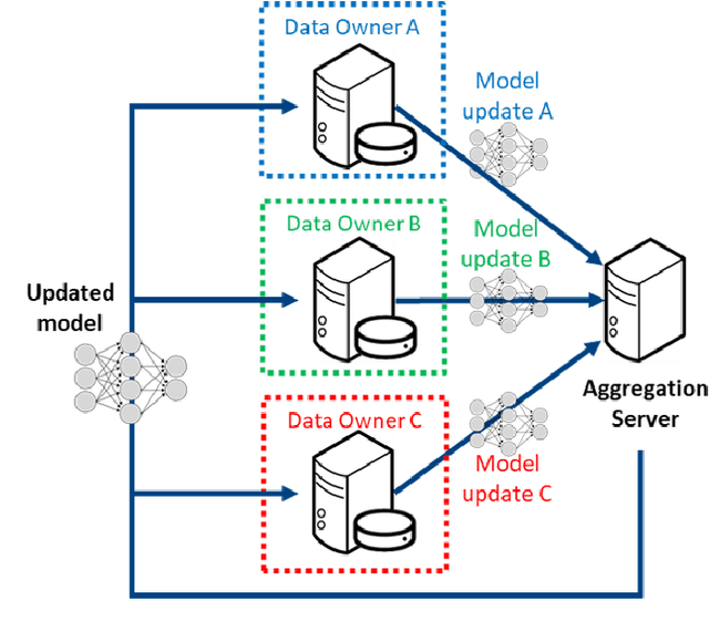
This manuscript describes the first challenge on Federated Learning, namely the Federated Tumor Segmentation (FeTS) challenge 2021. International challenges have become the standard for validation of biomedical image analysis methods. However, the actual performance of participating (even the winning) algorithms on "real-world" clinical data often remains unclear, as the data included in challenges are usually acquired in very controlled settings at few institutions. The seemingly obvious solution of just collecting increasingly more data from more institutions in such challenges does not scale well due to privacy and ownership hurdles. Towards alleviating these concerns, we are proposing the FeTS challenge 2021 to cater towards both the development and the evaluation of models for the segmentation of intrinsically heterogeneous (in appearance, shape, and histology) brain tumors, namely gliomas. Specifically, the FeTS 2021 challenge uses clinically acquired, multi-institutional magnetic resonance imaging (MRI) scans from the BraTS 2020 challenge, as well as from various remote independent institutions included in the collaborative network of a real-world federation (https://www.fets.ai/). The goals of the FeTS challenge are directly represented by the two included tasks: 1) the identification of the optimal weight aggregation approach towards the training of a consensus model that has gained knowledge via federated learning from multiple geographically distinct institutions, while their data are always retained within each institution, and 2) the federated evaluation of the generalizability of brain tumor segmentation models "in the wild", i.e. on data from institutional distributions that were not part of the training datasets.

Viaarxiv icon