 Add to Chrome
 Add to Firefox
 Add to Edge
Beyond Illumination: A Conditional Mutual Information-Guided Network for Low-Light Image Enhancement
Aug 03, 2026
Low-light image enhancement (LLIE) seeks to restore structural fidelity, natural color rendition, and proper exposure from images captured under inadequate lighting conditions. Recent state-of-the-art approaches, such as CIDNet, adopt a dual-branch architecture comprising a chrominance (HV) branch and an intensity (I) branch to separately model decoupled chromatic and luminance information within the HVI color space. However, these methods overlook the mutual interaction between intensity and chrominance components, which inherently limits their representational capacity and leads to suboptimal enhancement performance. To address this limitation, we propose the Conditional Mutual Information-Guided Network (CMIG-Net), which leverages conditional mutual information as a principled metric to quantitatively assess the contribution of chrominance features conditioned on the available intensity information. In particular, we design a Conditional Mutual Information Calibration (CMIC) module that generates a conditional information map, enabling region-adaptive recalibration of chrominance representations according to local illumination statistics. Furthermore, we introduce a Dynamic Dual-branch Information Restoration (D2IR) module, which adaptively governs bidirectional information flow between the intensity and chrominance branches, guided by both the conditional prior and the instantaneous restoration state. Extensive experiments on paired LLIE benchmarks demonstrate that CMIG-Net consistently outperforms CIDNet, achieving up to a 0.619 dB gain in PSNR, with a 0.382 dB improvement specifically on the challenging Sony-Total-Dark dataset.

The state-of-the-art in Cardiac MRI Reconstruction: Results of the CMRxRecon Challenge in MICCAI 2023
Apr 01, 2024

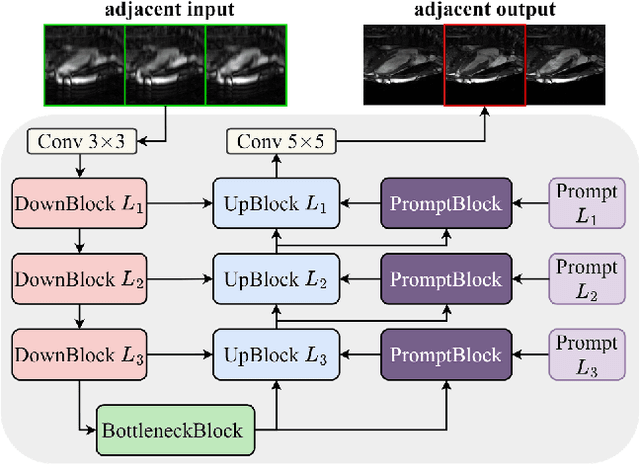

Cardiac MRI, crucial for evaluating heart structure and function, faces limitations like slow imaging and motion artifacts. Undersampling reconstruction, especially data-driven algorithms, has emerged as a promising solution to accelerate scans and enhance imaging performance using highly under-sampled data. Nevertheless, the scarcity of publicly available cardiac k-space datasets and evaluation platform hinder the development of data-driven reconstruction algorithms. To address this issue, we organized the Cardiac MRI Reconstruction Challenge (CMRxRecon) in 2023, in collaboration with the 26th International Conference on MICCAI. CMRxRecon presented an extensive k-space dataset comprising cine and mapping raw data, accompanied by detailed annotations of cardiac anatomical structures. With overwhelming participation, the challenge attracted more than 285 teams and over 600 participants. Among them, 22 teams successfully submitted Docker containers for the testing phase, with 7 teams submitted for both cine and mapping tasks. All teams use deep learning based approaches, indicating that deep learning has predominately become a promising solution for the problem. The first-place winner of both tasks utilizes the E2E-VarNet architecture as backbones. In contrast, U-Net is still the most popular backbone for both multi-coil and single-coil reconstructions. This paper provides a comprehensive overview of the challenge design, presents a summary of the submitted results, reviews the employed methods, and offers an in-depth discussion that aims to inspire future advancements in cardiac MRI reconstruction models. The summary emphasizes the effective strategies observed in Cardiac MRI reconstruction, including backbone architecture, loss function, pre-processing techniques, physical modeling, and model complexity, thereby providing valuable insights for further developments in this field.
CMRxRecon: An open cardiac MRI dataset for the competition of accelerated image reconstruction
Sep 19, 2023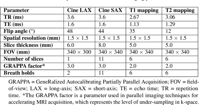
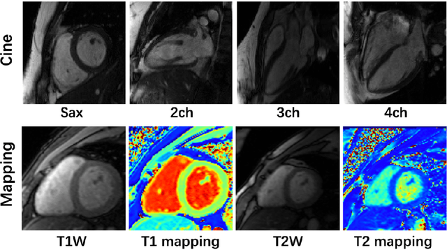
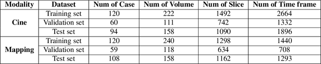
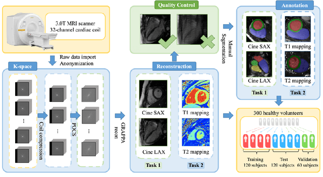
Cardiac magnetic resonance imaging (CMR) has emerged as a valuable diagnostic tool for cardiac diseases. However, a limitation of CMR is its slow imaging speed, which causes patient discomfort and introduces artifacts in the images. There has been growing interest in deep learning-based CMR imaging algorithms that can reconstruct high-quality images from highly under-sampled k-space data. However, the development of deep learning methods requires large training datasets, which have not been publicly available for CMR. To address this gap, we released a dataset that includes multi-contrast, multi-view, multi-slice and multi-coil CMR imaging data from 300 subjects. Imaging studies include cardiac cine and mapping sequences. Manual segmentations of the myocardium and chambers of all the subjects are also provided within the dataset. Scripts of state-of-the-art reconstruction algorithms were also provided as a point of reference. Our aim is to facilitate the advancement of state-of-the-art CMR image reconstruction by introducing standardized evaluation criteria and making the dataset freely accessible to the research community. Researchers can access the dataset at https://www.synapse.org/#!Synapse:syn51471091/wiki/.