 Add to Chrome
 Add to Firefox
 Add to Edge
Tropical Algebraic Geometry for Neuronal Representations: An Arakelov-Green Measure Based Descriptor for Graph Learning
Aug 05, 2026
The quantitative analysis of 3D neuronal morphologies requires capturing both graph topology and spatial geometry. Current message-passing Graph Neural Networks (GNNs) are bounded by the 1-Weisfeiler-Lehman (1-WL) test, limiting their ability to capture cycles induced by spatial proximities. To address this, we propose a training-free geometric prior based on tropical algebraic geometry. We apply the recently established tropical Abel-Jacobi transform and polarization distances to machine learning on tree-structured data. We introduce a structural transformation pipeline, comprising cycle space augmentation and quotient space construction, to convert spatial trees into cyclic metric graphs suitable for embedding into the Tropical Jacobian. Computing exact tropical polarization distances requires solving the NP-Hard Closest Vector Problem (CVP) on integer lattices. Instead of relying on explicit approximations with quantization errors (e.g., Babai's rounding), we adopt a continuous relaxation on the universal cover of the Albanese torus. We show that the discrete Arakelov-Green measure, computed in closed form via the graph Laplacian's generalized inverse, decomposes exactly into the intrinsic path metric minus the unquantized polarization distance on this cover, avoiding integer lattice searches. This metric yields two descriptors: eigenvectors provide node-level structural coordinates, and the permutation-invariant eigenvalue spectrum provides a graph-level signature. On the BREC benchmark, the eigenvector formulation demonstrates expressivity beyond the 1-WL limit. On 3D morphology datasets (ACT-4, JML-4, BIL-6), the spectrum seamlessly integrates into standard architectures (VAEs, GNNs, Tree-LSTMs) without additional trainable parameters, outperforming explicit lattice approximations and improving classification accuracy over existing spatial models.

AbdomenCT-1K: Is Abdominal Organ Segmentation A Solved Problem?
Oct 28, 2020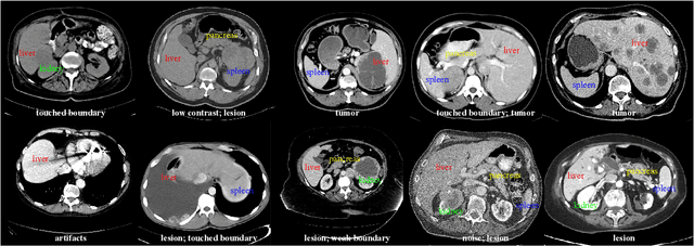
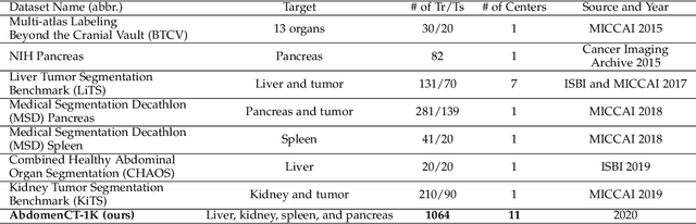
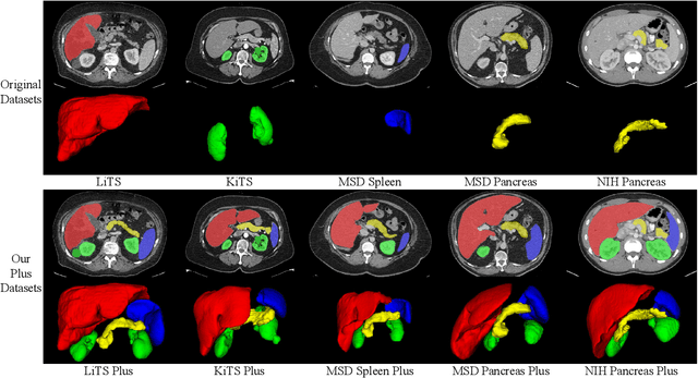
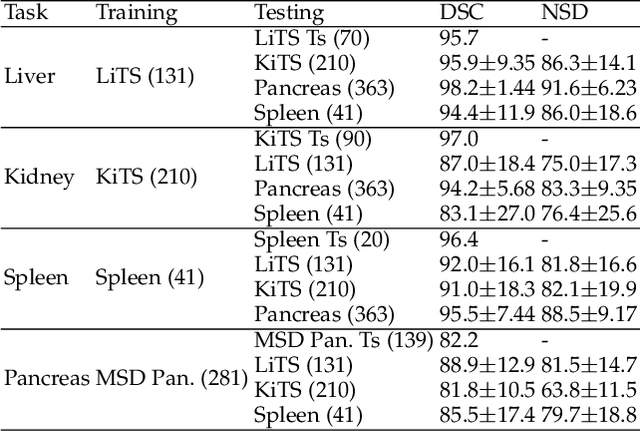
With the unprecedented developments in deep learning, automatic segmentation of main abdominal organs (i.e., liver, kidney, and spleen) seems to be a solved problem as the state-of-the-art (SOTA) methods have achieved comparable results with inter-observer variability on existing benchmark datasets. However, most of the existing abdominal organ segmentation benchmark datasets only contain single-center, single-phase, single-vendor, or single-disease cases, thus, it is unclear whether the excellent performance can generalize on more diverse datasets. In this paper, we present a large and diverse abdominal CT organ segmentation dataset, termed as AbdomenCT-1K, with more than 1000 (1K) CT scans from 11 countries, including multi-center, multi-phase, multi-vendor, and multi-disease cases. Furthermore, we conduct a large-scale study for liver, kidney, spleen, and pancreas segmentation, as well as reveal the unsolved segmentation problems of the SOTA method, such as the limited generalization ability on distinct medical centers, phases, and unseen diseases. To advance the unsolved problems, we build four organ segmentation benchmarks for fully supervised, semi-supervised, weakly supervised, and continual learning, which are currently challenging and active research topics. Accordingly, we develop a simple and effective method for each benchmark, which can be used as out-of-the-box methods and strong baselines. We believe the introduction of the AbdomenCT-1K dataset will promote future in-depth research towards clinical applicable abdominal organ segmentation methods. Moreover, the datasets, codes, and trained models of baseline methods will be publicly available at https://github.com/JunMa11/AbdomenCT-1K.