 Add to Chrome
 Add to Firefox
 Add to Edge
Acquire Precise and Comparable Fundus Image Quality Score: FTHNet and FQS Dataset
Nov 19, 2024The retinal fundus images are utilized extensively in the diagnosis, and their quality can directly affect the diagnosis results. However, due to the insufficient dataset and algorithm application, current fundus image quality assessment (FIQA) methods are not powerful enough to meet ophthalmologists` demands. In this paper, we address the limitations of datasets and algorithms in FIQA. First, we establish a new FIQA dataset, Fundus Quality Score(FQS), which includes 2246 fundus images with two labels: a continuous Mean Opinion Score varying from 0 to 100 and a three-level quality label. Then, we propose a FIQA Transformer-based Hypernetwork (FTHNet) to solve these tasks with regression results rather than classification results in conventional FIQA works. The FTHNet is optimized for the FIQA tasks with extensive experiments. Results on our FQS dataset show that the FTHNet can give quality scores for fundus images with PLCC of 0.9423 and SRCC of 0.9488, significantly outperforming other methods with fewer parameters and less computation complexity.We successfully build a dataset and model addressing the problems of current FIQA methods. Furthermore, the model deployment experiments demonstrate its potential in automatic medical image quality control. All experiments are carried out with 10-fold cross-validation to ensure the significance of the results.
UWAFA-GAN: Ultra-Wide-Angle Fluorescein Angiography Transformation via Multi-scale Generation and Registration Enhancement
May 01, 2024


Fundus photography, in combination with the ultra-wide-angle fundus (UWF) techniques, becomes an indispensable diagnostic tool in clinical settings by offering a more comprehensive view of the retina. Nonetheless, UWF fluorescein angiography (UWF-FA) necessitates the administration of a fluorescent dye via injection into the patient's hand or elbow unlike UWF scanning laser ophthalmoscopy (UWF-SLO). To mitigate potential adverse effects associated with injections, researchers have proposed the development of cross-modality medical image generation algorithms capable of converting UWF-SLO images into their UWF-FA counterparts. Current image generation techniques applied to fundus photography encounter difficulties in producing high-resolution retinal images, particularly in capturing minute vascular lesions. To address these issues, we introduce a novel conditional generative adversarial network (UWAFA-GAN) to synthesize UWF-FA from UWF-SLO. This approach employs multi-scale generators and an attention transmit module to efficiently extract both global structures and local lesions. Additionally, to counteract the image blurriness issue that arises from training with misaligned data, a registration module is integrated within this framework. Our method performs non-trivially on inception scores and details generation. Clinical user studies further indicate that the UWF-FA images generated by UWAFA-GAN are clinically comparable to authentic images in terms of diagnostic reliability. Empirical evaluations on our proprietary UWF image datasets elucidate that UWAFA-GAN outperforms extant methodologies. The code is accessible at https://github.com/Tinysqua/UWAFA-GAN.
UWAT-GAN: Fundus Fluorescein Angiography Synthesis via Ultra-wide-angle Transformation Multi-scale GAN
Jul 21, 2023


Fundus photography is an essential examination for clinical and differential diagnosis of fundus diseases. Recently, Ultra-Wide-angle Fundus (UWF) techniques, UWF Fluorescein Angiography (UWF-FA) and UWF Scanning Laser Ophthalmoscopy (UWF-SLO) have been gradually put into use. However, Fluorescein Angiography (FA) and UWF-FA require injecting sodium fluorescein which may have detrimental influences. To avoid negative impacts, cross-modality medical image generation algorithms have been proposed. Nevertheless, current methods in fundus imaging could not produce high-resolution images and are unable to capture tiny vascular lesion areas. This paper proposes a novel conditional generative adversarial network (UWAT-GAN) to synthesize UWF-FA from UWF-SLO. Using multi-scale generators and a fusion module patch to better extract global and local information, our model can generate high-resolution images. Moreover, an attention transmit module is proposed to help the decoder learn effectively. Besides, a supervised approach is used to train the network using multiple new weighted losses on different scales of data. Experiments on an in-house UWF image dataset demonstrate the superiority of the UWAT-GAN over the state-of-the-art methods. The source code is available at: https://github.com/Tinysqua/UWAT-GAN.
RFormer: Transformer-based Generative Adversarial Network for Real Fundus Image Restoration on A New Clinical Benchmark
Jan 03, 2022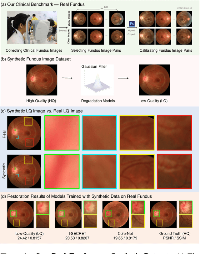
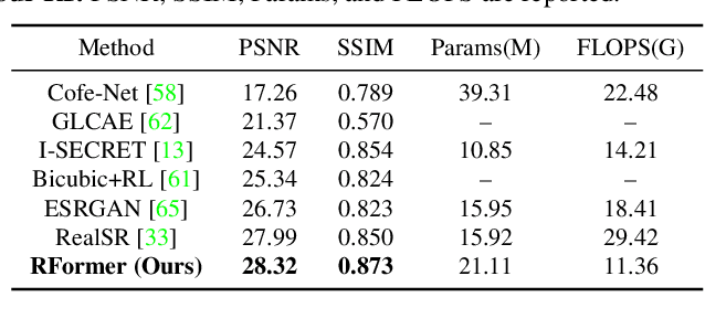
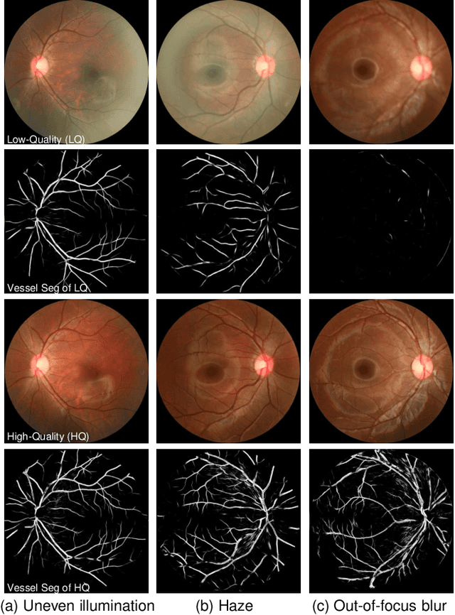
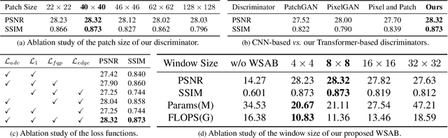
Ophthalmologists have used fundus images to screen and diagnose eye diseases. However, different equipments and ophthalmologists pose large variations to the quality of fundus images. Low-quality (LQ) degraded fundus images easily lead to uncertainty in clinical screening and generally increase the risk of misdiagnosis. Thus, real fundus image restoration is worth studying. Unfortunately, real clinical benchmark has not been explored for this task so far. In this paper, we investigate the real clinical fundus image restoration problem. Firstly, We establish a clinical dataset, Real Fundus (RF), including 120 low- and high-quality (HQ) image pairs. Then we propose a novel Transformer-based Generative Adversarial Network (RFormer) to restore the real degradation of clinical fundus images. The key component in our network is the Window-based Self-Attention Block (WSAB) which captures non-local self-similarity and long-range dependencies. To produce more visually pleasant results, a Transformer-based discriminator is introduced. Extensive experiments on our clinical benchmark show that the proposed RFormer significantly outperforms the state-of-the-art (SOTA) methods. In addition, experiments of downstream tasks such as vessel segmentation and optic disc/cup detection demonstrate that our proposed RFormer benefits clinical fundus image analysis and applications. The dataset, code, and models will be released.