 Add to Chrome
 Add to Firefox
 Add to Edge
Motion Artifact-Aware Self-Supervised Representation Learning for 3D Brain MRI Motion Artifact Reduction
Aug 10, 2026
Patient motion remains a source of image degradation in brain MRI, leading to signal loss, blurring, and geometric distortion that compromise quantitative analysis. Existing deep learning methods for motion correction typically rely on paired clean-corrupted data or k-space acquisitions, which are rarely available in clinical settings. We propose SSRL-MAR, a motion artifact-aware unpaired representation learning framework for motion artifact reduction that requires neither paired training data nor explicit motion labels. SSRL-MAR employed a three-stage training strategy: (1) contrastive learning on 3D patches to extract motion representations by contrasting clean and synthetically corrupted images, (2) a motion artifact-aware synthesis network to generate motion artifacts from clean scans, and (3) a motion artifact-aware generator to restore clean volumes using the learned degrader for self-supervised supervision. On in-silico dataset, SSRL-MAR achieved PSNR 23.81dB, SSIM 91.55%, and NMSE 0.79%. On in-vivo MR-ART dataset, the pretrained model reduced motion distortion, and unsupervised domain adaptation further improved anatomical fidelity. Against a source-only supervised model trained on the same simulated pairs, SSRL-MAR improved PSNR by up to 2.0 dB on MR-ART after unsupervised domain adaptation, and remained within 0.25-0.47 dB of an oracle supervised model that requires real paired data unavailable in practice. At the milder motion level, volumetric error in structures such as the corpus callosum and ventricular system decreased by more than 50%, confirming improved neuroanatomical consistency. These results indicate that SSRL-MAR provides a robust and scalable image-domain solution for 3D brain MRI motion correction, enabling reliable structural quantification in large-scale neuroimaging studies without requiring prospectively acquired pairs or acquisition-specific calibration.

MRI super-resolution in ten sampling steps using a diffusion bridge model
Aug 09, 2026Objective. MRI provides excellent soft-tissue contrast, but long acquisition times can cause patient discomfort and lead to motion artifacts, forcing a trade-off between spatial resolution and scan time. Diffusion-based super-resolution (SR) reconstructs high-resolution (HR) images from low-resolution (LR) inputs, but typically needs many sampling steps and initializes from a Gaussian prior ill-suited to image restoration. We developed an efficient diffusion framework that reconstructs HR MRI directly from LR data. Approach. We propose super-resolution diffusion bridge model (SR-DBM), a super-resolution diffusion bridge model that casts SR as a stochastic transport between the LR and HR image distributions. Through a Doob's h-transform of a mean-reverting stochastic differential equation, SR-DBM pins the process to the paired HR and LR images at its endpoints, initializing reconstruction from the measured anatomy rather than from Gaussian noise. The HR image is recovered by a deterministic reverse trajectory in which a network predicts the clean image at each of only ten sampling steps. We evaluated SR-DBM on ultra-high-field 7T brain T1 MP2RAGE maps and pelvic T2-weighted prostate images against nine comparison methods using PSNR, SSIM, GMSD, and LPIPS. Main results. SR-DBM attained the highest PSNR and SSIM and the lowest GMSD on both datasets (brain: 27.66+-1.52 dB, 0.96+-0.02, 7.96+-1.86$; prostate: 27.87+-2.29 dB, 0.80+-0.05, 8.38+- 1.44), with statistically significant gains over every comparison method (two-sided Wilcoxon signed-rank test with Holm correction, p<0.05). The strongest baseline, SR-EMamba, ranked second. Qualitatively, SR-DBM produced the smallest residual errors and best preserved fine structures and lesions.
Universal CT Representations from Anatomy to Disease Phenotype through Agglomerative Pretraining
May 21, 2026Computed tomography (CT) is a central to three-dimensional medical imaging, yet CT-based artificial intelligence remains fragmented across task-specific models for segmentation, classification, registration, and report analysis. Here we present FlexiCT, a family of CT foundation models trained by agglomerative continual pretraining on 266,227 CT volumes from 56 publicly available datasets, forming a large-scale public resource for CT representation learning. FlexiCT uses agglomerative pretraining across three stages: two-dimensional axial pretraining, three-dimensional anatomical pretraining and report-guided semantic alignment. This training strategy supports slice-level, volume-level and vision-language analysis. Across five downstream task families (segmentation, classification, registration, vision-language understanding and clinical retrieval), FlexiCT matches or exceeds prior task-specific approaches on multiple benchmarks. Its embeddings further organize CT scans along gradients associated with various tumor stages, suggesting that CT foundation models can capture imaging features relevant to disease phenotype characterization. Code is available at https://github.com/ricklisz/FlexiCT
MedLVR: Latent Visual Reasoning for Reliable Medical Visual Question Answering
Apr 10, 2026Medical vision--language models (VLMs) have shown strong potential for medical visual question answering (VQA), yet their reasoning remains largely text-centric: images are encoded once as static context, and subsequent inference is dominated by language. This paradigm is fundamentally limited in clinical scenarios, where accurate answers often depend on subtle, localized visual evidence that cannot be reliably preserved in static embeddings. We propose \textsc{MedLVR}, a latent visual reasoning framework that introduces an explicit visual evidence state into autoregressive decoding. Instead of relying solely on text-based intermediate reasoning, \textsc{MedLVR} interleaves a short latent reasoning segment within the decoder by reusing hidden states as continuous latent steps, enabling iterative preservation and refinement of query-relevant visual evidence before answer generation. To support effective visual supervision, we adopt a two-stage training strategy: region of interest (ROI)-supervised fine-tuning aligns latent states with clinically relevant image evidence, and Visual-Latent Policy Optimization (VLPO) further optimizes latent reasoning and answer generation under outcome-level rewards. Experiments on OmniMedVQA and five external medical VQA benchmarks show that \textsc{MedLVR} consistently outperforms recent reasoning baselines and improves the average score over the Qwen2.5-VL-7B backbone from 48.3\% to 53.4\%. These results show that latent visual reasoning provides an effective mechanism for preserving diagnostically relevant visual evidence and improving the reliability of medical VQA.
Evaluating GPT-5 as a Multimodal Clinical Reasoner: A Landscape Commentary
Mar 05, 2026The transition from task-specific artificial intelligence toward general-purpose foundation models raises fundamental questions about their capacity to support the integrated reasoning required in clinical medicine, where diagnosis demands synthesis of ambiguous patient narratives, laboratory data, and multimodal imaging. This landscape commentary provides the first controlled, cross-sectional evaluation of the GPT-5 family (GPT-5, GPT-5 Mini, GPT-5 Nano) against its predecessor GPT-4o across a diverse spectrum of clinically grounded tasks, including medical education examinations, text-based reasoning benchmarks, and visual question-answering in neuroradiology, digital pathology, and mammography using a standardized zero-shot chain-of-thought protocol. GPT-5 demonstrated substantial gains in expert-level textual reasoning, with absolute improvements exceeding 25 percentage-points on MedXpertQA. When tasked with multimodal synthesis, GPT-5 effectively leveraged this enhanced reasoning capacity to ground uncertain clinical narratives in concrete imaging evidence, achieving state-of-the-art or competitive performance across most VQA benchmarks and outperforming GPT-4o by margins of 10-40% in mammography tasks requiring fine-grained lesion characterization. However, performance remained moderate in neuroradiology (44% macro-average accuracy) and lagged behind domain-specific models in mammography, where specialized systems exceed 80% accuracy compared to GPT-5's 52-64%. These findings indicate that while GPT-5 represents a meaningful advance toward integrated multimodal clinical reasoning, mirroring the clinician's cognitive process of biasing uncertain information with objective findings, generalist models are not yet substitutes for purpose-built systems in highly specialized, perception-critical tasks.
Efficient Vision Mamba for MRI Super-Resolution via Hybrid Selective Scanning
Dec 22, 2025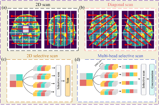
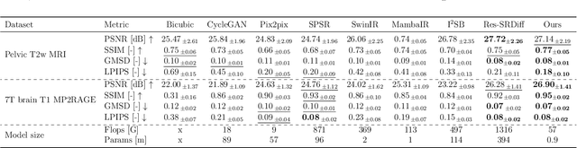
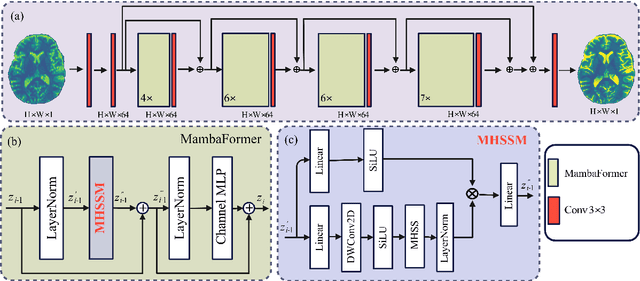
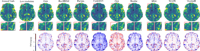
Background: High-resolution MRI is critical for diagnosis, but long acquisition times limit clinical use. Super-resolution (SR) can enhance resolution post-scan, yet existing deep learning methods face fidelity-efficiency trade-offs. Purpose: To develop a computationally efficient and accurate deep learning framework for MRI SR that preserves anatomical detail for clinical integration. Materials and Methods: We propose a novel SR framework combining multi-head selective state-space models (MHSSM) with a lightweight channel MLP. The model uses 2D patch extraction with hybrid scanning to capture long-range dependencies. Each MambaFormer block integrates MHSSM, depthwise convolutions, and gated channel mixing. Evaluation used 7T brain T1 MP2RAGE maps (n=142) and 1.5T prostate T2w MRI (n=334). Comparisons included Bicubic interpolation, GANs (CycleGAN, Pix2pix, SPSR), transformers (SwinIR), Mamba (MambaIR), and diffusion models (I2SB, Res-SRDiff). Results: Our model achieved superior performance with exceptional efficiency. For 7T brain data: SSIM=0.951+-0.021, PSNR=26.90+-1.41 dB, LPIPS=0.076+-0.022, GMSD=0.083+-0.017, significantly outperforming all baselines (p<0.001). For prostate data: SSIM=0.770+-0.049, PSNR=27.15+-2.19 dB, LPIPS=0.190+-0.095, GMSD=0.087+-0.013. The framework used only 0.9M parameters and 57 GFLOPs, reducing parameters by 99.8% and computation by 97.5% versus Res-SRDiff, while outperforming SwinIR and MambaIR in accuracy and efficiency. Conclusion: The proposed framework provides an efficient, accurate MRI SR solution, delivering enhanced anatomical detail across datasets. Its low computational demand and state-of-the-art performance show strong potential for clinical translation.
DINOv3 with Test-Time Training for Medical Image Registration
Aug 20, 2025Prior medical image registration approaches, particularly learning-based methods, often require large amounts of training data, which constrains clinical adoption. To overcome this limitation, we propose a training-free pipeline that relies on a frozen DINOv3 encoder and test-time optimization of the deformation field in feature space. Across two representative benchmarks, the method is accurate and yields regular deformations. On Abdomen MR-CT, it attained the best mean Dice score (DSC) of 0.790 together with the lowest 95th percentile Hausdorff Distance (HD95) of 4.9+-5.0 and the lowest standard deviation of Log-Jacobian (SDLogJ) of 0.08+-0.02. On ACDC cardiac MRI, it improves mean DSC to 0.769 and reduces SDLogJ to 0.11 and HD95 to 4.8, a marked gain over the initial alignment. The results indicate that operating in a compact foundation feature space at test time offers a practical and general solution for clinical registration without additional training.
Capabilities of GPT-5 on Multimodal Medical Reasoning
Aug 13, 2025Recent advances in large language models (LLMs) have enabled general-purpose systems to perform increasingly complex domain-specific reasoning without extensive fine-tuning. In the medical domain, decision-making often requires integrating heterogeneous information sources, including patient narratives, structured data, and medical images. This study positions GPT-5 as a generalist multimodal reasoner for medical decision support and systematically evaluates its zero-shot chain-of-thought reasoning performance on both text-based question answering and visual question answering tasks under a unified protocol. We benchmark GPT-5, GPT-5-mini, GPT-5-nano, and GPT-4o-2024-11-20 against standardized splits of MedQA, MedXpertQA (text and multimodal), MMLU medical subsets, USMLE self-assessment exams, and VQA-RAD. Results show that GPT-5 consistently outperforms all baselines, achieving state-of-the-art accuracy across all QA benchmarks and delivering substantial gains in multimodal reasoning. On MedXpertQA MM, GPT-5 improves reasoning and understanding scores by +29.26% and +26.18% over GPT-4o, respectively, and surpasses pre-licensed human experts by +24.23% in reasoning and +29.40% in understanding. In contrast, GPT-4o remains below human expert performance in most dimensions. A representative case study demonstrates GPT-5's ability to integrate visual and textual cues into a coherent diagnostic reasoning chain, recommending appropriate high-stakes interventions. Our results show that, on these controlled multimodal reasoning benchmarks, GPT-5 moves from human-comparable to above human-expert performance. This improvement may substantially inform the design of future clinical decision-support systems.
MRI motion correction via efficient residual-guided denoising diffusion probabilistic models
May 06, 2025Purpose: Motion artifacts in magnetic resonance imaging (MRI) significantly degrade image quality and impair quantitative analysis. Conventional mitigation strategies, such as repeated acquisitions or motion tracking, are costly and workflow-intensive. This study introduces Res-MoCoDiff, an efficient denoising diffusion probabilistic model tailored for MRI motion artifact correction. Methods: Res-MoCoDiff incorporates a novel residual error shifting mechanism in the forward diffusion process, aligning the noise distribution with motion-corrupted data and enabling an efficient four-step reverse diffusion. A U-net backbone enhanced with Swin-Transformer blocks conventional attention layers, improving adaptability across resolutions. Training employs a combined l1+l2 loss, which promotes image sharpness and reduces pixel-level errors. Res-MoCoDiff was evaluated on synthetic dataset generated using a realistic motion simulation framework and on an in-vivo dataset. Comparative analyses were conducted against established methods, including CycleGAN, Pix2pix, and MT-DDPM using quantitative metrics such as peak signal-to-noise ratio (PSNR), structural similarity index measure (SSIM), and normalized mean squared error (NMSE). Results: The proposed method demonstrated superior performance in removing motion artifacts across all motion severity levels. Res-MoCoDiff consistently achieved the highest SSIM and the lowest NMSE values, with a PSNR of up to 41.91+-2.94 dB for minor distortions. Notably, the average sampling time was reduced to 0.37 seconds per batch of two image slices, compared with 101.74 seconds for conventional approaches.
MRI super-resolution reconstruction using efficient diffusion probabilistic model with residual shifting
Mar 03, 2025Objective:This study introduces a residual error-shifting mechanism that drastically reduces sampling steps while preserving critical anatomical details, thus accelerating MRI reconstruction. Approach:We propose a novel diffusion-based SR framework called Res-SRDiff, which integrates residual error shifting into the forward diffusion process. This enables efficient HR image reconstruction by aligning the degraded HR and LR distributions.We evaluated Res-SRDiff on ultra-high-field brain T1 MP2RAGE maps and T2-weighted prostate images, comparing it with Bicubic, Pix2pix, CycleGAN, and a conventional denoising diffusion probabilistic model with vision transformer backbone (TM-DDPM), using quantitative metrics such as peak signal-to-noise ratio (PSNR), structural similarity index (SSIM), gradient magnitude similarity deviation (GMSD), and learned perceptual image patch similarity (LPIPS). Main results: Res-SRDiff significantly outperformed all comparative methods in terms of PSNR, SSIM, and GMSD across both datasets, with statistically significant improvements (p-values<<0.05). The model achieved high-fidelity image restoration with only four sampling steps, drastically reducing computational time to under one second per slice, which is substantially faster than conventional TM-DDPM with around 20 seconds per slice. Qualitative analyses further demonstrated that Res-SRDiff effectively preserved fine anatomical details and lesion morphology in both brain and pelvic MRI images. Significance: Our findings show that Res-SRDiff is an efficient and accurate MRI SR method, markedly improving computational efficiency and image quality. Integrating residual error shifting into the diffusion process allows for rapid and robust HR image reconstruction, enhancing clinical MRI workflows and advancing medical imaging research. The source at:https://github.com/mosaf/Res-SRDiff