 Add to Chrome
 Add to Firefox
 Add to Edge
Motion Artifact-Aware Self-Supervised Representation Learning for 3D Brain MRI Motion Artifact Reduction
Aug 10, 2026
Patient motion remains a source of image degradation in brain MRI, leading to signal loss, blurring, and geometric distortion that compromise quantitative analysis. Existing deep learning methods for motion correction typically rely on paired clean-corrupted data or k-space acquisitions, which are rarely available in clinical settings. We propose SSRL-MAR, a motion artifact-aware unpaired representation learning framework for motion artifact reduction that requires neither paired training data nor explicit motion labels. SSRL-MAR employed a three-stage training strategy: (1) contrastive learning on 3D patches to extract motion representations by contrasting clean and synthetically corrupted images, (2) a motion artifact-aware synthesis network to generate motion artifacts from clean scans, and (3) a motion artifact-aware generator to restore clean volumes using the learned degrader for self-supervised supervision. On in-silico dataset, SSRL-MAR achieved PSNR 23.81dB, SSIM 91.55%, and NMSE 0.79%. On in-vivo MR-ART dataset, the pretrained model reduced motion distortion, and unsupervised domain adaptation further improved anatomical fidelity. Against a source-only supervised model trained on the same simulated pairs, SSRL-MAR improved PSNR by up to 2.0 dB on MR-ART after unsupervised domain adaptation, and remained within 0.25-0.47 dB of an oracle supervised model that requires real paired data unavailable in practice. At the milder motion level, volumetric error in structures such as the corpus callosum and ventricular system decreased by more than 50%, confirming improved neuroanatomical consistency. These results indicate that SSRL-MAR provides a robust and scalable image-domain solution for 3D brain MRI motion correction, enabling reliable structural quantification in large-scale neuroimaging studies without requiring prospectively acquired pairs or acquisition-specific calibration.

MRI super-resolution in ten sampling steps using a diffusion bridge model
Aug 09, 2026Objective. MRI provides excellent soft-tissue contrast, but long acquisition times can cause patient discomfort and lead to motion artifacts, forcing a trade-off between spatial resolution and scan time. Diffusion-based super-resolution (SR) reconstructs high-resolution (HR) images from low-resolution (LR) inputs, but typically needs many sampling steps and initializes from a Gaussian prior ill-suited to image restoration. We developed an efficient diffusion framework that reconstructs HR MRI directly from LR data. Approach. We propose super-resolution diffusion bridge model (SR-DBM), a super-resolution diffusion bridge model that casts SR as a stochastic transport between the LR and HR image distributions. Through a Doob's h-transform of a mean-reverting stochastic differential equation, SR-DBM pins the process to the paired HR and LR images at its endpoints, initializing reconstruction from the measured anatomy rather than from Gaussian noise. The HR image is recovered by a deterministic reverse trajectory in which a network predicts the clean image at each of only ten sampling steps. We evaluated SR-DBM on ultra-high-field 7T brain T1 MP2RAGE maps and pelvic T2-weighted prostate images against nine comparison methods using PSNR, SSIM, GMSD, and LPIPS. Main results. SR-DBM attained the highest PSNR and SSIM and the lowest GMSD on both datasets (brain: 27.66+-1.52 dB, 0.96+-0.02, 7.96+-1.86$; prostate: 27.87+-2.29 dB, 0.80+-0.05, 8.38+- 1.44), with statistically significant gains over every comparison method (two-sided Wilcoxon signed-rank test with Holm correction, p<0.05). The strongest baseline, SR-EMamba, ranked second. Qualitatively, SR-DBM produced the smallest residual errors and best preserved fine structures and lesions.
Text-Guided Refinement of Multi-sequence Glioma Subregion Segmentation with a Vision-Language Foundation Model
Aug 05, 2026Background: Accurate glioma subregion delineation is important for radiotherapy planning and longitudinal monitoring, but manual contour correction is time-consuming. Models such as nnU-Net may generalize imperfectly and lack clinician-directed text correction. Purpose: We investigated adapting a three-dimensional (3D) vision-language foundation model for text-guided brain tumor segmentation refinement. Methods: We developed a lightweight VoxTell-based framework. Pretrained VoxTell generated initial masks. Oracle prompts derived from segmentation errors encoded target, action, location, imaging evidence, edit size, and preservation constraints. Frozen Qwen/VoxTell prompt embeddings were injected through trainable projections into its multiscale decoder conditioning; other weights remained frozen. Training, validation, and testing used 901, 100, and 250 BraTS-GLI cases. Cross-dataset transfer was evaluated on 100 meningioma, metastasis, pediatric tumor, and UPENN-GBM cases. Results: On the internal test set using post-contrast T1-weighted input, correct instructions improved subregion Dice similarity coefficient (DSC; enhancing tumor, edema, and necrotic/non-enhancing core) from $0.774\pm0.158$ to $0.796\pm0.137$. They outperformed blank prompts ($0.762\pm0.155$; Holm-adjusted $p<0.001$, $d_z=0.71$) and contradictory prompts ($0.770\pm0.163$; $p<0.001$, $d_z=0.48$). In cross-dataset testing, correct instructions improved DSC from $0.527\pm0.287$ to $0.550\pm0.278$ and outperformed contradictory instructions ($0.504\pm0.275$; $p<0.001$, $d_z=0.43$). Conclusion: A 3D vision-language foundation model can perform instruction-guided refinement of glioma subregion segmentations. Sensitivity to correct, blank, and contradictory prompts suggests text-dependent contour editing rather than nonspecific post-processing, supporting further evaluation as a clinician-in-the-loop tool.
Evaluating GPT-5 as a Multimodal Clinical Reasoner: A Landscape Commentary
Mar 05, 2026The transition from task-specific artificial intelligence toward general-purpose foundation models raises fundamental questions about their capacity to support the integrated reasoning required in clinical medicine, where diagnosis demands synthesis of ambiguous patient narratives, laboratory data, and multimodal imaging. This landscape commentary provides the first controlled, cross-sectional evaluation of the GPT-5 family (GPT-5, GPT-5 Mini, GPT-5 Nano) against its predecessor GPT-4o across a diverse spectrum of clinically grounded tasks, including medical education examinations, text-based reasoning benchmarks, and visual question-answering in neuroradiology, digital pathology, and mammography using a standardized zero-shot chain-of-thought protocol. GPT-5 demonstrated substantial gains in expert-level textual reasoning, with absolute improvements exceeding 25 percentage-points on MedXpertQA. When tasked with multimodal synthesis, GPT-5 effectively leveraged this enhanced reasoning capacity to ground uncertain clinical narratives in concrete imaging evidence, achieving state-of-the-art or competitive performance across most VQA benchmarks and outperforming GPT-4o by margins of 10-40% in mammography tasks requiring fine-grained lesion characterization. However, performance remained moderate in neuroradiology (44% macro-average accuracy) and lagged behind domain-specific models in mammography, where specialized systems exceed 80% accuracy compared to GPT-5's 52-64%. These findings indicate that while GPT-5 represents a meaningful advance toward integrated multimodal clinical reasoning, mirroring the clinician's cognitive process of biasing uncertain information with objective findings, generalist models are not yet substitutes for purpose-built systems in highly specialized, perception-critical tasks.
Efficient Vision Mamba for MRI Super-Resolution via Hybrid Selective Scanning
Dec 22, 2025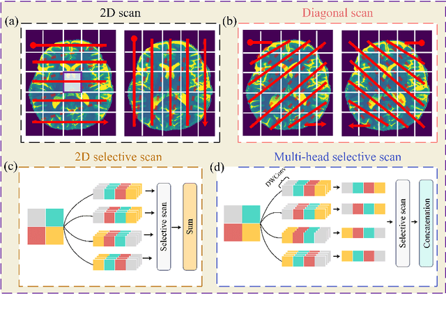
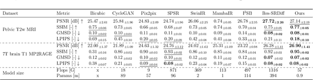
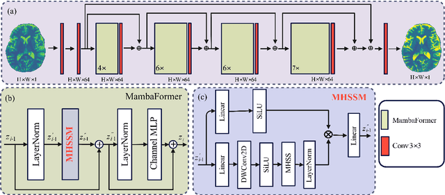
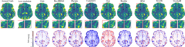
Background: High-resolution MRI is critical for diagnosis, but long acquisition times limit clinical use. Super-resolution (SR) can enhance resolution post-scan, yet existing deep learning methods face fidelity-efficiency trade-offs. Purpose: To develop a computationally efficient and accurate deep learning framework for MRI SR that preserves anatomical detail for clinical integration. Materials and Methods: We propose a novel SR framework combining multi-head selective state-space models (MHSSM) with a lightweight channel MLP. The model uses 2D patch extraction with hybrid scanning to capture long-range dependencies. Each MambaFormer block integrates MHSSM, depthwise convolutions, and gated channel mixing. Evaluation used 7T brain T1 MP2RAGE maps (n=142) and 1.5T prostate T2w MRI (n=334). Comparisons included Bicubic interpolation, GANs (CycleGAN, Pix2pix, SPSR), transformers (SwinIR), Mamba (MambaIR), and diffusion models (I2SB, Res-SRDiff). Results: Our model achieved superior performance with exceptional efficiency. For 7T brain data: SSIM=0.951+-0.021, PSNR=26.90+-1.41 dB, LPIPS=0.076+-0.022, GMSD=0.083+-0.017, significantly outperforming all baselines (p<0.001). For prostate data: SSIM=0.770+-0.049, PSNR=27.15+-2.19 dB, LPIPS=0.190+-0.095, GMSD=0.087+-0.013. The framework used only 0.9M parameters and 57 GFLOPs, reducing parameters by 99.8% and computation by 97.5% versus Res-SRDiff, while outperforming SwinIR and MambaIR in accuracy and efficiency. Conclusion: The proposed framework provides an efficient, accurate MRI SR solution, delivering enhanced anatomical detail across datasets. Its low computational demand and state-of-the-art performance show strong potential for clinical translation.
Systematic Review and Meta-analysis of AI-driven MRI Motion Artifact Detection and Correction
Sep 05, 2025Background: To systematically review and perform a meta-analysis of artificial intelligence (AI)-driven methods for detecting and correcting magnetic resonance imaging (MRI) motion artifacts, assessing current developments, effectiveness, challenges, and future research directions. Methods: A comprehensive systematic review and meta-analysis were conducted, focusing on deep learning (DL) approaches, particularly generative models, for the detection and correction of MRI motion artifacts. Quantitative data were extracted regarding utilized datasets, DL architectures, and performance metrics. Results: DL, particularly generative models, show promise for reducing motion artifacts and improving image quality; however, limited generalizability, reliance on paired training data, and risk of visual distortions remain key challenges that motivate standardized datasets and reporting. Conclusions: AI-driven methods, particularly DL generative models, show significant potential for improving MRI image quality by effectively addressing motion artifacts. However, critical challenges must be addressed, including the need for comprehensive public datasets, standardized reporting protocols for artifact levels, and more advanced, adaptable DL techniques to reduce reliance on extensive paired datasets. Addressing these aspects could substantially enhance MRI diagnostic accuracy, reduce healthcare costs, and improve patient care outcomes.
MRI motion correction via efficient residual-guided denoising diffusion probabilistic models
May 06, 2025Purpose: Motion artifacts in magnetic resonance imaging (MRI) significantly degrade image quality and impair quantitative analysis. Conventional mitigation strategies, such as repeated acquisitions or motion tracking, are costly and workflow-intensive. This study introduces Res-MoCoDiff, an efficient denoising diffusion probabilistic model tailored for MRI motion artifact correction. Methods: Res-MoCoDiff incorporates a novel residual error shifting mechanism in the forward diffusion process, aligning the noise distribution with motion-corrupted data and enabling an efficient four-step reverse diffusion. A U-net backbone enhanced with Swin-Transformer blocks conventional attention layers, improving adaptability across resolutions. Training employs a combined l1+l2 loss, which promotes image sharpness and reduces pixel-level errors. Res-MoCoDiff was evaluated on synthetic dataset generated using a realistic motion simulation framework and on an in-vivo dataset. Comparative analyses were conducted against established methods, including CycleGAN, Pix2pix, and MT-DDPM using quantitative metrics such as peak signal-to-noise ratio (PSNR), structural similarity index measure (SSIM), and normalized mean squared error (NMSE). Results: The proposed method demonstrated superior performance in removing motion artifacts across all motion severity levels. Res-MoCoDiff consistently achieved the highest SSIM and the lowest NMSE values, with a PSNR of up to 41.91+-2.94 dB for minor distortions. Notably, the average sampling time was reduced to 0.37 seconds per batch of two image slices, compared with 101.74 seconds for conventional approaches.
MRI super-resolution reconstruction using efficient diffusion probabilistic model with residual shifting
Mar 03, 2025Objective:This study introduces a residual error-shifting mechanism that drastically reduces sampling steps while preserving critical anatomical details, thus accelerating MRI reconstruction. Approach:We propose a novel diffusion-based SR framework called Res-SRDiff, which integrates residual error shifting into the forward diffusion process. This enables efficient HR image reconstruction by aligning the degraded HR and LR distributions.We evaluated Res-SRDiff on ultra-high-field brain T1 MP2RAGE maps and T2-weighted prostate images, comparing it with Bicubic, Pix2pix, CycleGAN, and a conventional denoising diffusion probabilistic model with vision transformer backbone (TM-DDPM), using quantitative metrics such as peak signal-to-noise ratio (PSNR), structural similarity index (SSIM), gradient magnitude similarity deviation (GMSD), and learned perceptual image patch similarity (LPIPS). Main results: Res-SRDiff significantly outperformed all comparative methods in terms of PSNR, SSIM, and GMSD across both datasets, with statistically significant improvements (p-values<<0.05). The model achieved high-fidelity image restoration with only four sampling steps, drastically reducing computational time to under one second per slice, which is substantially faster than conventional TM-DDPM with around 20 seconds per slice. Qualitative analyses further demonstrated that Res-SRDiff effectively preserved fine anatomical details and lesion morphology in both brain and pelvic MRI images. Significance: Our findings show that Res-SRDiff is an efficient and accurate MRI SR method, markedly improving computational efficiency and image quality. Integrating residual error shifting into the diffusion process allows for rapid and robust HR image reconstruction, enhancing clinical MRI workflows and advancing medical imaging research. The source at:https://github.com/mosaf/Res-SRDiff
A Physics-Informed Deep Learning Model for MRI Brain Motion Correction
Feb 13, 2025Background: MRI is crucial for brain imaging but is highly susceptible to motion artifacts due to long acquisition times. This study introduces PI-MoCoNet, a physics-informed motion correction network that integrates spatial and k-space information to remove motion artifacts without explicit motion parameter estimation, enhancing image fidelity and diagnostic reliability. Materials and Methods: PI-MoCoNet consists of a motion detection network (U-net with spatial averaging) to identify corrupted k-space lines and a motion correction network (U-net with Swin Transformer blocks) to reconstruct motion-free images. The correction is guided by three loss functions: reconstruction (L1), perceptual (LPIPS), and data consistency (Ldc). Motion artifacts were simulated via rigid phase encoding perturbations and evaluated on IXI and MR-ART datasets against Pix2Pix, CycleGAN, and U-net using PSNR, SSIM, and NMSE. Results: PI-MoCoNet significantly improved image quality. On IXI, for minor artifacts, PSNR increased from 34.15 dB to 45.95 dB, SSIM from 0.87 to 1.00, and NMSE reduced from 0.55% to 0.04%. For moderate artifacts, PSNR improved from 30.23 dB to 42.16 dB, SSIM from 0.80 to 0.99, and NMSE from 1.32% to 0.09%. For heavy artifacts, PSNR rose from 27.99 dB to 36.01 dB, SSIM from 0.75 to 0.97, and NMSE decreased from 2.21% to 0.36%. On MR-ART, PI-MoCoNet achieved PSNR gains of ~10 dB and SSIM improvements of up to 0.20, with NMSE reductions of ~6%. Ablation studies confirmed the importance of data consistency and perceptual losses, yielding a 1 dB PSNR gain and 0.17% NMSE reduction. Conclusions: PI-MoCoNet effectively mitigates motion artifacts in brain MRI, outperforming existing methods. Its ability to integrate spatial and k-space information makes it a promising tool for clinical use in motion-prone settings. Code: https://github.com/mosaf/PI-MoCoNet.git.
Advancing MRI Reconstruction: A Systematic Review of Deep Learning and Compressed Sensing Integration
Jan 24, 2025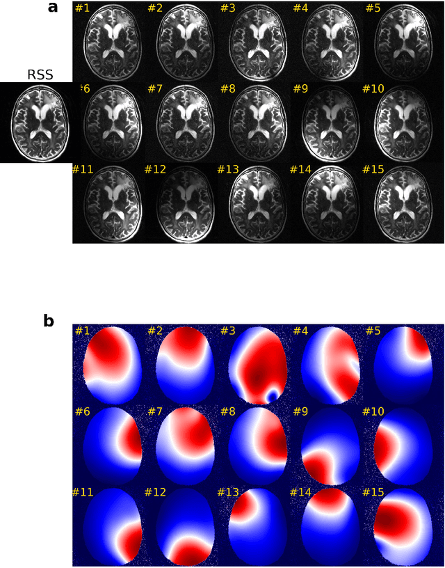
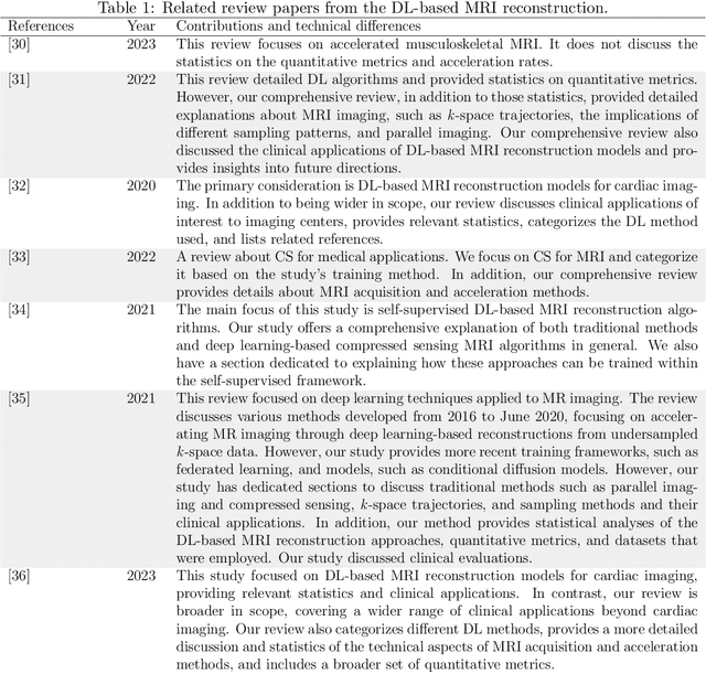
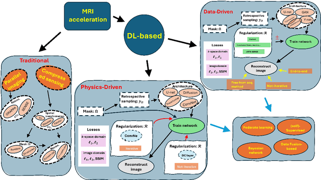
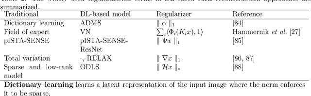
Magnetic resonance imaging (MRI) is a non-invasive imaging modality and provides comprehensive anatomical and functional insights into the human body. However, its long acquisition times can lead to patient discomfort, motion artifacts, and limiting real-time applications. To address these challenges, strategies such as parallel imaging have been applied, which utilize multiple receiver coils to speed up the data acquisition process. Additionally, compressed sensing (CS) is a method that facilitates image reconstruction from sparse data, significantly reducing image acquisition time by minimizing the amount of data collection needed. Recently, deep learning (DL) has emerged as a powerful tool for improving MRI reconstruction. It has been integrated with parallel imaging and CS principles to achieve faster and more accurate MRI reconstructions. This review comprehensively examines DL-based techniques for MRI reconstruction. We categorize and discuss various DL-based methods, including end-to-end approaches, unrolled optimization, and federated learning, highlighting their potential benefits. Our systematic review highlights significant contributions and underscores the potential of DL in MRI reconstruction. Additionally, we summarize key results and trends in DL-based MRI reconstruction, including quantitative metrics, the dataset, acceleration factors, and the progress of and research interest in DL techniques over time. Finally, we discuss potential future directions and the importance of DL-based MRI reconstruction in advancing medical imaging. To facilitate further research in this area, we provide a GitHub repository that includes up-to-date DL-based MRI reconstruction publications and public datasets-https://github.com/mosaf/Awesome-DL-based-CS-MRI.