 Add to Chrome
 Add to Firefox
 Add to Edge
Benchmarking and Explaining Deep Learning Cortical Lesion MRI Segmentation in Multiple Sclerosis
Jul 16, 2025Cortical lesions (CLs) have emerged as valuable biomarkers in multiple sclerosis (MS), offering high diagnostic specificity and prognostic relevance. However, their routine clinical integration remains limited due to subtle magnetic resonance imaging (MRI) appearance, challenges in expert annotation, and a lack of standardized automated methods. We propose a comprehensive multi-centric benchmark of CL detection and segmentation in MRI. A total of 656 MRI scans, including clinical trial and research data from four institutions, were acquired at 3T and 7T using MP2RAGE and MPRAGE sequences with expert-consensus annotations. We rely on the self-configuring nnU-Net framework, designed for medical imaging segmentation, and propose adaptations tailored to the improved CL detection. We evaluated model generalization through out-of-distribution testing, demonstrating strong lesion detection capabilities with an F1-score of 0.64 and 0.5 in and out of the domain, respectively. We also analyze internal model features and model errors for a better understanding of AI decision-making. Our study examines how data variability, lesion ambiguity, and protocol differences impact model performance, offering future recommendations to address these barriers to clinical adoption. To reinforce the reproducibility, the implementation and models will be publicly accessible and ready to use at https://github.com/Medical-Image-Analysis-Laboratory/ and https://doi.org/10.5281/zenodo.15911797.
ConfLUNet: Multiple sclerosis lesion instance segmentation in presence of confluent lesions
May 28, 2025Accurate lesion-level segmentation on MRI is critical for multiple sclerosis (MS) diagnosis, prognosis, and disease monitoring. However, current evaluation practices largely rely on semantic segmentation post-processed with connected components (CC), which cannot separate confluent lesions (aggregates of confluent lesion units, CLUs) due to reliance on spatial connectivity. To address this misalignment with clinical needs, we introduce formal definitions of CLUs and associated CLU-aware detection metrics, and include them in an exhaustive instance segmentation evaluation framework. Within this framework, we systematically evaluate CC and post-processing-based Automated Confluent Splitting (ACLS), the only existing methods for lesion instance segmentation in MS. Our analysis reveals that CC consistently underestimates CLU counts, while ACLS tends to oversplit lesions, leading to overestimated lesion counts and reduced precision. To overcome these limitations, we propose ConfLUNet, the first end-to-end instance segmentation framework for MS lesions. ConfLUNet jointly optimizes lesion detection and delineation from a single FLAIR image. Trained on 50 patients, ConfLUNet significantly outperforms CC and ACLS on the held-out test set (n=13) in instance segmentation (Panoptic Quality: 42.0% vs. 37.5%/36.8%; p = 0.017/0.005) and lesion detection (F1: 67.3% vs. 61.6%/59.9%; p = 0.028/0.013). For CLU detection, ConfLUNet achieves the highest F1[CLU] (81.5%), improving recall over CC (+12.5%, p = 0.015) and precision over ACLS (+31.2%, p = 0.003). By combining rigorous definitions, new CLU-aware metrics, a reproducible evaluation framework, and the first dedicated end-to-end model, this work lays the foundation for lesion instance segmentation in MS.
Explainability of AI Uncertainty: Application to Multiple Sclerosis Lesion Segmentation on MRI
Apr 07, 2025


Trustworthy artificial intelligence (AI) is essential in healthcare, particularly for high-stakes tasks like medical image segmentation. Explainable AI and uncertainty quantification significantly enhance AI reliability by addressing key attributes such as robustness, usability, and explainability. Despite extensive technical advances in uncertainty quantification for medical imaging, understanding the clinical informativeness and interpretability of uncertainty remains limited. This study introduces a novel framework to explain the potential sources of predictive uncertainty, specifically in cortical lesion segmentation in multiple sclerosis using deep ensembles. The proposed analysis shifts the focus from the uncertainty-error relationship towards relevant medical and engineering factors. Our findings reveal that instance-wise uncertainty is strongly related to lesion size, shape, and cortical involvement. Expert rater feedback confirms that similar factors impede annotator confidence. Evaluations conducted on two datasets (206 patients, almost 2000 lesions) under both in-domain and distribution-shift conditions highlight the utility of the framework in different scenarios.
Cortical lesions, central vein sign, and paramagnetic rim lesions in multiple sclerosis: emerging machine learning techniques and future avenues
Jan 19, 2022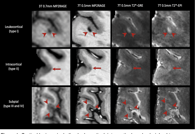
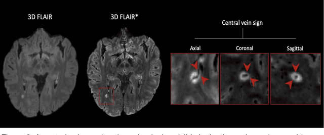
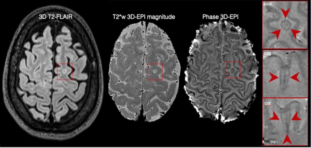
The current multiple sclerosis (MS) diagnostic criteria lack specificity, and this may lead to misdiagnosis, which remains an issue in present-day clinical practice. In addition, conventional biomarkers only moderately correlate with MS disease progression. Recently, advanced MS lesional imaging biomarkers such as cortical lesions (CL), the central vein sign (CVS), and paramagnetic rim lesions (PRL), visible in specialized magnetic resonance imaging (MRI) sequences, have shown higher specificity in differential diagnosis. Moreover, studies have shown that CL and PRL are potential prognostic biomarkers, the former correlating with cognitive impairments and the latter with early disability progression. As machine learning-based methods have achieved extraordinary performance in the assessment of conventional imaging biomarkers, such as white matter lesion segmentation, several automated or semi-automated methods have been proposed for CL, CVS, and PRL as well. In the present review, we first introduce these advanced MS imaging biomarkers and their imaging methods. Subsequently, we describe the corresponding machine learning-based methods that were used to tackle these clinical questions, putting them into context with respect to the challenges they are still facing, including non-standardized MRI protocols, limited datasets, and moderate inter-rater variability. We conclude by presenting the current limitations that prevent their broader deployment and suggesting future research directions.