 Add to Chrome
 Add to Firefox
 Add to Edge
Cells are Actors: Social Network Analysis with Classical ML for SOTA Histology Image Classification
Jul 10, 2021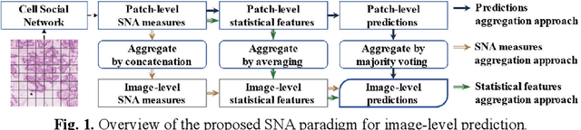
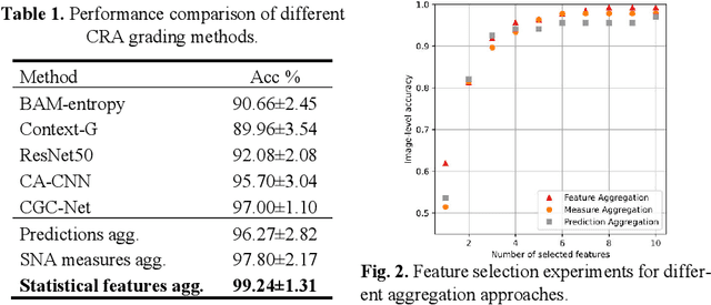
Digitization of histology images and the advent of new computational methods, like deep learning, have helped the automatic grading of colorectal adenocarcinoma cancer (CRA). Present automated CRA grading methods, however, usually use tiny image patches and thus fail to integrate the entire tissue micro-architecture for grading purposes. To tackle these challenges, we propose to use a statistical network analysis method to describe the complex structure of the tissue micro-environment by modelling nuclei and their connections as a network. We show that by analyzing only the interactions between the cells in a network, we can extract highly discriminative statistical features for CRA grading. Unlike other deep learning or convolutional graph-based approaches, our method is highly scalable (can be used for cell networks consist of millions of nodes), completely explainable, and computationally inexpensive. We create cell networks on a broad CRC histology image dataset, experiment with our method, and report state-of-the-art performance for the prediction of three-class CRA grading.
Semantic annotation for computational pathology: Multidisciplinary experience and best practice recommendations
Jun 25, 2021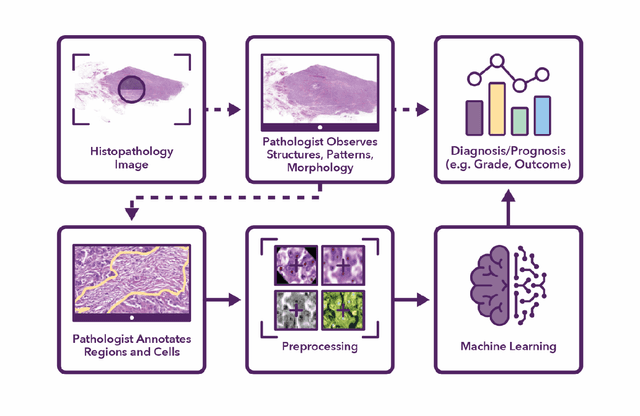
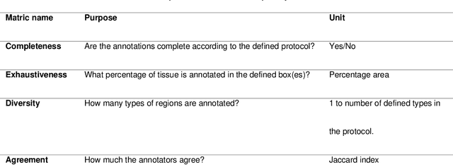
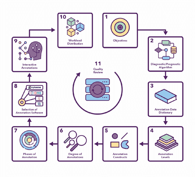
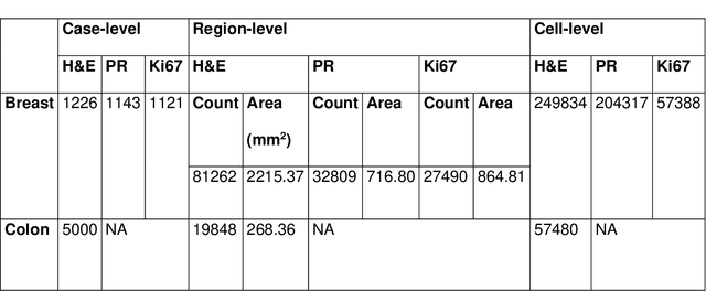
Recent advances in whole slide imaging (WSI) technology have led to the development of a myriad of computer vision and artificial intelligence (AI) based diagnostic, prognostic, and predictive algorithms. Computational Pathology (CPath) offers an integrated solution to utilize information embedded in pathology WSIs beyond what we obtain through visual assessment. For automated analysis of WSIs and validation of machine learning (ML) models, annotations at the slide, tissue and cellular levels are required. The annotation of important visual constructs in pathology images is an important component of CPath projects. Improper annotations can result in algorithms which are hard to interpret and can potentially produce inaccurate and inconsistent results. Despite the crucial role of annotations in CPath projects, there are no well-defined guidelines or best practices on how annotations should be carried out. In this paper, we address this shortcoming by presenting the experience and best practices acquired during the execution of a large-scale annotation exercise involving a multidisciplinary team of pathologists, ML experts and researchers as part of the Pathology image data Lake for Analytics, Knowledge and Education (PathLAKE) consortium. We present a real-world case study along with examples of different types of annotations, diagnostic algorithm, annotation data dictionary and annotation constructs. The analyses reported in this work highlight best practice recommendations that can be used as annotation guidelines over the lifecycle of a CPath project.
Automatic Recognition of the Supraspinatus Tendinopathy from Ultrasound Images using Convolutional Neural Networks
Nov 23, 2020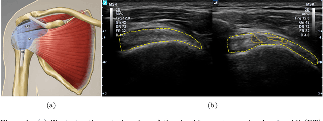
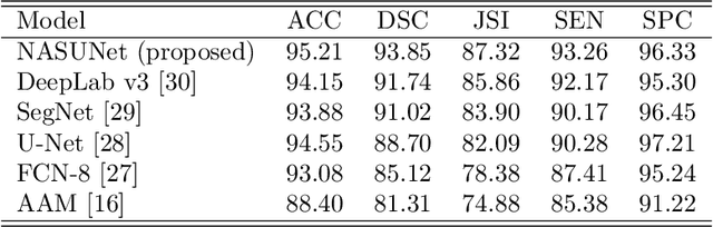
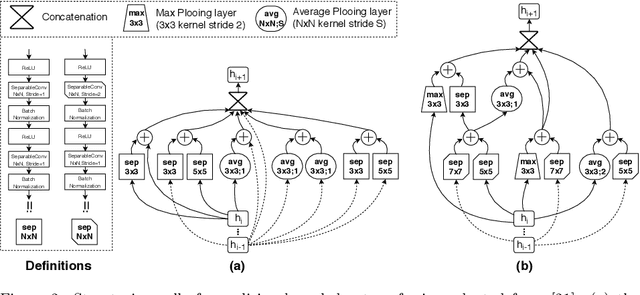
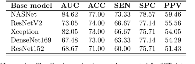
Tendon injuries like tendinopathies, full and partial thickness tears are prevalent, and the supraspinatus tendon (SST) is the most vulnerable ones in the rotator cuff. Early diagnosis of SST tendinopathies is of high importance and hard to achieve using ultrasound imaging. In this paper, an automatic tendinopathy recognition framework based on convolutional neural networks has been proposed to assist the diagnosis. This framework has two essential parts of tendon segmentation and classification. Tendon segmentation is done through a novel network, NASUNet, which follows an encoder-decoder architecture paradigm and utilizes a multi-scale Enlarging cell. Moreover, a general classification pipeline has been proposed for tendinopathy recognition, which supports different base models as the feature extractor engine. Two feature maps comprising positional information of the tendon region have been introduced as the network input to make the classification network spatial-aware. To evaluate the tendinopathy recognition system, a data set consisting of 100 SST ultrasound images have been acquired, in which tendinopathy cases are double-verified by magnetic resonance imaging. In both segmentation and classification tasks, lack of training data has been compensated by incorporating knowledge transferring, transfer learning, and data augmentation techniques. In cross-validation experiments, the proposed tendinopathy recognition model achieves 91% accuracy, 86.67% sensitivity, and 92.86% specificity, showing state-of-the-art performance against other models.
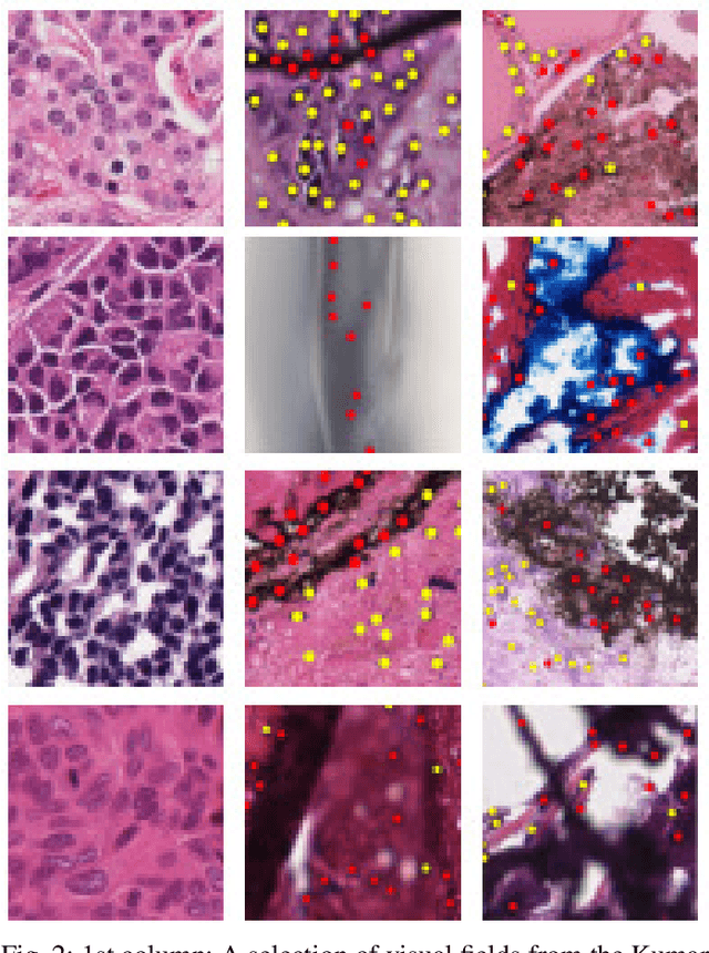
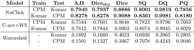
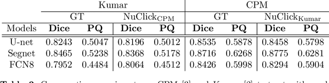
NuClick: A Deep Learning Framework for Interactive Segmentation of Microscopy Images
May 29, 2020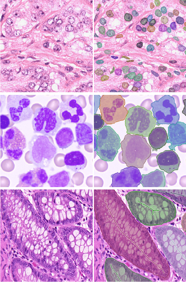
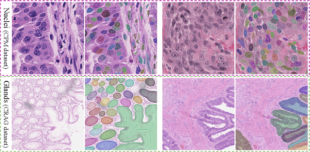
Object Segmentation is an important step in the work-flow of computational pathology. Deep learning based models as the best forming models require huge amount of labeled data for precise and reliable prediction. However, collecting labeled data is expensive, because it necessarily involves expert knowledge. This is perhaps best illustrated by medical tasks where measurements call for expensive machinery and labels are the fruit of a time-consuming analysis that draws from multiple human experts. As nuclei, cells and glands are fundamental objects for downstream analysis in histology, in this paper we propose a simple CNN-based approach to speed up collecting segmentation annotation for these objects by utilizing minimum input from an annotator. We show for nuclei and cells as small objects, one click inside objects is enough to have precise annotation. For glands as large objects, providing a squiggle to show the extend of gland can guide the model to outline the exact boundaries. This supervisory signals are fed to network as an auxiliary channels along with RGB channels. With detailed experiments, we show that our approach is generalizable, robust against variations in the user input and that it can be used to obtain annotations for completely different domains. Practically, a model trained on the masks generated by NuClick could achieve first rank in LYON19 challenge. Furthermore, as the output of our framework, we release two data-sets: 1) a dataset of lymphocyte annotations within IHC images and 2) a dataset of WBCs annotated in blood sample images.
PanNuke Dataset Extension, Insights and Baselines
Apr 22, 2020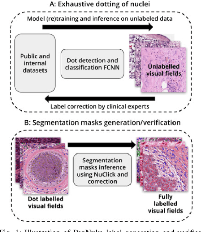
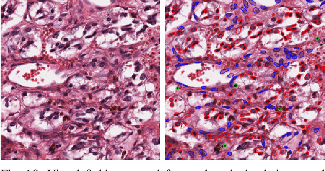
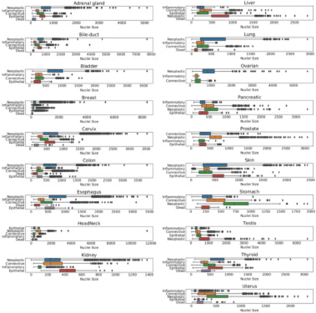
The emerging area of computational pathology (CPath) is ripe ground for the application of deep learning (DL) methods to healthcare due to the sheer volume of raw pixel data in whole-slide images (WSIs) of cancerous tissue slides. However, it is imperative for the DL algorithms relying on nuclei-level details to be able to cope with data from `the clinical wild', which tends to be quite challenging. We study, and extend recently released PanNuke dataset consisting of ~200,000 nuclei categorized into 5 clinically important classes for the challenging tasks of segmenting and classifying nuclei in WSIs. Previous pan-cancer datasets consisted of only up to 9 different tissues and up to 21,000 unlabeled nuclei and just over 24,000 labeled nuclei with segmentation masks. PanNuke consists of 19 different tissue types that have been semi-automatically annotated and quality controlled by clinical pathologists, leading to a dataset with statistics similar to the clinical wild and with minimal selection bias. We study the performance of segmentation and classification models when applied to the proposed dataset and demonstrate the application of models trained on PanNuke to whole-slide images. We provide comprehensive statistics about the dataset and outline recommendations and research directions to address the limitations of existing DL tools when applied to real-world CPath applications.
NuClick: From Clicks in the Nuclei to Nuclear Boundaries
Sep 07, 2019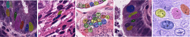
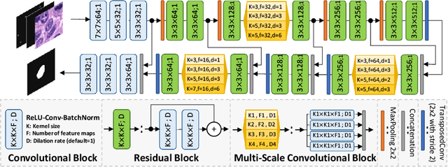
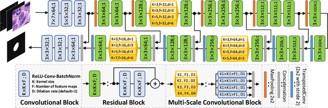
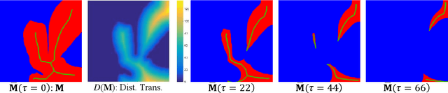
Best performing nuclear segmentation methods are based on deep learning algorithms that require a large amount of annotated data. However, collecting annotations for nuclear segmentation is a very labor-intensive and time-consuming task. Thereby, providing a tool that can facilitate and speed up this procedure is very demanding. Here we propose a simple yet efficient framework based on convolutional neural networks, named NuClick, which can precisely segment nuclei boundaries by accepting a single point position (or click) inside each nucleus. Based on the clicked positions, inclusion and exclusion maps are generated which comprise 2D Gaussian distributions centered on those positions. These maps serve as guiding signals for the network as they are concatenated to the input image. The inclusion map focuses on the desired nucleus while the exclusion map indicates neighboring nuclei and improve the results of segmentation in scenes with nuclei clutter. The NuClick not only facilitates collecting more annotation from unseen data but also leads to superior segmentation output for deep models. It is also worth mentioning that an instance segmentation model trained on NuClick generated labels was able to rank first in LYON19 challenge.
Nuclear Instance Segmentation using a Proposal-Free Spatially Aware Deep Learning Framework
Aug 27, 2019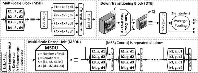
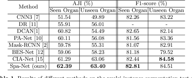
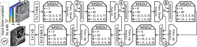
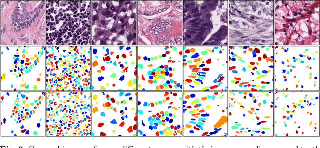
Nuclear segmentation in histology images is a challenging task due to significant variations in the shape and appearance of nuclei. One of the main hurdles in nuclear instance segmentation is overlapping nuclei where a smart algorithm is needed to separate each nucleus. In this paper, we introduce a proposal-free deep learning based framework to address these challenges. To this end, we propose a spatially-aware network (SpaNet) to capture spatial information in a multi-scale manner. A dual-head variation of the SpaNet is first utilized to predict the pixel-wise segmentation and centroid detection maps of nuclei. Based on these outputs, a single-head SpaNet predicts the positional information related to each nucleus instance. Spectral clustering method is applied on the output of the last SpaNet, which utilizes the nuclear mask and the Gaussian-like detection map for determining the connected components and associated cluster identifiers, respectively. The output of the clustering method is the final nuclear instance segmentation mask. We applied our method on a publicly available multi-organ data set and achieved state-of-the-art performance for nuclear segmentation.
Leveraging Transfer Learning for Segmenting Lesions and their Attributes in Dermoscopy Images
Sep 23, 2018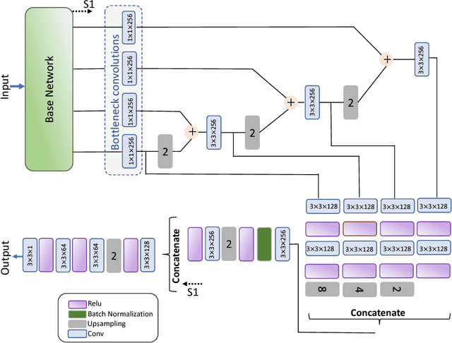
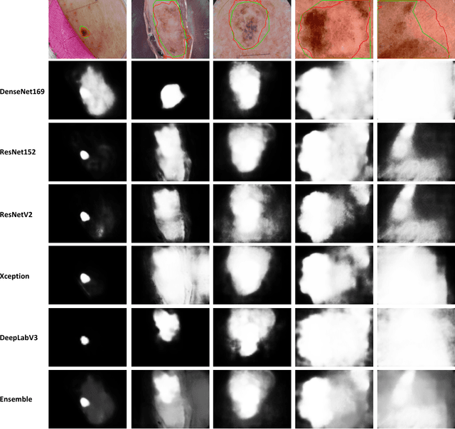
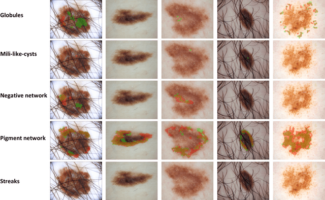
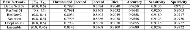
Computer-aided diagnosis systems for classification of different type of skin lesions have been an active field of research in recent decades. It has been shown that introducing lesions and their attributes masks into lesion classification pipeline can greatly improve the performance. In this paper, we propose a framework by incorporating transfer learning for segmenting lesions and their attributes based on the convolutional neural networks. The proposed framework is inspired by the well-known UNet architecture. It utilizes a variety of pre-trained networks in the encoding path and generates the prediction map by combining multi-scale information in decoding path using a pyramid pooling manner. To circumvent the lack of training data and increase the proposed model generalization, an extensive set of novel augmentation routines have been applied during the training of the network. Moreover, for each task of lesion and attribute segmentation, a specific loss function has been designed to obviate the training phase difficulties. Finally, the prediction for each task is generated by ensembling the outputs from different models. The proposed approach achieves promising results on the cross-validation experiments on the ISIC2018- Task1 and Task2 data sets.
Nuclei Detection Using Mixture Density Networks
Aug 22, 2018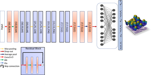
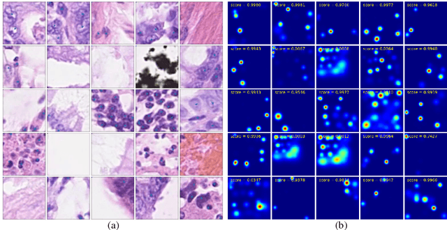
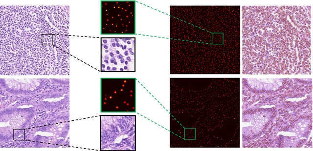
Nuclei detection is an important task in the histology domain as it is a main step toward further analysis such as cell counting, cell segmentation, study of cell connections, etc. This is a challenging task due to the complex texture of histology image, variation in shape, and touching cells. To tackle these hurdles, many approaches have been proposed in the literature where deep learning methods stand on top in terms of performance. Hence, in this paper, we propose a novel framework for nuclei detection based on Mixture Density Networks (MDNs). These networks are suitable to map a single input to several possible outputs and we utilize this property to detect multiple seeds in a single image patch. A new modified form of a cost function is proposed for training and handling patches with missing nuclei. The probability maps of the nuclei in the individual patches are next combined to generate the final image-wide result. The experimental results show the state-of-the-art performance on complex colorectal adenocarcinoma dataset.
Supervised Saliency Map Driven Segmentation of the Lesions in Dermoscopic Images
Jun 07, 2018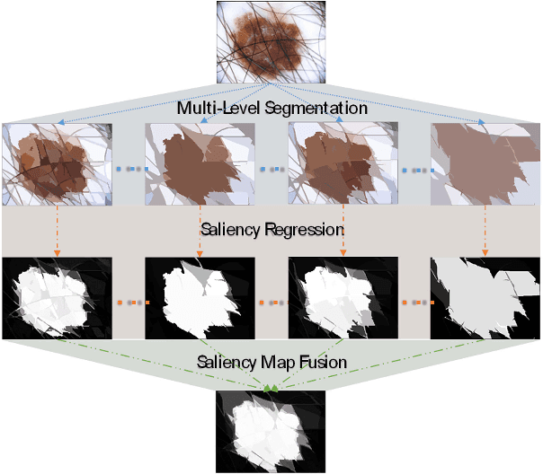
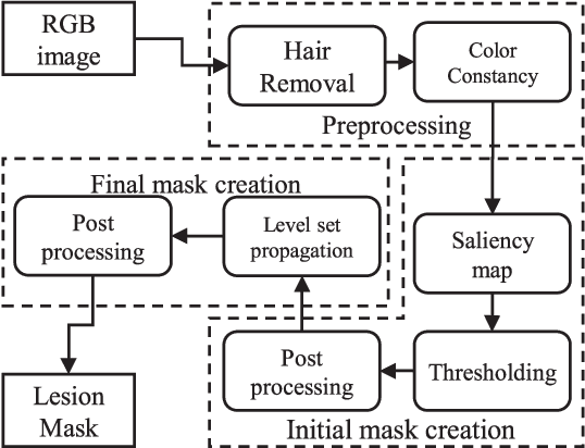
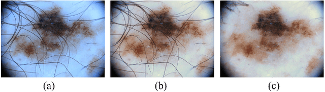
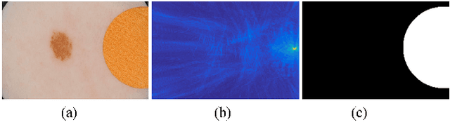
Lesion segmentation is the first step in most automatic melanoma recognition systems. Deficiencies and difficulties in dermoscopic images such as color inconstancy, hair occlusion, dark corners and color charts make lesion segmentation an intricate task. In order to detect the lesion in the presence of these problems, we propose a supervised saliency detection method tailored for dermoscopic images based on the discriminative regional feature integration (DRFI). DRFI method incorporates multi-level segmentation, regional contrast, property, background descriptors, and a random forest regressor to create saliency scores for each region in the image. In our improved saliency detection method, mDRFI, we have added some new features to regional property descriptors. Also, in order to achieve more robust regional background descriptors, a thresholding algorithm is proposed to obtain a new pseudo-background region. Findings reveal that mDRFI is superior to DRFI in detecting the lesion as the salient object in dermoscopic images. The proposed overall lesion segmentation framework uses detected saliency map to construct an initial mask of the lesion through thresholding and post-processing operations. The initial mask is then evolving in a level set framework to fit better on the lesion's boundaries. The results of evaluation tests on three public datasets show that our proposed segmentation method outperforms the other conventional state-of-the-art segmentation algorithms and its performance is comparable with most recent approaches that are based on deep convolutional neural networks.