 Add to Chrome
 Add to Firefox
 Add to Edge
Generative AI and Foundation Models in Medical Image
Aug 03, 2026
In recent years, generative AI has attracted significant public attention, and its use has been rapidly expanding across a wide range of domains. From creative tasks such as text summarization, idea generation, and source code generation, to the streamlining of medical support tasks like diagnostic report generation and summarization, AI is now deeply involved in many areas. Today's breadth of AI applications is clearly distinct from what was seen before generative AI gained widespread recognition. Representative generative AI services include DALL-E 3 (OpenAI, California, USA) and Stable Diffusion (Stability AI, London, England, UK) for image generation, ChatGPT (OpenAI, California, USA), and Gemini (Google, California, USA) for text generation. The rise of generative AI has been influenced by advances in deep learning models and the scaling up of data, models, and computational resources based on the scaling laws. Moreover, the emergence of foundation models, which are trained on large-scale datasets and possess general-purpose knowledge applicable to various downstream tasks, is creating a new paradigm in AI development. These shifts brought about by generative AI and foundation models also profoundly impact medical image processing, fundamentally changing the framework for AI development in healthcare. This paper provides an overview of diffusion models used in image generation AI and large language models (LLMs) used in text generation AI, and introduces their applications in medical support. This paper also discusses foundation models, which are gaining attention alongside generative AI, including their construction methods and applications in the medical field. Finally, the paper explores how to develop foundation models and high-performance AI for medical support by fully utilizing national data and computational resources.
* Review Article

Classification of COVID-19 cases from chest CT volumes using hybrid model of 3D CNN and 3D MLP-Mixer
Jul 31, 2026This paper proposes an automated classification method of COVID-19 chest CT volumes using improved 3D MLP-Mixer. Novel coronavirus disease 2019 (COVID-19) spreads over the world, causing a large number of infected patients and deaths. Sudden increase in the number of COVID-19 patients causes a manpower shortage in medical institutions. Computer-aided diagnosis (CAD) system provides quick and quantitative diagnosis results. CAD system for COVID-19 enables efficient diagnosis workflow and contributes to reduce such manpower shortage. In image-based diagnosis of viral pneumonia cases including COVID-19, both local and global image features are important because viral pneumonia cause many ground glass opacities and consolidations in large areas in the lung. This paper proposes an automated classification method of chest CT volumes for COVID-19 diagnosis assistance. MLP-Mixer is a recent method of image classification using Vision Transformer-like architecture. It performs classification using both local and global image features. To classify 3D CT volumes, we developed a hybrid classification model that consists of both a 3D convolutional neural network (CNN) and a 3D version of the MLP-Mixer. Classification accuracy of the proposed method was evaluated using a dataset that contains 1205 CT volumes and obtained 79.5% of classification accuracy. The accuracy was higher than that of conventional 3D CNN models consists of 3D CNN layers and simple MLP layers.
* Accepted as a poster presentation in SPIE Medical Imaging 2023
Automated classification method of COVID-19 cases from chest CT volumes using 2D and 3D hybrid CNN for anisotropic volumes
Jul 31, 2026This paper proposes an automated classification method of chest CT volumes based on likelihood of COVID-19 cases. Novel coronavirus disease 2019 (COVID-19) spreads over the world, causing a large number of infected patients and deaths. Sudden increase in the number of COVID-19 patients causes a manpower shortage in medical institutions. Computer-aided diagnosis (CAD) system provides quick and quantitative diagnosis results. CAD system for COVID-19 enables efficient diagnosis workflow and contributes to reduce such manpower shortage. This paper proposes an automated classification method of chest CT volumes for COVID-19 diagnosis assistance. We propose a COVID-19 classification convolutional neural network (CNN) that has a 2D/3D hybrid feature extraction flows. The 2D/3D hybrid feature extraction flows are designed to effectively extract image features from anisotropic volumes such as chest CT volumes for diagnosis. The flows extract image features on three mutually perpendicular planes in CT volumes and then combine the features to perform classification. Classification accuracy of the proposed method was evaluated using a dataset that contains 1288 CT volumes. An averaged classification accuracy was 83.3%. The accuracy was higher than that of a classification CNN which does not have 2D and 3D hybrid feature extraction flows.
* Oral Presentation in SPIE Medical Imaging 2022
Point Tracking in Surgery--The 2025 Surgical Tattoos in Infrared Challenge (STIRC2025)
Jul 14, 2026Point tracking in surgery is crucial to enable applications in downstream tasks such as segmentation, 3D reconstruction, virtual tissue landmarking, autonomous probe-based scanning, and subtask autonomy. This paper introduces the 2025 iteration of a point tracking challenge to address this, wherein participants submit their algorithms for quantification. Their algorithms are evaluated using a dataset named surgical tattoos in infrared (STIR), with the challenge named the STIR Challenge 2025 (STIRC2025). The STIR Challenge 2025 comprises two quantitative components: accuracy and efficiency. The accuracy component tests the accuracy of algorithms on in vivo and ex vivo sequences. The efficiency component tests algorithm inference latency. The challenge was conducted as a part of MICCAI EndoVis 2025, and seven teams participated in this challenge. In this paper we summarize the challenge results and participant methods. The challenge dataset is available at: https://zenodo.org/records/20191078, and the code for baseline models and metrics calculation is available here: https://github.com/athaddius/STIRMetrics
OSS: Open Suturing Skills Vision-Based Assessment Challenge 2024-2025
May 21, 2026Achieving high levels of surgical skill through effective training is essential for optimal patient outcomes. Automated, data-driven skill assessment holds significant potential to improve surgical training. While machine learning-based methods are increasingly popular for assessing skills in minimally invasive surgery, their application to open surgery remains limited. We present the results of a dedicated MICCAI challenge designed to benchmark and advance vision-based skill assessment in open surgery. The challenge dataset comprises videos of an open suturing training task recorded with a static GoPro camera in a dry-lab setting, with instrument trajectories available in addition to the primary video modality. The OSS Challenge was hosted over two consecutive years, comprising two and three independent tasks, respectively: (1) classifying skill level into four classes, (2) predicting the full Objective Structured Assessment of Technical Skills across eight categories, and (3) tracking hands and surgical tools. Participants submitted diverse solutions including deep learning-based video models, tracking-driven methods, and hybrid approaches. General-purpose spatiotemporal video models consistently achieved the strongest performance, though conceptually diverse approaches reached competitive levels when well-executed. Predicting fine-grained OSATS scores remains challenging but benefits substantially from increased training data. Keypoint tracking proves difficult given frequent occlusions and out-of-frame instances, limiting current applicability for motion-based skill analysis. This work benchmarks innovative and diverse solutions for surgical skill assessment, highlighting both the promise and current limitations of video-based evaluation in open surgery and identifying critical directions for advancing automated skill assessment toward clinical impact.
WSI-INR: Implicit Neural Representations for Lesion Segmentation in Whole-Slide Images
Mar 04, 2026Whole-slide images (WSIs) are fundamental for computational pathology, where accurate lesion segmentation is critical for clinical decision making. Existing methods partition WSIs into discrete patches, disrupting spatial continuity and treating multi-resolution views as independent samples, which leads to spatially fragmented segmentation and reduced robustness to resolution variations. To address the issues, we propose WSI-INR, a novel patch-free framework based on Implicit Neural Representations (INRs). WSI-INR models the WSI as a continuous implicit function mapping spatial coordinates directly to tissue semantics features, outputting segmentation results while preserving intrinsic spatial information across the entire slide. In the WSI-INR, we incorporate multi-resolution hash grid encoding to regard different resolution levels as varying sampling densities of the same continuous tissue, achieving a consistent feature representation across resolutions. In addition, by jointly training a shared INR decoder, WSI-INR can capture general priors across different cases. Experimental results showed that WSI-INR maintains robust segmentation performance across resolutions; at Base/4, our resolution-specific optimization improves Dice score by +26.11%, while U-Net and TransUNet decrease by 54.28% and 36.18%, respectively. Crucially, this work enables INRs to segment highly heterogeneous pathological lesions beyond structurally consistent anatomical tissues, offering a fresh perspective for pathological analysis.
Federated Learning with Partially Labeled Data: A Conditional Distillation Approach
Dec 25, 2024



In medical imaging, developing generalized segmentation models that can handle multiple organs and lesions is crucial. However, the scarcity of fully annotated datasets and strict privacy regulations present significant barriers to data sharing. Federated Learning (FL) allows decentralized model training, but existing FL methods often struggle with partial labeling, leading to model divergence and catastrophic forgetting. We propose ConDistFL, a novel FL framework incorporating conditional distillation to address these challenges. ConDistFL enables effective learning from partially labeled datasets, significantly improving segmentation accuracy across distributed and non-uniform datasets. In addition to its superior segmentation performance, ConDistFL maintains computational and communication efficiency, ensuring its scalability for real-world applications. Furthermore, ConDistFL demonstrates remarkable generalizability, significantly outperforming existing FL methods in out-of-federation tests, even adapting to unseen contrast phases (e.g., non-contrast CT images) in our experiments. Extensive evaluations on 3D CT and 2D chest X-ray datasets show that ConDistFL is an efficient, adaptable solution for collaborative medical image segmentation in privacy-constrained settings.
A Bayesian Approach to Weakly-supervised Laparoscopic Image Segmentation
Oct 11, 2024In this paper, we study weakly-supervised laparoscopic image segmentation with sparse annotations. We introduce a novel Bayesian deep learning approach designed to enhance both the accuracy and interpretability of the model's segmentation, founded upon a comprehensive Bayesian framework, ensuring a robust and theoretically validated method. Our approach diverges from conventional methods that directly train using observed images and their corresponding weak annotations. Instead, we estimate the joint distribution of both images and labels given the acquired data. This facilitates the sampling of images and their high-quality pseudo-labels, enabling the training of a generalizable segmentation model. Each component of our model is expressed through probabilistic formulations, providing a coherent and interpretable structure. This probabilistic nature benefits accurate and practical learning from sparse annotations and equips our model with the ability to quantify uncertainty. Extensive evaluations with two public laparoscopic datasets demonstrated the efficacy of our method, which consistently outperformed existing methods. Furthermore, our method was adapted for scribble-supervised cardiac multi-structure segmentation, presenting competitive performance compared to previous methods. The code is available at https://github.com/MoriLabNU/Bayesian_WSS.
ConDistFL: Conditional Distillation for Federated Learning from Partially Annotated Data
Aug 08, 2023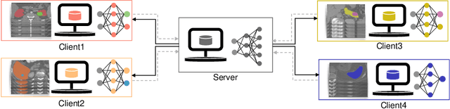
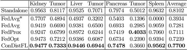
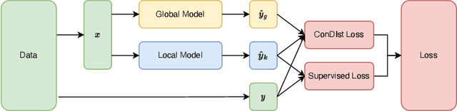
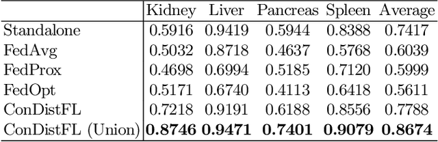
Developing a generalized segmentation model capable of simultaneously delineating multiple organs and diseases is highly desirable. Federated learning (FL) is a key technology enabling the collaborative development of a model without exchanging training data. However, the limited access to fully annotated training data poses a major challenge to training generalizable models. We propose "ConDistFL", a framework to solve this problem by combining FL with knowledge distillation. Local models can extract the knowledge of unlabeled organs and tumors from partially annotated data from the global model with an adequately designed conditional probability representation. We validate our framework on four distinct partially annotated abdominal CT datasets from the MSD and KiTS19 challenges. The experimental results show that the proposed framework significantly outperforms FedAvg and FedOpt baselines. Moreover, the performance on an external test dataset demonstrates superior generalizability compared to models trained on each dataset separately. Our ablation study suggests that ConDistFL can perform well without frequent aggregation, reducing the communication cost of FL. Our implementation will be available at https://github.com/NVIDIA/NVFlare/tree/dev/research/condist-fl.
Identifying Suspicious Regions of Covid-19 by Abnormality-Sensitive Activation Mapping
Mar 27, 2023This paper presents a fully-automated method for the identification of suspicious regions of a coronavirus disease (COVID-19) on chest CT volumes. One major role of chest CT scanning in COVID-19 diagnoses is identification of an inflammation particular to the disease. This task is generally performed by radiologists through an interpretation of the CT volumes, however, because of the heavy workload, an automatic analysis method using a computer is desired. Most computer-aided diagnosis studies have addressed only a portion of the elements necessary for the identification. In this work, we realize the identification method through a classification task by using a 2.5-dimensional CNN with three-dimensional attention mechanisms. We visualize the suspicious regions by applying a backpropagation based on positive gradients to attention-weighted features. We perform experiments on an in-house dataset and two public datasets to reveal the generalization ability of the proposed method. The proposed architecture achieved AUCs of over 0.900 for all the datasets, and mean sensitivity $0.853 \pm 0.036$ and specificity $0.870 \pm 0.040$. The method can also identify notable lesions pointed out in the radiology report as suspicious regions.