 Add to Chrome
 Add to Firefox
 Add to Edge
Point Tracking in Surgery--The 2025 Surgical Tattoos in Infrared Challenge (STIRC2025)
Jul 14, 2026
Point tracking in surgery is crucial to enable applications in downstream tasks such as segmentation, 3D reconstruction, virtual tissue landmarking, autonomous probe-based scanning, and subtask autonomy. This paper introduces the 2025 iteration of a point tracking challenge to address this, wherein participants submit their algorithms for quantification. Their algorithms are evaluated using a dataset named surgical tattoos in infrared (STIR), with the challenge named the STIR Challenge 2025 (STIRC2025). The STIR Challenge 2025 comprises two quantitative components: accuracy and efficiency. The accuracy component tests the accuracy of algorithms on in vivo and ex vivo sequences. The efficiency component tests algorithm inference latency. The challenge was conducted as a part of MICCAI EndoVis 2025, and seven teams participated in this challenge. In this paper we summarize the challenge results and participant methods. The challenge dataset is available at: https://zenodo.org/records/20191078, and the code for baseline models and metrics calculation is available here: https://github.com/athaddius/STIRMetrics

A deep learning-based method for prostate segmentation in T2-weighted magnetic resonance imaging
Jan 27, 2019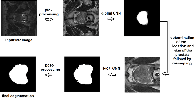

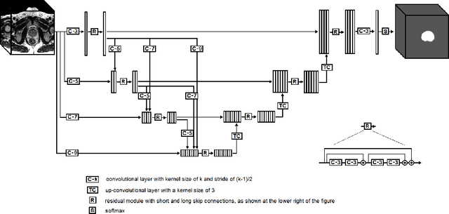
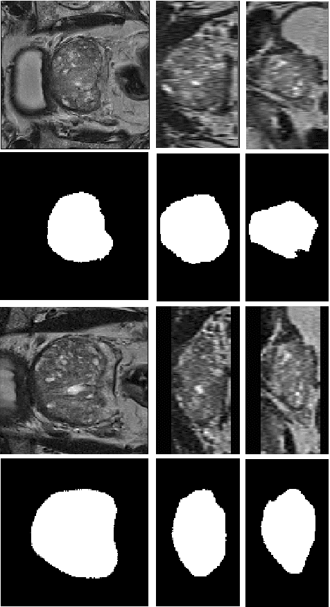
We propose a novel automatic method for accurate segmentation of the prostate in T2-weighted magnetic resonance imaging (MRI). Our method is based on convolutional neural networks (CNNs). Because of the large variability in the shape, size, and appearance of the prostate and the scarcity of annotated training data, we suggest training two separate CNNs. A global CNN will determine a prostate bounding box, which is then resampled and sent to a local CNN for accurate delineation of the prostate boundary. This way, the local CNN can effectively learn to segment the fine details that distinguish the prostate from the surrounding tissue using the small amount of available training data. To fully exploit the training data, we synthesize additional data by deforming the training images and segmentations using a learned shape model. We apply the proposed method on the PROMISE12 challenge dataset and achieve state of the art results. Our proposed method generates accurate, smooth, and artifact-free segmentations. On the test images, we achieve an average Dice score of 90.6 with a small standard deviation of 2.2, which is superior to all previous methods. Our two-step segmentation approach and data augmentation strategy may be highly effective in segmentation of other organs from small amounts of annotated medical images.